
Geert I. Pagenstert and Beat Hintermann
DEFINITION
 The plantaris tendon is an ideal source for soft tissue augmentation for ligament reconstructions or tendon repair.1,14
The plantaris tendon has high tensile strength with the structured collagen characteristic of physiologic tendons.
Harvest of the plantaris tendon rarely creates appreciable donor site morbidity.
The goal of this chapter is to provide the foot and ankle surgeon with a simple and reliable method to harvest readily available local tissue for plantaris tendon grafting even if its harvest was not originally planned.
ANATOMY
The plantaris muscle originates from the lateral femoral condyle, courses along the lower leg's superficial posterior compartment, and has its musculotendinous junction just distal to the level of the knee joint.
The proximal plantaris tendon is situated between the gastrocnemius and the soleus muscle. Distally, it lies immediately adjacent to the Achilles tendon in the distal third of the lower leg and typically inserts in the calcaneal tuberosity (FIG 1A).
The length of the plantaris tendon ranges from 30 to 45 cm.11
The plantaris tendon insertion is variable.3,4,11
 In addition to inserting on the calcaneal tuberosity, it may insert at the bursa calcanei, retinaculum flexorum, ankle capsule, plantar aponeurosis, or blend with the Achilles tendon or intermuscular septum (FIG 1B).
Because of this variability, distal harvesting procedures are unsuccessful in about 12% to 20% of cases.4,9,12
In about 6% to 7% of individuals, the plantaris tendon is absent. In humans, the plantaris muscle and tendon serve little function; however, early in evolution, it was far more developed in monkeys.3,8,10,11Therefore, harvesting the plantaris tendon in humans causes no appreciable donor site morbidity.2,7–9
Bohnsack et al1 compared highest tensile strength per cubic millimeter (N/mm3) of commonly used autografts:
Peroneus longus, 61 N/mm3
Peroneus brevis, 41 N/mm3
Plantaris, 94 N/mm3
Achilles split, 36 N/mm3
Fascia lata, 27 N/mm3
Periosteal flap, 2 N/mm3
Corium, 12 N/mm3
Anterior talofibular ligament, 8 N/mm3
They found the plantaris tendon to have the highest N/mm3.
PATHOGENESIS
Chronic joint instability and chronic tendon ruptures develop over months to years.
In joint instability, acutely torn ligaments eventually scar in an elongated state.
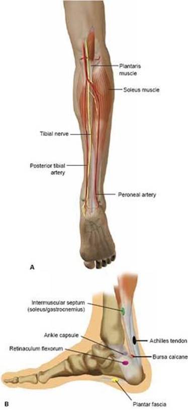
FIG 1 • Anatomy of the plantaris tendon. A. The only tubular structure between the soleus and the gastrocnemius muscles is the plantaris tendon. Therefore, blind dissection between these muscles does not risk nerves and vessels. Major vessels run underneath the soleus muscle. B. Insertions of the plantaris tendon are variable and may complicate the successful harvesting procedure.
Acute untreated tendon ruptures, similarly, eventually demonstrate degeneration and elongation.
Both chronic ligament and tendon injuries eventually may become so degenerated that surgical reconstruction is not possible with residual local tissue, especially in the case of chronically attenuated ligaments or tendons that have been subjected to repeated trauma or microtrauma. These situations lend themselves well to plantaris autografting.
PATIENT HISTORY AND PHYSICAL FINDINGS
Although isolated plantaris tendon ruptures have been reported, no specific physical examination technique has been developed that can isolate the plantaris tendon to confirm in advance that it is present when plantaris tendon harvest is being considered.
IMAGING AND OTHER DIAGNOSTIC STUDIES
Ultrasound is useful in identifying the presence of a plantaris tendon and has a high specificity.10
When MRI is used to evaluate an injury to the ankle ligament or tendon, the imaging study also may prove useful in identifying the presence of an ispsilateral plantaris tendon that may be used for tissue augmentation.7,8,13
SURGICAL MANAGEMENT
The plantaris tendon is ideal tissue for most ligament or tendon augmentations in the foot and ankle, especially because it typically is in the sterile field for virtually every foot and ankle procedure.
Preoperative Planning
If an MRI scan was obtained to determine the initial pathology, it can be used to identify the existence of the plantaris tendon before surgery.8,13
If not, a preoperative ultrasound examination is helpful.10,13
Positioning
Harvest of the plantaris tendon is relatively simple, regardless of which patient position is dictated by the particular foot and ankle procedure.
No tourniquet is needed for plantaris tendon harvesting.
Approach
The proximal approach is a 2-cm longitudinal incision about 30 cm proximal to the medial malleolus (FIG 2).
The distal approach is a 2-cm longitudinal incision at the medial Achilles insertion on the medial calcaneal tuberosity (FIG 3).

FIG 2 • The authors' preferred technique of proximal plantaris harvesting. A 2-cm incision is made at the proximal quarter of the medial calf.

FIG 3 • Distal plantaris harvesting using a 2-cm skin incision at the medial border of the Achilles tendon insertion at the medial calcaneal tuberosity.
TECHNIQUES
 The plantaris may be harvested from a proximal or distal approach; for both approaches, only a minimally invasive, 2-cm incision is required.
AUTHORS' PREFERRED TECHNIQUE FOR PROXIMAL PLANTARIS HARVESTING 6,7
Subcutaneous blunt dissection to the fascia is performed with care taken to protect the saphenous nerve and vein (TECH FIG 1A). A 2-cm longitudinal incision is made in the fascia to enable the surgeon's finger to enter the intermuscular space (TECH FIG 1B). The only tubular structure that runs between the gastrocnemius and the soleus muscle bellies is the plantaris tendon. No nerves or vessels are at risk.13
The tendon is mobilized with the finger or a nerve retractor (TECH FIG 1C).
The plantaris tendon is isolated in a distal direction using a blunt tendon stripper. The tendon stripper is advanced while maintaining the tendon under tension (TECH FIG 1D).
At the level of the calcaneus, the inner cylinder of the stripper is rotated to transect the tendon. The tendon is stored in a wet sponge for later use (TECH FIG 1E).
The fascia is reapproximated with absorbable suture, and wound closure is performed with a subcuticular running stitch (TECH FIG 1F). We routinely use adhesive strips to promote an optimal cosmetic appearance to the healed wound.

TECH FIG 1 • A. Blunt spreading of the subcutaneous fat down to the fascia without damage to the saphenous nerve or vein. B. A 2-cm fascial incision is made, and blunt dissection is carried out further down between the gastrocnemius and soleus muscles with the surgeon's finger. C. The plantaris tendon is developed with a nerve retractor. D. Introduction of the tendon stripper. E. The harvested autograft usually is about 30 cm long.
DISTAL PLANTARIS HARVEST
A 2-cm skin incision is made at the medial border of the Achilles tendon insertion at the calcaneal tuberosity (see Fig 3).
Blunt dissection is carried down to the Achilles tendon and the plantaris tendon. The variability of the plantaris insertions may necessitate a slightly more involved dissection to identify the distal extent of the plantaris tendon (see Fig 1B).
The tendon is mobilized and harvested with a blunt tendon stripper from distal to proximal (TECH FIG 2A).
At the level of the popliteal fossa, the inner cylinder of the stripper is rotated to transect the tendon (TECH FIG 2B). The tendon is stored in a wet sponge until it is needed for ligament or tendon augmentation.
The skin is closed with interrupted sutures and adhesive strips.

TECH FIG 2 • A. The plantaris tendon is located by blunt dissection. B. The tendon stripper is advanced from distal to proximal up to the popliteal fossa. Then the inner cylinder of the stripper is rotated to cut the tendon.

POSTOPERATIVE CARE
Adhesive wound strips can reduce skin tension and broadening of the scar.
After wound healing the patient may use scar massage to reduce subcutaneous adhesions.
OUTCOMES
The proximal harvesting procedure of the plantaris tendon was reported in a clinical study of plantaris tendon autograft for lateral ligament reconstruction of chronic ankle instability.7 In 52 cases (93%), a strong 25- to 35-cm tendon graft was harvested. In one case (2%) the plantaris tendon was deemed too weak to serve as appropriate donor material. In three (5%) of 56 ankle reconstructions the plantaris tendon could not be located during surgery without preoperative imaging or ultrasound. This observation was consistent with incidence studies of the plantaris tendon in cadavers (absence ranged between 6% and 7%).8,10,13
Use of the distal approach to harvest the plantaris tendon also has been reported. Investigators failed to locate the plantaris tendon distally in 12% to 20% of cases in these studies.4,9,12,13
COMPLICATIONS
We have performed proximal plantaris harvesting in 102 cases with only one case of mild dysesthesia at a broadened scar. This did not create any functional deficits for the patient.
Despite the close proximity of the saphenous nerve and vein, we have not observed any saphenous nerve or vein injuries.
In 36 patients we have performed a distal approach plantaris harvest. Four of these patients (11% of cases) developed a hypertrophic or hypersensitive scar that created shoe irritation.
REFERENCES
1. Bohnsack M, Surie B, Kirsch IL, et al. Biomechanical properties of commonly used autogenous transplants in the surgical treatment of chronic lateral ankle instability. Foot Ankle Int 2002;23:661–664.
2. Brunner R, Gaechter A. Repair of fibular ligaments: Comparison of reconstructive techniques using plantaris and peroneal tendons. Foot Ankle 1991;11:359–367.
3. Daseler EH, Anson BH. The plantaris muscle. An anatomical study of 750 specimens. J Bone Joint Surg 1943;25:822–827.
4. Harvey FJ, Chu G, Harvey PM. Surgical availability of the plantaris tendon. J Hand Surg Am 1983;8:243–247.
5. Hintermann B. Biomechanics of the unstable ankle joint and clinical implications. Med Sci Sports Exerc 1999;31:459–469.
6. Pagenstert GI, Hintermann B, Knupp M. Operative management of chronic ankle instability: Plantaris graft. Foot Ankle Clin 2006; 11:567–583.
7. Pagenstert GI, Valderrabano V, Hintermann B. Lateral ankle ligament reconstruction with free plantaris tendon graft. Tech Foot Ankle Surg 2005;4:104–112.
8. Saxena A, Bareither D. Magnetic resonance and cadaveric findings of the incidence of plantaris tendon. Foot Ankle Int 2000;21:570–572.
9. Segesser B, Goesele A. [Weber fibular ligament-plasty with plantar tendon with Segesser modification]. Sportverletz Sportschaden 1996;10:88–93.
10. Simpson SL, Hertzog MS, Barja RH. The plantaris tendon graft: An ultrasound study. J Hand Surg Am 1991;16:708–711.
11. Tillmann B, Toendury G. Flexorengruppe der unteren Extremität. In: Leonhardt H, Tillmann B, Toendury G, et al., eds. Bewegungsapparat, 3rd ed. Stuttgart-New York: Thieme, 1987:584–793.
12. Weber BG, Hupfauer W. Zur Behandlung der frischen fibularen Bandruptur und der chronischen fibularen Bandinsuffizienz. Arch Orthop Trauma Surg 1969;65:251–257.
13. Wening JV, Katzer A, Phillips F, et al. [Detection of the tendon of the musculus plantaris longus—diagnostic imaging and anatomic correlate]. Unfallchir 1996;22:30–35.
14. White WL. The unique, accessible and useful plantaris tendon. Plast Reconstr Surg 1960;25:133–141.