
Morgan N. Chen, Sheeraz A. Qureshi, and Andrew C. Hecht
DEFINITION
 The anterior approach can be used to access the thoracic spine for decompression, deformity correction, and stabilization. This approach allows for access to treat conditions such as intervertebral disc herniation, infection, tumor, and trauma.
ANATOMY
The thoracic spinal cord may have a tenuous blood supply, particularly in patients with congenital anomalies and kyphosis.
The midthoracic cord represents a watershed zone for vascularity. The artery of Adamkiewicz supplies the thoracic cord but can have a variable origin. Its origin is usually (80%) from the left side at the T10 level but can vary from T5 to L5.1
SURGICAL MANAGEMENT
Preoperative Planning
Radiographs of the thoracic spine and chest should be obtained to determine the level of surgery and help in “rib counting.”
 It is often helpful to obtain lumbar radiographs also to determine the number of lumbar segments below the most distal thoracic rib. Knowing this information preoperatively helps in counting “up” from the sacrum intraoperatively if needed.
In the absence of obvious bony pathology such as fractures, infections, or tumors, it is very easy to inadvertently localize the wrong level in the thoracic spine. The surgeon should be sure to have a strategy for intraoperative level identification based on careful scrutiny of radiographs and MRI or CT scans before surgery, understanding that the quality of portable films obtained intraoperatively may not be optimal.
When obtaining an MRI to better understand the nature of the pathology in relation to the thoracic spinal cord, the surgeon should ask for a topogram to be performed so that there is no question as to the level or levels of involvement.
On CT scan or MRI, the surgeon should pay close attention to the position of the aorta and inferior vena cava, especially on the axial cuts, as this may affect the side from which the spine is approached, especially if a corpectomy will be performed.
Anesthesia considerations include the use of an oral gastric tube and double-lumen endotracheal tube, which allows for collapse of the ipsilateral lung.
If the surgical site is T10 or distal, selective deflation of the ipsilateral lung is usually not necessary.
If the surgical site is proximal to T10, selective deflation is helpful in keeping the lung out of the field, but it may lead to more postoperative issues with atelectasis.
Neurologic monitoring is frequently used when performing thoracic operations.
Positioning
The patient should be in the lateral decubitus position with the arms in prayer position.
The thorax vertex should be positioned over the break of the bed, all pressure points should be padded, pillows should be placed between legs and arms, and an axillary roll should be used to prevent compression of the axillary vessels (FIG 1).
The operating surgeon typically stands behind the patient during the exposure. However, it may be helpful to stand in front of the patient when performing the decompression, as the line of sight into the spinal canal is better from that vantage point.
Approach (Right Versus Left)
Considerations for thoracic approaches include.
Approach from the side of herniation in cases of posterolateral or lateral herniation.
Look at the axial CT or MRI scans to determine the location of the heart and great vessels. In most thoracic cases, these structures are either on the left or central. Thus, all other factors being equal, a right-sided approach is favored in most cases.
In the distal thoracic spine (eg, T10–12), the liver may be in the way of a right-sided approach. Because it is a bit more difficult to retract the liver than the kidney or spleen, a leftsided approach may be favorable.
Considerations for thoracolumbar approaches include:
The left-sided approach is generally favored, as it is easier to mobilize the great arteries (aorta, iliacs) from their left-central position to the right, rather than mobilizing the great veins (which tend to be further to the right) toward the left.
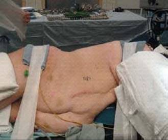
FIG 1 • Patient placed in the lateral decubitus position. It is important to ensure that all bony prominences are well padded.
TECHNIQUES
ANTERIOR THORACIC APPROACH FROM T1 TO T4
 For upper thoracic exposures a right-sided approach is preferred to avoid the heart.
The surgeon makes a curved skin incision below the tip of the scapula (TECH FIG 1A).
This incision is carried down to the latissimus dorsi muscle and then the latissimus is incised, leaving a cuff of the muscle on the scapula for later closure (TECH FIG 1B).
A large retractor (ie, Richardson retractor) can then be held by the assistant while the surgeon incises the periosteum over the appropriate rib and then resects the rib as far anteriorly and posteriorly as possible (TECH FIG 1C).
At this point, the chest is entered through the rib bed and a Finochietto or Omni retractor can be placed, with one of the blades holding the scapula up and out of the way.
 Now the lung can be deflated and retracted anteriorly and inferiorly (TECH FIG 1D).
The pleura overlying the spine is now sharply incised. Placing suture into the edges of the pleura makes subsequent closure easier.
Segmental vessels are identified and ligated as needed and the vertebral bodies (the “valleys”) and disc spaces (the “hills”) are identified.

TECH FIG 1 • Anterior thoracic approach from T1 to T4. A. A curved incision should be made just under the tip of the scapula. B. The incision is carried down to the latissimus dorsi. A cuff of muscle is left attached to the scapula for repair upon closure. C. The surgeon incises the periosteum over the rib. D. The deflated lung is retracted anteriorly and inferiorly while protecting the esophagus and great vessels.
ANTERIOR THORACIC APPROACH FROM T5 TO T12
The surgeon should plan the incision directly over the desired rib (ie, 10th rib for T9-10 disc). A curvilinear skin incision is made along the path of the rib from the anterior border of the latissimus dorsi to the costochondral junction anteriorly (TECH FIG 2A).
Due to the downslope of the ribs, it is generally preferable to make an incision that is more proximal rather than distal. If the incision is too distal, the ribs may impede access to the more proximal segment, necessitating a second thoracotomy. In contrast, it is easier to access levels that are distal to the rib that is resected. Thus, if in doubt as to the exact rib to be resected, the incision should be made more proximal.
Skin and subcutaneous fat are incised to expose the trapezius and latissimus dorsi.
The trapezius and latissimus dorsi are divided in line with the incision using electrocautery. The rhomboids may need to be split to gain more exposure cephalad.
Once the correct rib is identified, the surgeon divides the periosteum over the upper border of the rib to avoid injury to the intercostal nerve and vessels (TECH FIG 2B).
The rib is stripped subperiosteally anteriorly to the costochondral angle and as far posteriorly as possible (TECH FIG 2C).
The rib is removed with a rib cutter and passed off the field. The rib is cut at the midaxillary line anteriorly and as far posteriorly as possible. The rib can be used as a strut graft or autologous bone graft.
The periosteal rib sleeve and parietal pleura are incised to enter the thorax. A rib spreader is placed to hold the ribs apart (TECH FIG 2D).
The ipsilateral lung is deflated and retracted medially to expose the parietal pleura overlying the spine.
The parietal pleura overlying the spine is incised and retracted medially. Stitches can be placed in the parietal


TECH FIG 2 • Anterior thoracic approach from T5 to T12. (In these images, the patient's head is to the upper left and the patient's back is toward the surgeon.) A. The incision in planned directly over the rib. Injecting the subcutaneous tissues with a combination of anesthetic and epinephrine aids in hemostasis. B. The skin and subcutaneous tissues have been divided, exposing the desired rib. C. Subperiosteal exposure of the rib before excision. Note the thin parietal pleura beneath the rib bed. D. After excision of the rib the parietal pleura is entered, exposing the ipsilateral lung. E. The parietal pleura and the underlying segmental vessels. F. The vertebral bodies and intervertebral discs are exposed after segmental arteries are ligated and the overlying soft tissues are removed. Once the costotransverse and costovertebral articulations are excised (G), the rib head can be removed with a high-speed burr (H). pleura to make closure easier. The underlying segmental vessels are visualized (TECH FIG 2E).
The segmental arteries arising from the aorta can run in an ascending, recurrent, horizontal, or descending direction depending on the level of involvement.
The surgeon carefully ligates as few segmental vessels as possible to gain adequate exposure to the spine. Ligating more segmental vessels than necessary places the spinal cord at increased risk for ischemia because the thoracic spinal cord has a tenuous blood supply (TECH FIG 2F).
In cases of suspected vascular anomalies, such as congenital kyphosis, the surgeon should consider temporary occlusion of the segmental vessels and check evoked potentials before vessel ligation. If a patient has had a prior spine exposure on one side, the surgeon should be wary of ligating the contralateral segmental vessels. Instead, the surgery should be performed through the previously exposed side, or a preoperative angiogram should be obtained to identify the important arterial feeders to the spinal cord.
The intrathoracic vertebral bodies and intervertebral discs are now exposed. To gain access to the posterior intervertebral disc, the rib head may need to be removed.
The costotransverse and costovertebral articulations are removed to excise the rib head (TECH FIG 2G).
The soft tissues overlying the transverse process, pedicle, and vertebral body are removed.
The superior edge of the pedicle is identified and followed back to the intervertebral space.
The superior edge of pedicle is burred to expose the posterior intervertebral disc and lateral margin of the dura (TECH FIG 2H).
Detaching the Diaphragm
Exposure of T12-L1 may require detaching the diaphragm.
The diaphragm inserts and originates from the xiphoid and the inferior six ribs.
The lateral arcuate ligament arises from the transverse process of L1.
The crura extend more distally on the right.
The diaphragm is innervated centrally by the phrenic nerves.
The surgeon starts at the costal angle and incises the costodiaphragmatic reflection until extraperitoneal fat is visualized.
The diaphragm is divided off the anterior chest wall (TECH FIG 3). The surgeon should leave a 1to 2-cm cuff of diaphragm on the anterior chest wall to allow for diaphragm repair at closure. To avoid diaphragm denervation, the diaphragm should be incised only at its periphery. The diaphragm is split up to the lateral arcuate ligament.
The medial and lateral crura are detached, exposing the underlying peritoneum.
The peritoneum is swept medially until the retroperitoneal space is visualized.
The surgeon bluntly dissects and sweeps the fascia of Gerota medially to expose the spine and the overlying parietal pleura.
The aorta and vena cava are identified.
The surgeon can elevate the psoas muscle if needed.
The parietal pleura is incised to expose the spine.
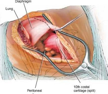
TECH FIG 3 • The diaphragm is incised circumferentially 2 cm from its peripheral attachment to the chest wall. Marker stitches should be placed for resuturing upon closure.
THORACOABDOMINAL RETROPERITONEAL LUMBAR SPINE APPROACH FROM T10 TO L3
The patient is positioned in the lateral decubitus position with the right side down. The approach should be made from the left side to avoid the liver and inferior vena cava.
The crura of the diaphragm are detached as described above.
An oblique incision is made from the quadratus lumborum to the lateral border of the rectus abdominis (TECH FIG 4).
This approach can be extended to L5 in most patients and even to S1 in those with low-riding iliac crests.
The subcutaneous tissue is incised and the fascia of the external oblique is divided.
The external and internal obliques, transverse abdominis, and transversalis fascia are incised.
The peritoneum is exposed and bluntly reflected anteriorly.
The ureter is identified and reflected anteriorly with retroperitoneal fat.
The vertebral bodies, psoas, and great vessels are identified.
The genitofemoral nerve lies on anterior psoas muscle, and excessive traction should be avoided.
The segmental vessels that lie over the middle of the vertebral bodies are identified and ligated.
The psoas is bluntly dissected off the vertebrae and retracted laterally.
The vertebral body, pedicle, and neuroforamen can be visualized.
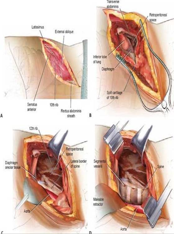
TECH FIG 4 • Thoracoabdominal approach. A. A curvilinear incision is made over the 10th rib and the muscle layers are identified. B. The retroperitoneal space is entered through the costal cartilage after removing the 10th rib. C. The light areolar tissue that signifies the retroperitoneal space is identified, and the peritoneum is mobilized from the undersurface of the diaphragm and abdominal wall as well as the aorta. D. Exposure of the spine is done after ligation of segmental vessels.
PEARLS AND PITFALLS

POSTOPERATIVE CARE
Chest tubes are left in place until output is less than 150 mL over 24 hours.
COMPLICATIONS
The exiting nerve root can be injured while removing the pedicle.
Vascular injury
Intercostal neuralgia
Atelectasis
Neurologic injury
Wrong-level surgery
Significant bleeding can be encountered when entering the epidural space.
Visceral injury
REFERENCE
1. Grace RR, Mattox KL. Anterior spinal artery syndrome following abdominal aortic aneurysmectomy: case report and review of the literature. Arch Surg 1977;112:813–815.