
James B. Carr
DEFINITION
 An intra-articular calcaneus fracture is an injury that involves the joint surfaces of the calcaneus, usually with displacement.
A fracture-dislocation of the calcaneus occurs when the posterior facet dislocates from beneath the talus and ends up displaced beneath the fibula. It carries a poor prognosis if treated nonoperatively.
“Soft tissue” damage refers to the injury to the skin, adipose, tendinous, muscular, and nerve structures that surround the calcaneus and ranges from mild bruising to near-amputation in open fractures.
 Fracture blisters and varying degrees of skin contusion occur most commonly.
“Wrinkle sign” refers to the skin wrinkles that appear when the injury swelling response is resolving.
A primary fracture line is one that occurs early in the mechanism of the calcaneal fracture. There are two that occur, and if their pathogenesis is understood, this can explain the majority of the pathology observed. This will be defined further in the Pathogenesis section.
ANATOMY
The calcaneus is the largest bone in the foot. It has a complex shape that makes exact surgical reconstruction difficult.
The calcaneus functions to transmit weight-bearing forces of the leg into the foot.
The calcaneus has a shock absorber function by assisting in mobility of the ankle and subtalar joints, thus allowing the foot to accommodate to variations in terrain.
The calcaneus has four articular facets that produce this mobility: posterior, anterior, middle, and cuboid. Exact articular alignment is required for full function of this four-joint complex.
The internal structure of the calcaneus reflects its weight-bearing role.
There is particularly dense trabecular bone in the juxtaarticular regions, especially below the posterior facet (the thalamic trabecular system).
The tendo Achilles insertion also has dense trabecular bone.
Cortical bone of 3 to 4 mm in thickness occurs in the superior-medial region (sustentaculum area) and in the superior-lateral strut of bone that runs between the cuboid and posterior facets (anterolateral fragment). These regions of cortical bone will come into play when discussing the internal fixation of the calcaneus (FIG 1).
The soft tissues of the calcaneus are easily damaged by trauma. Management of this injury component is essential to avoid iatrogenic surgical complications.
PATHOGENESIS
Despite the seemingly infinite varieties of fractures that occur, stereotypic fracture lines, fragments, and displacements can be recognized.
The calcaneus is fractured by a combination of shear and compression forces generated by the talus descending upon the calcaneus.
Two primary fracture lines occur.
The first occurs in the angle of Gissane and divides the calcaneus into anterior and posterior fragments. It can split either the middle or anterior facet, and the fracture continues on the lateral wall in an inverted Y shape (FIG 2).
The second fracture divides the calcaneus into medial and lateral halves and shears the posterior facet into two or more fragments.
 As the talus continues to compress the calcaneus, the lateral half of the posterior facet is impacted into the body of the calcaneus, with the recoil producing a step-off in the posterior facet.
This same fracture line commonly continues into the cuboid facet and, in combination with the first primary fracture line, produces the anterolateral fragment and superomedial fragment.
In this way, these two fracture lines produce fracture components that include the superomedial fragment, anterolateral fragment, posterior facet, and tuberosity.
Characteristic displacements of these components occur.
The tuberosity is driven up between the pieces of the posterior facet, can tilt into valgus or varus, and is usually translated laterally.
The lateral posterior facet fragments are impacted and rotated plantarly into the body of the calcaneus (FIG 3A).
The posterior facet breaks into one of three patterns, which form the basis of the Sanders classification:
Sanders II: two main pieces (FIG 3B)
Sanders III: three main pieces (FIG 3C)
Sanders IV: multifragmentary
The superomedial fragment retains alignment to the talus by means of its ligamentous attachments but can be subtly displaced by overlap with the anterior process. This overlap occurs along the primary fracture line that occurs in the sinus tarsi.
The anterolateral fragment displaces superiorly a variable amount. It typically extends into the cuboid facet, with varying degrees of displacement (FIG 3D).
The lateral calcaneal wall is displaced outward in the area of the trochlear tubercle. This, in combination with tuberosity translation, accounts for the heel widening and peroneal impingement that occur.
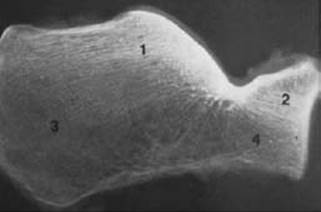
FIG 1 • A lateral radiograph of a calcaneus specimen sectioned in the sagittal plane. The trabecular systems are visualized and numbered 1 through 4. The densest bone is in the juxta-articular regions. Thick cortical bone also is present in the anterolateral fragment and medial wall in the sustentacular region. 1, thalamic trabecular system; 2, anterior apophyseal trabecular system; 3, anterior plantar trabecular system; 4, posterior plantar.

FIG 2 • Pattern of calcaneal fracture-dislocation and reduction.
The first fracture types recognized were the joint depression and tongue-type patterns, which are readily identified on a lateral heel radiograph.
The tongue fracture maintains a connection between the tuberosity and the posterior facet, while the joint depression separates the fractured joint surface from the tuberosity (FIG 4).
Because of this anatomy, certain tongue fractures have a large portion, or even the entire posterior facet, in continuity with the tuberosity (AO-OTA 73 C1). Thus, reduction of the tuberosity will reduce indirectly the posterior facet and restore the angle of Bohler. This particular pattern is well suited for small incision or percutaneous techniques.
FIG 3 • A. The posterior facet displaces and rotates in a plantar direction (arrow). B,C. CT scans showing a Sanders II fracture with a large superomedial fragment (B) and a Sanders III fracture (C). D. The primary fracture line extends into the calcaneal cuboid facet. The anterolateral fragment is represented by the most lateral piece (arrow). (D: Courtesy of Paul Tornetta III, MD.)

FIG 4 • Calcaneus fracture patterns: tongue (A) and joint depression (B).
Reduction of a joint depression pattern is best performed with an open reduction.
NATURAL HISTORY
An intra-articular fracture of the calcaneus is a serious injury that will diminish foot function.
Nonoperative treatment is with early motion and delayed weight bearing 6 to 8 weeks after injury. This method has the least chance of iatrogenic injury.
In a classic review by Lindsay and Dewar, only 17% of patients had no foot symptoms with long-term follow-up.
The loss of ability to perform manual labor is common, with an average time off work of 4 to 6 months for laborers.
Loss of subtalar motion to varying extents will occur.
Tibiotalar impingement and anterior ankle pain can be produced if the crush deformity is severe enough.
It can take 18 to 24 months for the foot symptoms to maximally improve after this injury. Most improvement occurs in the first 12 months.
The key concept here is that patients who continue to improve symptomatically can be observed until maximum improvement occurs.
A recent randomized, prospective study found that the need for late subtalar arthrodesis is five to six times greater if nonoperative treatment is used on all injuries. The overall rate was approximately 17%.
PATIENT HISTORY AND PHYSICAL FINDINGS
The history is typically one of a fall or vehicle crash. Occasionally in a diabetic, a seemingly trivial ankle sprain-type mechanism can occur. Important risk factors for operative treatment complications include smoking, diabetes, peripheral vascular disease, and steroid use. The foot and ankle are visually inspected.
Swelling is graded as mild, moderate, or severe.
Operative treatment in the face of severe soft tissue swelling is prone to wound healing complications.
Fracture blisters are graded as fluid-filled or blood-filled. If unhealed, fracture blisters are a source of skin bacterial colonization. Blood-filled blisters denote a deeper dermal injury.
Skin contusion is noted.
If present, the wrinkle sign is noted. It means the swelling is resolving and surgical incisions are less likely to experience complications.
Open wounds are noted.
The physician palpates the foot and ankle, looks for spine injuries or ipsilateral fractures, and performs a secondary survey for other injuries.
Spine injuries are said to accompany up to 10% of all calcaneal fractures.
The physician assesses for compartment syndrome, including passive flexion and extension of the toes, tenseness of the foot swelling, and compartment pressure measurement.
Positive results are degree of pain elicited and pressures within 30 mm of diastolic blood pressure.
Compartment syndrome can occur in 5% to 10% of all calcaneal fractures.
The physician performs a neurologic examination to check the sensory function of foot and toes, including light touch and pinprick.
Calcaneal fractures can damage the posterior tibial nerve and occasionally sensory nerves. Findings may be altered with compartment syndrome.
IMAGING AND OTHER DIAGNOSTIC STUDIES
Anteroposterior and lateral (FIG 5A,B) foot radiographs are the initial screening study.
The axial (Harris) view should also be obtained (FIG 5C). This view will demonstrate the medial wall and show the relation of the superomedial fragment to the tuberosity.
Broden views are radiographs that focus on the subtalar joint. They are taken with the foot internally rotated, and the x-ray beam angled to varying degrees cephalad (FIG 5D,E). By using different degrees of cephalad angulation, different parts of the posterior facet may be imaged. They are best used intraoperatively to judge the reduction of the posterior facet and the medial wall of the calcaneus.

FIG 5 • A,B. Lateral radiographs showing a severely displaced tongue-type Sanders II fracture of the calcaneus with midfoot dislocation (A) and a Sanders III fracture (B). C. The axial Harris view demonstrates displacement of the medial wall (arrow). Reduction of this pathoanatomy is the basis for the medial approach. D. Broden views are obtained by centering the x-ray beam on the posterior facet, internally rotating the foot, and directing the beam cephalad. E. As the beam is aimed cephalad, more anterior portions of the posterior facet are visualized. Also note the medial wall on profile. As the x-ray is tilted more, the view becomes a true Harris axial view (if the internal rotation is removed).
If the fracture is displaced, a computed tomography (CT) scan is recommended to define the anatomy (see Fig 3B,C).
A CT scan with biplanar cuts and reconstructions is recommended. This will best delineate the fragments and displacements.
DIFFERENTIAL DIAGNOSIS
Fracture of the talus
Lateral process of talus fracture
Fracture of the midfoot (eg, navicular fracture)
Severe ankle sprain
Subtalar dislocation
Stress fractures of the calcaneus can masquerade as a soft tissue disorder of the hindfoot (eg, plantar fasciitis).
NONOPERATIVE MANAGEMENT
The indications for nonoperative treatment include posterior facet displacement less than 2 mm, and medical conditions such as peripheral vascular disease or diabetes.
Some surgeons consider smoking a relative contraindication; it certainly predisposes to a higher wound complication rate.
Severe fracture blisters or closed soft tissue injury can preclude operative treatment, although open reduction and internal fixation can be performed as late as 4 weeks after injury.
The recommended nonoperative treatment is compression wrapping, early motion, and delayed weight bearing at 6 to 8 weeks after injury. This offers the least iatrogenic risk to the patient while optimizing chances for subtalar motion.
Once weight bearing is started, the patient continues with range-of-motion exercises.
Strengthening of the foot and ankle muscles is added as fracture consolidation progresses.
A well-cushioned shoe usually offers the best pain relief. Two different shoe sizes may be needed in extreme cases.
A rocker-bottom sole can be added to assist with the toe-off stance phase of gait.
A double upright brace with a rocker-bottom-soled shoe can assist ambulation in patients with severe injury who have impairment upon fracture healing.
Nonoperative treatment is not recommended for calcaneal fracture dislocations, as a painful deformed foot is practically guaranteed if it is left unreduced.
SURGICAL MANAGEMENT
The displaced intra-articular calcaneal fracture presents a difficult challenge.
Foot pain and stiffness are common even with the best of treatment, and iatrogenic problems such as infection can result in loss of limb in extreme circumstances, and at the least predispose to a poor result.
Thus, a careful, individualized approach is recommended, with a priority on avoiding iatrogenic problems while attaining an anatomic alignment of the calcaneus.
Indications include displacement of the posterior facet of more than 2 mm, and calcaneus fracture dislocation.
Research shows that certain patient groups, such as those receiving worker's compensation, are predisposed to a poor result with operative treatment, but that does not obviate the benefits of obtaining anatomic foot alignment and lessening the chances of late subtalar fusion.
Operative restoration of at least the calcaneal shape should be considered for fractures with severe displacement (eg, tuberosity displaced superiorly behind the ankle joint), as late reconstructions can be difficult.
The choice of any surgical approach or technique should always have the goal of total anatomic restoration, although extreme comminution can compromise attainment of this goal.
Preoperative Planning
Once operative treatment has been elected, the surgical approach is chosen based on a number of factors, including the surgeon's training and experience and the pathoanatomy present.
The timing is in general when the wrinkle sign develops, typically 7 to 14 days after injury. Fracture blisters should be epithelialized.
The injury pathoanatomy is analyzed first by looking at the posterior facet pattern (Sanders II, III, or IV), displacement, and location of the primary fracture line in the posterior facet.
Fractures that are more medial are more difficult to visualize, and more fragments involving the posterior facet are more difficult to fixate anatomically.
Fractures that separate the entire posterior facet and have a tongue pattern are amenable to percutaneous Essex-Lopresti techniques.
Conversely, joint depression fractures require open reduction of the posterior facet.
A highly comminuted Sanders IV fracture may alter the goals to restoration of the calcaneal body shape and primary fusion.
The other fracture components to be analyzed for displacement are the superomedial fragment, anterolateral fragment, and tuberosity. The surgical plan should address each of these pathologies for reduction strategy and fixation.
The typical reduction order is first to correct any superomedial fragment subluxation.
Next, the superomedial fragment is reduced and held to the tuberosity.
The posterior facet is then reduced and fixed.
Finally, the anterolateral fragment is reduced and fixed.
The size and integrity of the superomedial fragment is critical, as fixation techniques largely center on screw placement into its substance. A small or comminuted superomedial fragment makes rigid fixation harder to achieve and may call for alternative techniques.
Restoration of the superomedial fragment to the tuberosity will restore the calcaneal shape and make room for reduction of the displaced posterior facet fragments.
The superomedial fragment may be incarcerated in the sinus tarsi and subtly subluxated. This is recognized by the preoperative CT scan on the sagittal reconstructions, and by the lack of congruence of the superomedial fragment with the undersurface of the talus.
Failure to correct this subluxation makes posterior facet reduction very difficult.
The anterolateral fragment should key into location just in front of the reduced posterior facet and restores lateral column length.
It can be fixed with either lag screws into the superomedial fragment, or a mini-fragment plate. Some of the perimeter plates have a small extension to pull this fragment into place.
The fixation chosen depends on the approach taken. Fractures splitting the posterior facet will require lag screws inserted from lateral to medial; they range in size from 2 to 4 mm, depending on the fractures present.
Sanders III fractures are converted into two major pieces with the use of countersunk mini-fragment screws that fix the intermediate piece to the more medial piece.
Extra-long mini-fragment screws are desirable to reach the medial cortical bone.
The plate chosen depends on the approach.
The extensile lateral approach will require some type of low-profile “perimeter” plate.
Strategic placement of small and mini-fragment plates, and occasionally lag screws alone, is used in small-incision techniques.
Plans must be made for imaging, most typically fluoroscopy. This will allow control of the AP, lateral, axial, and Broden views intraoperatively.
Arthroscopy can also help visualize the posterior facet, especially in its anterior portions.
Positioning
The extensile lateral approach is performed in the lateral decubitus position with the injured foot on top. A thigh tourniquet is applied.
The fluoroscope is brought in from the side opposite the surgeon regardless of the surgical approach.
The same position can be used for percutaneous manipulations of tongue fractures. This allows conversion to the extensile lateral approach, as recommended by Tornetta.
Small-incision approaches are performed supine with a bump under the ipsilateral hip. A tourniquet is placed but not routinely inflated.
For small-incision techniques, the patient is pulled down to the end of the table. The point of the heel should project slightly beyond the end of the bed. This allows for placement of axially directed implants.
If used, the arthroscope is placed with the monitor on the same side as the C-arm, toward the head of the bed.
Approach
Small-incision techniques will address most calcaneal pathologies but require a firm understanding of the fragments, displacements, and deforming forces present.
They are ideally suited to Sanders II fractures with a large superomedial fragment and only mild to moderate posterior facet displacement.
The extensile lateral approach is applicable to all fracture patterns and displacements. Its use in open fractures warrants caution with respect to soft tissue complications.
TECHNIQUES
PERCUTANEOUS REDUCTION AND FIXATION OF TONGUE FRACTURE
 If performed acutely, percutaneous reductions can be technically easier and do not increase the risk of infection in my experience.
 If there is any question, the surgeon should use the presence of the wrinkle sign and healing of fracture blisters.
This technique is ideally indicated for tongue patterns that have a large percentage of the posterior facet connected to the tuberosity (Sanders IIC) (TECH FIG 1A).
The technique can be used for Sanders IIA and IIB patterns, but the facet reduction is more difficult if done percutaneously.
I prefer to perform this surgery supine, with the addition of a sinus tarsi incision for failure of the percutaneous reduction.
Tornetta prefers the lateral position, with conversion to the extensile lateral approach if percutaneous manipulations are unsuccessful.

TECH FIG 1 • A. A displaced tongue fracture demonstrates the typical displacement and location of an injury amenable to percutaneous reduction. The anterior process is not comminuted. The medial fragment was small. B.Introduction of a large smooth pin to manipulate the fracture. Note the incomplete reduction in the angle of Gissane, and posteriorly where the tongue fracture exits. C. Multiple wires for cannulated screws have been placed. The anterior process is not comminuted and can be used for screw purchase. D. Reduction obtained and screw placement. E–G. For this Sanders II fracture, after repair of the navicular fracture, a small incision was made in the plantar lateral foot, localizing the area beneath the angle of Gissane. E. A 5-mm pin was placed on a Synthes (Paoli, PA) T-handled chuck. The pin was then placed through the stab incision and advanced just inside the lateral wall of the calcaneus. At the same time, a cannulated screw guidewire was placed in the plantar portion of the calcaneus. F. With the manipulating pin applying a firm upward pressure with the foot plantarflexed, the wire was driven from the tuberosity into the tongue fragment. This was replaced with a 4.0-mm cannulated screw. G. A second cannulated screw was placed from the lateral calcaneus into the superomedial fragment.
The patient is placed supine with a generous bump under the ipsilateral hip to assist access to the heel.
0.25% Marcaine with epinephrine is injected into the fracture hematoma and soft tissues. A popliteal block is also placed by the anesthesia team. The combination of these two blocks will allow for outpatient surgery management of this injury.
A 1/8 Steinmann pin is introduced into the calcaneus from the posterior tuberosity into the region just beneath the posterior facet.
The pin is then used as a levering tool to restore the Bohler angle of the fractured calcaneus (TECH FIG 1B).
Taking a lateral view of the normal heel and saving it on the fluoroscope provides a comparison to judge reduction.
Once the fracture is reduced, one or two cannulated screws are introduced from the tuberosity into the anterior process of the calcaneus (TECH FIG 1C,D).
Alternative or adjunctive fixation strategies include placing a 4.0-mm screw from the plantar tuberosity into the dorsal calcaneus surface. This resists plantar displacement of the tongue fragment.
Another lag screw possibility is one directed from the lateral calcaneus into the superomedial fragment (TECH FIG 1E–G). This is more difficult in this pattern because by definition it has a small superomedial fragment.
OPEN REDUCTION
If the Bohler angle is not reducible or if a step-off remains in the posterior facet, an open reduction is performed.
I prefer a small sinus tarsi incision approach to aid in the reduction.
A 4- to 6-cm sinus tarsus incision is made to expose the posterior facet, the anterolateral fragment, and a portion of the lateral calcaneal wall (TECH FIG 2).
The posterior facet is reduced under direct vision, and the reduction is confirmed with fluoroscopy. An arthroscope is helpful as well.
A traction pin in the tuberosity can help restore calcaneal height.
Lateral to medially directed lag screws are placed across the posterior facet. A mini-fragment plate is used to bridge the posterior facet to the anterolateral fragment.
The lateral wall should be manually compressed at this point.
Consideration can be given to adding a calcium phosphate cold hardening composite to provide extra support.
Layered closure is performed.
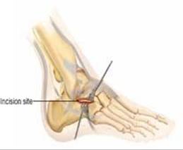
TECH FIG 2 • Lateral approach for the small incision technique.
SIMULTANEOUS MEDIAL AND LATERAL APPROACHES
In general, except for open fractures, timing should be guided by the presence of the wrinkle sign and healing of fracture blisters.
This technique is ideally indicated for Sanders II fractures with 2 to 10 mm of displacement of the posterior facet, and a large superomedial fragment. It can be applied to nearly any fracture pattern, but the limited exposure makes posterior facet reduction more difficult for Sanders III and IV patterns.
A generous bump is placed under the ipsilateral hip. The heel is left slightly off the end of the bed to facilitate the placement of axially directed fixation.
0.25% Marcaine with epinephrine is injected into the fracture hematoma and soft tissues. A popliteal block is also placed by the anesthesia team.
The combination of these two blocks will allow for outpatient surgery management of this injury.
Incisions and Dissection
The medial approach is posterior and parallel to the neurovascular bundle (TECH FIG 3A).
The medial calcaneal sensory branch is identified deep to the flexor retinaculum and preserved. This directly exposes the superomedial fragment and keeps the neurovascular bundle in the anterior flap.
The lateral approach extends anteriorly 4 to 6 cm from the tip of the fibula (see TECH FIG 2).
This will provide exposure of the posterior facet and anterolateral fragment.
It is performed after the medial approach.
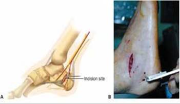
TECH FIG 3 • A. Medial approach for the small-incision technique. B. A tensioned 1.6-mm smooth Kirschner wire is placed in the inferior tuberosity and is used to apply traction to the calcaneus. The heel is slightly off the end of the bed to facilitate placement of axial fixation.
At this point, all fracture fragments are identified and cleaned of debris.
The posterior facet is partly reduced to avoid obstruction of the superomedial fragment and tuberosity reduction.
A 1.6-mm Steinmann pin is introduced into the tuberosity of the calcaneus in a medial-to-lateral direction. Tensioned with a Kirschner bow, it allows for correction of shortening (TECH FIG 3B).
The medial fracture fragments are cleaned of debris, and landmarks for reduction are identified.
Medial Reduction and Fixation
Reduction and fixation can be done with one of two strategies.
The first is with an antiglide 2.7-mm plate.
One can predrill a hole on the tuberosity fragment next to the fracture site and to the length measured.
With use of distraction and manipulation, an approximate reduction of the superomedial fragment and tuberosity is obtained, particularly with respect to length.
A 2.7-mm five-hole T plate is then placed on the bone and the premeasured screw inserted. As the plate tightens to the bone, it will help reduce any tuberosity translation (TECH FIG 4).
The reduction is checked by fluoroscopy in all planes.
If satisfactory, additional screws can be inserted, taking care to avoid the posterior facet.
The second method is to obtain a reduction by traction and translation of the tuberosity.
One can then introduce axial cannulated screws—one up the inside of the medial wall and the other as a lag screw from the inferior lateral tuberosity into the superomedial fragment.
This latter lag screw is a useful adjunct to a medial antiglide plate.
Lateral Reduction and Fixation
Once the medial side is reduced, the lateral side is addressed.
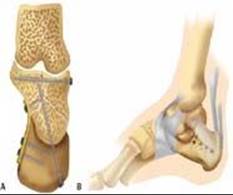
TECH FIG 4 • Axial (A) and medial (B) views of fixation with a medial plate and lateral screws.
The posterior facet is manipulated and reduced. The reduction is checked with Broden views and the arthroscope (TECH FIG 5).
It is common to approximate one portion of the facet, only to have another portion malreduced.
Once an anatomic reduction is obtained, provisional fixation with Kirschner wires is performed. Two lateral-tomedial–directed lag screws are then inserted just beneath the articular surface of the posterior facet.
The anterolateral fragment will now reduce anatomically to the posterior facet. It can be fixated with either a mini-fragment plate that bridges from the posterior facet to the anterolateral fragment, or a lag screw from the anterolateral fragment to the superomedial fragment.
The lateral wall should be manually compressed at this point.
Consideration can be given to adding a calcium phosphate cold hardening composite to provide extra support.
Layered closure is performed.

TECH FIG 5 • The arthroscope is introduced through the open lateral wound to provide magnification and visualization of the subtalar surface.
EXTENSILE LATERAL APPROACH
Timing should be guided by the presence of the wrinkle sign and healing of fracture blisters. This can take up to 3 weeks.
This technique is indicated for all types of calcaneal fractures with intra-articular displacement. Open fractures are best approached in a staged fashion.
The lateral decubitus position is used, with the injured side up. The C-arm comes in opposite the surgeon.
0.25% bupivacaine with epinephrine is injected into the fracture hematoma and soft tissues. A popliteal block is also placed by the anesthesia team. Tourniquet control is required.
An L-shaped incision is made parallel to the Achilles tendon and curves anteriorly along the border of the plantar skin of the heel (TECH FIG 6A).
The incision is made to bone, with a subperiosteal and periosteal flap raised along its entire extent. In the region of the peroneal tendon sheath attachment, the tendons are retracted anteriorly and the dissection is continued on the deep layer, above the abductor fascia.
Once the entire lateral side of the calcaneus is exposed, two Kirschner wires can be driven into the talus to provide retraction of the flap (TECH FIG 6B).
The lateral wall is entered and the posterior facet elevated from its displaced position (TECH FIG 6C).
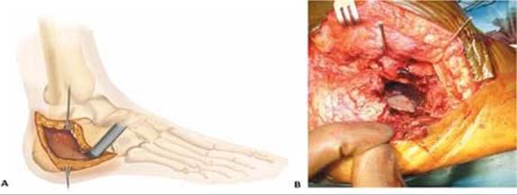

TECH FIG 6 • A. Incision for the extensile lateral approach. B. The lateral flap has been raised. The Kirschner wires in the talus serve to retract the soft tissues and assist exposure. C. After removal of the lateral wall, the depressed posterior facet is clearly seen. D. Kirschner wires provide provisional fixation, and a plate is contoured to the superior calcaneus, behind the posterior facet. The remainder of the plate is left flat to prevent varus displacement of the tuberosity. E. Intraoperative lateral radiograph with the Kirschner wires and plate in place. F. Lateral view of plate showing screw placement. G. Final postoperative lateral radiograph. The screws are aimed into the juxta-articular regions of the calcaneus.
If the superomedial fragment is subluxated by the anterior process, this is corrected by leverage and a Kirschner wire driven across the reduced fragments.
A Schanz screw is driven into the tuberosity to facilitate manipulation of the fragments. Alternatively, a tensioned wire can be used.
Attention is turned to the medial wall, where the superomedial fragment is identified.
With traction and manipulation of the pieces, the medial wall is reduced and pinned with axial Kirschner wires that travel just inside the medial wall of the calcaneus.
The posterior facet can now be reduced and provisionally fixated with Kirschner wires, and lateral-to-medial–directed lag screws are placed. Broden views are essential to ensure anatomic reduction.
The anterolateral fragment is reduced to the posterior facet and pinned into the superomedial fragment.
A perimeter plate is now applied to the lateral surface of the calcaneus (TECH FIG 6D,E). Contouring of the plate is not recommended, except in the area next to the curved posterior portion of the posterior facet if needed. It functions in a sense as a giant washer, serving to compress the calcaneus from lateral to medial.
Screws are directed into the tuberosity and the juxta-articular areas of the calcaneus (TECH FIG 6F,G). The superomedial fragment usually offers the best bone for screw fixation, along with the region beneath the posterior facet.
Care is taken to avoid penetrating the medial cuboid facet (which curves toward the tuberosity) and the medial posterior facet (which curves toward the plantar surface).
Careful layered closure is performed on the wound.
Hemostasis is obtained before closure and consideration is given to placing a deep drain or wound vacuum over the suture line. Simple running stitch is sufficient for most cases, although some surgeons prefer Allgöwer-Donati–type mattress skin closure.
A well-padded short-leg cast is applied and split to allow for swelling.
POSTOPERATIVE CARE
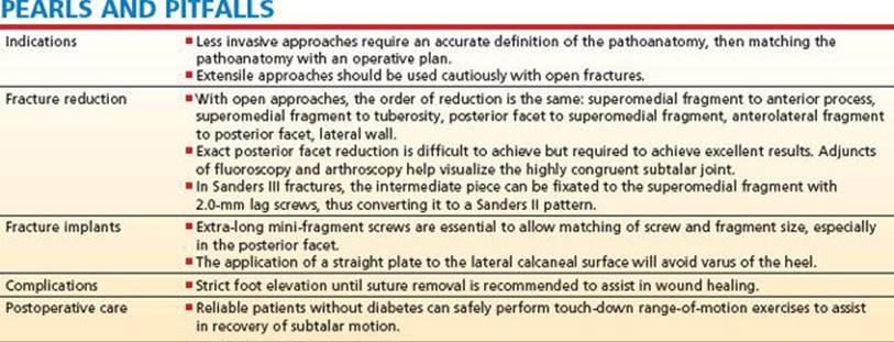
A well-padded short-leg cast is applied, split, and over-wrapped with a loosely applied elastic wrap.
The patient is instructed to maintain strict elevation as much as possible until the sutures are removed.
The same cast is retained until the sutures are removed 2 weeks postoperatively.
In reliable nondiabetic patients, the cast can be discontinued and range-of-motion exercises begun.
Touch-down weight bearing to promote ankle and subtalar motion can be started at the same time.
Physical therapy is prescribed on an individualized basis.
At 6 weeks, a radiograph is obtained and weight bearing progressed as pain allows. Full weight bearing is expected by 12 weeks postoperatively.
OUTCOMES
Despite appropriate care, most patients with a calcaneal fracture will lose some degree of foot function and have permanent symptoms.
While nonoperative treatment yields the fewest iatrogenic complications, it accepts malunion in nearly 100% and a higher incidence of later subtalar fusion.
Symptom improvement can take up to a year to plateau.
In a recent randomized study, visual analog pain scores between nonoperative and operative groups were similar, but nonoperative treatment resulted in a 5.5 times greater incidence of late subtalar fusion.
In that same study, females, non-workmen's-compensation cases, and nonmanual laborers had improved results with operative treatment.
Better results were also seen with an anatomic reduction versus a nonanatomic one.
Soft tissue complications frequently lead to a poor result.
Amputations have been reported with extensile lateral approaches.
Open techniques should be used cautiously in diabetic patients, although injuries such as fracture-dislocation are best treated with operative reduction and fixation.
COMPLICATIONS
The complications of nonoperative treatment include malunion, persistent foot pain, and a higher chance of later subtalar fusion.
Severe crush deformities affect not only the subtalar joint, but the midfoot and ankle as well. They can be difficult to reconstruct, so initial management to avoid such a malunion is recommended.
Smoking, diabetes, and open fracture are the most significant risk factors for soft tissue complications.
Infection occurs in about 2% of fractures treated operatively with open incisions.
Flap necrosis can occur with any incision but is most likely with the extensile lateral approach. Débridement and closure by secondary intention is often successful for minor flap losses. If a large portion of the flap is lost, consultation with a plastic surgeon is recommended.
Deep infection is managed with débridement and intravenous antibiotics based on culture results.
Retention of hardware (if providing bone stability) until bone healing is optimal.
Removal of the hardware to eradicate the infection once the bone is healed is sometimes needed.
Posterior tibial nerve injury can result from the fracture and commonly presents with severe pain nonresponsive to narcotics in the postinjury period.
Administration of medications aimed at neuropathic pain is recommended, and consultation with a pain specialist is considered.
Cushioned shoe inserts are often comforting to individuals with postfracture plantar heel pain. A rocker-bottom shoe can also reduce discomfort.
Late implant-related symptoms are rare with percutaneous or small-incision techniques. They are lessened by the use of low-profile “perimeter” plates with the extensile lateral approach.
REFERENCES
1. Abidi N, Dhawan S, Gruen G, et al. Wound-healing risk factors after open reduction and internal fixation of calcaneal fractures. Foot Ankle 1998;19:856–861.
2. Benirschke S, Sangeorzan B. Extensive intraarticular fractures of the foot: surgical management of calcaneal fractures. Clin Orthop Relat Res 1993;290:128–134.
3. Bézes H, Massart PL, Delvaux D, et al. The operative treatment of intraarticular calcaneal fractures: indications, technique and results in 257 cases. Clin Orthop Relat Res 1993;290:55–59.
4. Buckley R, Tough S, McCormack R, et al. Operative compared with nonoperative treatment of displaced intra-articular calcaneal fractures: a prospective, randomized, controlled multicenter trial. J Bone Joint Surg Am 2002;84A:1733–1744.
5. Burdeaux B. Fractures of the calcaneus: open reduction and internal fixation from the medial side: a 21-year prospective study. Foot Ankle 1997;18:685–692.
6. Burdeaux B. Reduction of calcaneal fractures by the McReynolds medial approach technique and its experimental basis. Clin Orthop Relat Res 1983;177:87–103.
7. Carr JB. Mechanism and pathoanatomy of the intraarticular calcaneal fracture. Clin Orthop Relat Res 1993;290:36–40.
8. Carr JB. Surgical treatment of intra-articular calcaneal fractures: a review of small incision approaches. J Orthop Trauma 2005;19: 109–117.
9. Carr JB. Surgical treatment of intra-articular calcaneal fractures: a review of small incision approaches. J Orthop Trauma 2005;19: 109–117.
10. Carr JB, Hamilton J, Bear L. Experimental intra-articular calcaneal fractures: anatomic basis for a new classification. Foot Ankle 1989; 10:81–87.
11. Carr JB, Scherl J. Small incision approach for intraarticular calcaneal fractures. Presented at: Orthopaedic Trauma Association annual meeting; 1998; Toronto, Ontario, Canada.
12. Carr JB, Tigges R, Wayne J, et al. Internal fixation of experimental calcaneal fractures: a biomechanical analysis of two fixation methods. J Orthop Trauma 1997;11:425–429.
13. Ebraheim N, Elgafy H, Sabry F, et al. Sinus tarsi approach with trans-articular fixation for displaced intra-articular fractures of the calcaneus. Foot Ankle 2000;21:105–113.
14. Folk J, Starr A, Early J. Early wound complications of operative treatment of calcaneus fractures: analysis of 190 fractures. J Orthop Trauma 1999;13:369–372.
15. Fernandez D, Koella C. Combined percutaneous and “minimal” internal fixation for displaced articular fractures of the calcaneus. Clin Orthop Relat Res 1993;290:108–116.
16. Gupta A, Ghalambor N, Nihal A, Trepman E. The modified Palmer lateral approach for calcaneal fractures: wound healing and postoperative computed tomographic evaluation of fracture reduction. Foot Ankle 2003;24:744–753.
17. Johnson E, Gebhardt J. Surgical management of calcaneal fractures using bilateral incisions and minimal internal fixation. Clin Orthop Relat Res 1993;290:117–124.
18. Koski A, Koukkanen H, Tukiainen E. Postoperative wound complications after internal fixation of closed calcaneal fractures: a retrospective analysis of 126 consecutive patients with 148 fractures. Scand J Surg 2005;94:243–245.
19. Letrounel E. Open treatment of acute calcaneal fractures. Clin Orthop Relat Res 1993;290:60–67.
20. Levine D, Helfet D. An introduction of the minimally invasive osteosynthesis of intra-articular calcaneal fractures. Injury 2001;32: S-A51–54.
21. Lindsay WR, Dewar FP. Fractures of the os calcis. Am J Surg 1958;95(4):555–576.
22. McReynolds J. The surgical treatment of fractures of the os calcis. Orthop Trans 1982;3:415.
23. Rammelt S, Amlang M, Barthel S, et al. Minimally-invasive treatment of calcaneal fractures. Injury 2004;35:S-B55–63.
24. Rammelt S, Gavlik J, Barthel S, et al. The value of subtalar arthroscopy in the management of intra-articular calcaneus fractures. Foot Ankle 2002;23:906–916.
25. Raymakers J, Dekkers G, Brink P. Results after operative treatment of intra-articular calcaneal fractures with a minimum follow-up of 2 years. Injury 1998;29:593–599.
26. Sanders R. Displaced intra-articular fractures of the calcaneus. J Bone Joint Surg Am 2000;82A:225–250.
27. Sanders R, Fortin P, DiPasquale T, et al. Operative treatment in 120 displaced intraarticular calcaneal fractures: results using a prognostic computed tomography scan classification. Clin Orthop Relat Res 1993;290:87–95.
28. Stephenson J. Surgical treatment of displaced intraarticular fractures of the calcaneus: a combined lateral and medial approach. Clin Orthop Relat Res 1993;290:68–75.
29. Thordarson D, Krieger L. Operative vs. nonoperative treatment of intra-articular fractures of the calcaneus: a prospective randomized trial. Foot Ankle 1996;17:2–9.
30. Thordarson D, Latteier M. Open reduction and internal fixation of calcaneal fractures with a low profile titanium calcaneal perimeter plate. Foot Ankle 2003;24:217–221.
31. Tornetta P. Open reduction and internal fixation of the calcaneus using minifragment plates. J Orthop Trauma 1996;10:63–67.
32. Tornetta P. Percutaneus treatment of calcaneal fractures. Clin Orthop Relat Res 2000;375:91–96.
33. Tornetta P III. The Essex-Lopresti reduction for calcaneal fractures revisited. J Orthop Trauma 1998;12:469–473.
34. Zwipp H, Tscherne H, Therman H, et al. Osteosynthesis of displaced intraarticular fractures of the calcaneus: results in 123 cases. Clin Orthop Relat Res 1993;290:76–86.