
W. GRANT STEVENS, ANDREA E. VAN PELT, AND ADRIAN M. PRZYBYLA
INTRODUCTION/HISTORY
According to ASAPS national statistical data, over 115,000 mastopexies were performed in 2008, a 394% increase from 1997. Mastopexy currently ranks number eight on the list of the most commonly performed aesthetic surgical procedures.1
Although mastopexy techniques have evolved in tandem with innovations in breast reduction, mastopexy involves lifting and shaping by redistributing the tissue without reducing volume. The challenge lies in choosing the right technique to maximize correction of ptosis, minimize scars, and slow the recurrence of ptosis over time.2
Most women seeking mastopexy have a relative deficiency of breast parenchyma within a larger, ptotic skin envelope. Age, gravity, weight fluctuation, pregnancy, and lactation all contribute to the development of breast ptosis and loss of a youthful shape. Loss of elasticity results in stretching and lengthening of skin and glandular attachments. As glandular tissue settles, the upper pole of the breast loses its convexity and appears deflated. Other reasons women seek mastopexy include correction of congenital deformities such as tubular breasts, or to achieve symmetry of the contralateral breast in post-ablative breast reconstruction.3
In the setting of ptosis with considerable glandular atrophy or when the woman desires a larger breast size, mastopexy may be combined with implant augmentation. In some cases, loss of upper pole fullness can be corrected with an implant only. The increased volume from implant augmentation fills the ptotic skin envelope and decreases the amount of skin resection. Other situations such as congenital or acquired breast asymmetries and tuberous deformities may require combined augmentation and mastopexy for optimal aesthetic outcomes.
DEFINITION AND CLASSIFICATION OF PTOSIS
Several classification systems have been devised to better define ptosis. Regnault’s classification is the most widely employed, establishing grades of ptosis based on nipple position in relation to the inframammary fold and the skin envelope. She defines pseudoptosis, partial ptosis, and three degrees of true ptosis4 (Table 54.1and Figure 54.1).
There have been a number of modifications to Regnault’s classification that take into account components such as skin elasticity, glandular volume, and parenchymal distribution.3 Brink describes a ptotic breast that “pirouettes around its adherent base,” as the nipple rotates inferiorly and descends below the immobilized fold resulting in elongated distances from clavicle to nipple, nipple to inframammary fold, and clavicle to inframammary fold.
PATIENT SELECTION
Women are evaluated in terms of breast volume, the size and quality of the skin envelope, nipple position, areolar size, the degree of ptosis, presence of asymmetry, and their expectations regarding shape and surgical scars.5 The length of the incisions, the amount of skin to be excised, and whether the woman desires modification of breast size are considered. Decreasing the glandular mass with a small reduction can decrease the effects of gravity and recurrence of ptosis. Augmentation may permit shorter incisions by filling the skin envelope but may not satisfactorily address nipple descent.3,6 A flexible surgical plan takes all these variables into account.
Issues related to scar length and breast shape take on added importance in mastopexy compared with reduction. General guidelines based on the degree of ptosis will help determine the best approach. When the nipple has descended 1 to 2 cm below the inframammary fold, surgical elevation of the nipple–areola complex is required for correction. When the nipple is pointing downward and the breast is low, correction requires major reduction of skin and upward position of the nipple–areolar complex. Glandular ptosis can be improved with augmentation or mastopexy, depending on the patient’s desired breast size.3
When evaluating a patient for any type of breast surgery, diligent cancer screening and baseline examinations are performed, including a review of previous mammograms, biopsies, breast scars, breast tissue, and family history of breast cancer. Baseline mammograms are obtained for patients older than 35 years, or earlier in high risk patients. In regard to future breast cancer detection, simple operations with less manipulation and internal repositioning of parenchyma are preferable to limit internal scarring.3
ALGORITHM FOR CHOOSING THE RIGHT PROCEDURE
The senior author (WGS) uses an algorithm based on the degree of required nipple elevation and the woman’s desired postoperative volume.
Pseudoptosis
• For women who desire larger breasts, a biplanar augmentation will provide superior fullness to match inferior fullness.
• For women who desire the same breast size, a small resection may be performed in conjunction with biplanar augmentation.
• Women who desire smaller breasts can undergo an inframammary wedge excision.
Grade I ptosis requiring no more than 2 cm nipple elevation
• For women who desire larger breasts, an augmentation (preferably biplanar) can be performed with a circumareolar mastopexy.
• If the patient desires the same breast volume, a circumareolar mastopexy is performed.
• For women who desire smaller breasts, a small reduction may be performed.
Grade II ptosis requiring 3 to 4 cm of nipple elevation
• For women who desire larger breasts, an augmentation–mastopexy is performed. This may be performed in one or two operative stages depending on the surgeon’s preference and experience. Circumvertical mastopexy is preferred and may require a horizontal wedge excision depending on the nipple-to-fold distance. Typical distances are 7 to 8 cm for a B cup, 9 to 10 cm for a C cup, and 10 to 11 cm for a D cup breast. Skin and flap undermining are kept to a minimum. The patient should be counseled preoperatively that the resulting shape of the breast takes priority over the presence or absence of a horizontal scar.


FIGURE 54.1. Breast ptosis classification. A. Normal. B. Minor or first degree. C. Moderate or second degree. D. Severe or third degree. E. Glandular ptosis.
• For women who want no change in volume, a vertical or Wise pattern mastopexy is performed.
• For women who desire smaller breasts, a small glandular reduction is performed.
Grade III ptosis requiring >4 cm of nipple elevation
• For women who desire larger breasts, a Wise pattern mastopexy–augmentation is performed in one stage. Massive weight loss patients often require a secondary procedure due to poor tissue quality.
• For women who desire the same volume, a Wise pattern mastopexy is utilized. For women who desire smaller breasts, a Wise pattern reduction is performed.6
TECHNIQUES
The goal in mastopexy is to restore a firm and youthful breast by reshaping the parenchyma and tightening the ptotic skin envelope, while maintaining nipple–areolar vascularity and minimizing the extent of scarring. Many authors have proposed algorithms to match a certain technique with the degree of ptosis.3,5-7 There is no ideal technique and the shortest scar may not necessarily be the best one. Scar reduction at the expense of breast shape, position, or longevity of correction is a poor trade-off; however, some women opt for this.2,8
Skin only mastopexies tend to lose shape over time and accelerate secondary ptosis, particularly in large, heavy breasts. Suturing the gland itself may result in a more durable shape.9,10 Some surgeons advocate suturing the superficial fascial system with permanent sutures.11 Others pass an inferiorly based flap under a pectoralis muscle loop to help maintain suspension.12 In general, smaller corrections require shorter scars, less skin excision, and tissue rearrangement. Skin excisions should allow tightening both vertically and horizontally and encourage reshaping into a more conical shape, while permitting elevation of nipple–areolar complex.3
Although techniques are continuously evolving, three basic scarring patterns remain, each with several variations: circumareolar, circumvertical, and inverted T.
Circumareolar
Circumareolar techniques can be either concentric or eccentric. Removal of a crescent of skin at the upper areolar border provides only minimal elevation of the nipple–areola. This is reserved for 1 cm lifts and eccentric areolae. Concentric periareolar mastopexies can be done with or without remodeling of the gland and are usually limited to small lifts. Circumareolar incisions offer the shortest possible scar pattern with the advantage of scar camouflage at the areolar border. Most surgeons employ circumareolar techniques for correction of grade I ptosis. Features common to all circumareolar techniques include the following: the new areola is circumscribed, points around the areola are connected to form a circle or oval pattern that is larger in diameter than the original areola; skin between the inner and outer diameters is de-epithelialized. Temporary wrinkling and pleating is common postoperatively, which improves over several months. A wider skin excision is associated with a greater degree of skin pleating and flattening of the breast mound, as well as the potential for scar and areolar widening.Flattening can be advantageous in correcting tuberous breast deformity. Spear proposed a series of rules to minimize this tendency for periareolar tension, wrinkling, and complications. As a general guideline, the ratio of outer to inner diameter circumareolar markings should ideally be less than or equal to 2:1, with a maximum ratio of 3:1.13
Because of the criticisms associated with skin-only techniques, several variations of the circumareolar mastopexy were designed to improve breast projection, create upper pole fullness, and prolong the correction of ptosis via parenchymal remodeling. Circumareolar pursestring sutures were added to prevent areolar distortion and scar widening.14-16
Benelli developed the round block technique to increase projection and prevent areolar tension. One of two glandular reshaping techniques is used, depending on the degree of support needed. For simple ptosis in small breasts, the base of the breast is plicated and invaginated. He otherwise performs a criss-cross glandular overlap of lateral and medial flaps to increase projection and decrease the base width. Shape is maintained by fixating the glandular cone to pectoralis fascia. Thick skin at the base of the breast is preserved so that it can maintain its supportive function. The round block involves a nonabsorbable pursestring cerclage around the areola. Benelli notes that this technique is not suitable for all mastopexies and that patients must be willing to accept a less than perfect shape in favor of a reduced scar.14,16
Goes developed another circumareolar method of reshaping the breast parenchyma by implanting mesh as an internal brassiere. After creating skin flaps and reshaping the gland via rotation and plication techniques, a combination absorbable/nonabsorbable mesh is sandwiched between the de-epithelialized periareolar dermis and the redraped skin flap. The periareolar pursestring suture is removed 6 to 9 months postoperatively.15
Vertical Techniques
Vertical, or circumvertical, techniques add a vertical or oblique limb to the periareolar scar. They can be used to correct all grades of ptosis, but are predominantly employed in mild to moderate ptosis (Figure 54.2). As an alternative to the traditional inverted T approach, vertical techniques were designed to decrease scarring, improve projection and upper pole fullness, and maintain a long-lasting shape. Skin markings around the areola may be oval or dome shaped or resemble an ice cream cone or parachute. “VOQ” is sometimes used to describe a periareolar “O” atop a vertical “V” that closes to resemble a “Q.”3 In glandular remodeling, the conical shape is often overcorrected, allowing the breast to settle in its final position over several months. Vertical techniques have been criticized for being technically challenging and frequently requiring revision, which has prompted various modifications to make them easier to learn. Lassus, Lejour, Hall-Findlay, and Hammond are the pioneers of vertical scar techniques.

FIGURE 54.2. Circumvertical mastopexy. A–C. Before circumvertical mastopexy. D–F. After circumvertical mastopexy.
Lassus first published his technique in 1969 and again in 1970.17 Since his original design, Lassus has made modifications to decrease vertical scar length and improve nipple–areolar blood flow. He prefers a superiorly based pedicle for preservation of nipple sensation. In more ptotic breasts requiring greater than 10 cm of nipple–areolar elevation, a lateral or medial pedicle is used. The skin pattern resembles an oval, similar to that of a periareolar mastopexy. A glandular flap is dissected perpendicular to the chest wall medially and laterally, and inferiorly down to the inframammary fold. The inferior portion of the glandular flap is elevated, folded under, and anchored to pectoralis fascia to create fullness and projection. A wedge of skin, with or without gland, is excised from the lower breast. The conical shape is created by centrally coapting the medial and lateral glandular pillars. Lassus describes the postoperative breast shape as “the nose of a Concord,” noting it takes approximately 2 to 2.5 months for the breast to acquire a satisfactory appearance. Immediately post-op the vertical scar may be visible below the inframammary fold, but within 2 to 3 months with descent of the breast, the scar is usually no longer apparent. Lassus would occasionally excise a small horizontal wedge, but later adjusted his technique by elevating the lower marking above the inframammary fold to avoid the horizontal scar.17-19
Lejour further modified Lassus’ technique to shorten the vertical scar. She described a superior pedicle and added extensive inferior skin undermining with gathering of the vertical scar to keep it above the inframammary fold. Any necessary reduction is accomplished with liposuction immediately prior to the mastopexy. Thin, ½ cm skin flaps facilitate draping and gathering of excess skin on the inferior pole and create a wider central glandular pedicle. Women with particularly heavy or ptotic breasts may have persistent skin redundancy in the lower breast requiring subsequent revision.10
Hall-Findlay’s vertical technique (Chapter 56) begins with markings that resemble a modified Wise pattern without medial and lateral extensions. Markings curve toward each other in the lower breast to meet 2 to 4 cm above the inframammary fold. She prefers a medial pedicle, but may use a superior pedicle for small mastopexies. In general, the pedicle is not undermined, but if needed, it may be released from pectoralis fascia to facilitate upward rotation. Skin flaps are beveled, but the inferior skin flap is kept uniformly thin. Medially based glandular flaps are mobilized and rotated up under the pedicle as an auto-augmentation to provide upper pole fullness. Medial and lateral pillars are sutured together inferiorly with deep sutures, narrowing the base of the breast, elevating the inframammary fold, and producing the conical shape. Skin is gathered to help it retract during healing.9,20
Hammond developed the SPAIR technique (short scar periareolar inferior pedicle reduction mammoplasty), which is easily adapted for mastopexy. Hammond designs an 8 cm inferior pedicle centered on the breast meridian. On either side of the pedicle, marks are placed 8 to 10 cm up from the inframammary fold and connected with a curvilinear line. A variable distance of 4 to 6 cm above the inframammary fold denotes the superior part of the pattern and the top of the transposed areola. The breast is pushed up, medially, and laterally to mark medial and lateral limits of dissection and form an elongated oval-shaped pattern. The inferior pedicle and a 5 mm rim of periareolar dermis are de-epithelialized. Medial and superior skin flaps are beveled, while the lateral skin flap is maintained at a uniform 2 cm thickness. Medial and superior flaps are undermined for 2 to 3 cm along the pectoralis fascia. The lower edge of the undermined superior flap is plicated to pectoralis fascia to restore upper pole fullness. The edge of the medially undermined flap is sutured to itself. The deep base of the inferior pedicle is then plicated to pectoralis fascia to secure an elevated position. The redundant inferior skin envelope is temporarily tailor tacked with staples to create a smooth, rounded inferior contour. New excision lines are marked along the staples, and the intervening skin is de-epithelialized. The vertical incision is closed by draping medial and lateral skin flaps over the inferior pedicle. A nonabsorbable interlocking periareolar pursestring is cinched down to the desired areolar size. In patients who do not need aggressive reshaping, Hammond uses a simple circumvertical skin resection without undermining skin or glandular flaps that can be accomplished under local anesthesia.21-23
Inverted T Technique
The inverted T mastopexy (Chapter 55) may be used for all grades of ptosis but is most useful for grade III, where the nipple–areolar complex requires more than 4 cm of elevation. It consists of periareolar, vertical, and horizontal components. In general, longer horizontal and wider vertical skin excisions allow for maximum correction of ptosis (Figure 54.3). As with other techniques, there are many different incision patterns and glandular rearrangements that produce an inverted T or anchor-shaped scar. The Wise pattern is the most commonly used, both for breast reduction and for mastopexy.8 Results are predictable but result in longer scars. Bottoming out, particularly with inferior pedicles, is a long-term problem. There have been many attempts to shorten or eliminate the horizontal component. Vertical techniques combined with short horizontal elliptical excisions at the inframammary fold help keep the horizontal scar short. The lower ellipse tightens the skin envelope, shortens the nipple to fold distance, and diminishes skin pleating and dogears.2-3,8,24-27 If the horizontal component is drawn as part of the original design, the pattern resembles an owl, where the periareolar and vertical components represent the head and body, and the horizontal wedge represents the owl’s feet.28,29
MASTOPEXY COMPLICATIONS AND REVISIONS
Post-mastectomy complications include hematoma or seroma, infection, asymmetry or nipple malposition, poor scarring, loss of nipple sensation, necrosis of the areola, nipple or skin flaps, and recurrent or persistent ptosis. Loss of sensation and necrosis are among the most devastating complications. Persistent ptosis implies inadequate correction of preoperative ptosis, whereas recurrent ptosis occurs later after an initially adequate correction. In a retrospective study of 150 consecutive patients undergoing mastopexy (298 breasts), the most common reason for revision after primary mastopexy was poor scarring (6%). Other common complications were seroma (2.7%), hematoma (3%), dogear formation (3%), and minor infections (2%). Persistent ptosis, asymmetry, and radial nerve weakness were observed in 1% of the patient population. Nipple and flap necrosis did not occur in this series, as flap undermining was minimized as much as possible. Seventy-five percent of the revision procedures were for scar-related issues, which was statistically significant. Other revisions were for recurrent or persistent ptosis and asymmetry. There was no significant difference in complication and revision rates between inverted T and vertical pattern mastopexies, or between primary mastopexies and those with previous breast surgery.30

FIGURE 54.3. T mastopexy. A–C. Before T mastopexy. D–F. After T mastopexy.
MASTOPEXY–AUGMENTATION
Combining skin envelope reduction with breast volume expansion requires careful planning. Some experts warn that the risk of the combined procedure is greater than the risk of the individual procedures alone.31,32However, plastic surgeons have been safely utilizing the combined approach for decades.33,34,36,40,42When compared with the option of a staged breast augmentation and mastopexy that necessitates a second procedure in every case, many patients and surgeons desire a one-stage procedure that results in less time, cost, and recovery for the patient. The decision regarding staging is based on the clinical scenario, surgeon’s experience and level of comfort, and the patient’s choice. Women with high risk of wound-healing complications, such as diabetics, smokers, and immunocompromised patients, are not good candidates for the one-stage procedure.13,31,35,36 When staged, the mastopexy is typically performed first, although some surgeons prefer the reverse—allowing the implant to stretch out the skin.37
Breast augmentation may be performed in conjunction with any of the described mastopexy techniques: crescent, circumareolar, circumvertical, or inverted T (Figures 54.4–54.6). Preoperative skin excision markings are drawn conservatively and used as a guideline, keeping in mind that skin envelope dimensions and nipple position will change after the implant is placed. Augmentation alone will elevate nipple position somewhat and increase the nipple-to-fold distance.Skin flap undermining is kept to a minimum to preserve perfusion to the nipple–areolar complex and skin flaps. When possible, the parenchymal incision for implant placement should be oriented in a different direction than the overlying skin incision to diffuse tension. Most recommend placing the implant in a submuscular pocket to minimize disruption of pectoralis musculocutaneous perforators and reduce the risk of capsular contracture. Glandular tissue is securely reapproximated with one or two deep layers of suture to completely cover the implant. Skin is frequently tacked around the implant before committing to the originally planned skin excision. If the nipple-to-fold distance is too long, a transverse inferior wedge excision within the fold may be necessary.13,36,38-40

FIGURE 54.4. Circumareolar mastopexy with augmentation. A–C. Before circumareolar mastopexy with augmentation. D–F. After circumareolar mastopexy and placement of 300 cc cohesive gel implants.
SECONDARY MASTOPEXY–AUGMENTATION
Women often present to the surgeon’s office desiring augmentation/mastopexy after having undergone previous breast surgery such as augmentation (67% to 70%), lumpectomy or biopsy (15%), mastopexy/augmentation (13%), reduction (5%), mastopexy (3%), and mastectomy with implant reconstruction (2%). Indications for secondary surgery include recurrent ptosis, capsular contracture, and a desire to change implant size, correct asymmetries, or revise poor scarring.36,41,42
Secondary surgery may involve combinations of capsular work, implant removal or exchange, and mastopexy.31,42 Technical complexity and risk of complications are greater with secondary augmentation/mastopexy.Preservation of blood supply to the nipple–areolar complex and skin envelope is of utmost importance. Previously augmented patients have some degree of thinning of the tissues from the implant, and capsulotomy or capsulectomy may thin tissues even more. If the patient has undergone a prior mastopexy or reduction, it is imperative to know the original pedicle orientation13 (Figure 54.7).

FIGURE 54.5. Circumvertical mastopexy with augmentation. A–C. Before circumvertical mastopexy with augmentation. D–F. After circumvertical mastopexy and placement of 330 cc cohesive gel implants.
There are several surgical options for augmented patients who present with ptosis. If the patient opts for explantation alone, the breast typically resumes its pre-augmentation degree of ptosis. There is debate over whether or not to perform an aggressive capsulectomy. Some fear that residual capsule leads to seroma and hinders reattachment of breast tissue to the underlying chest wall.31 The senior author has not found this to be the case when the implants are intact and the capsule is soft. If the patient desires mastopexy following removal of implants, it may be performed simultaneously or delayed. In the setting of tissue atrophy and thinning, it may be safer to avoid capsulectomy to prevent devascularization of the nipple and skin.
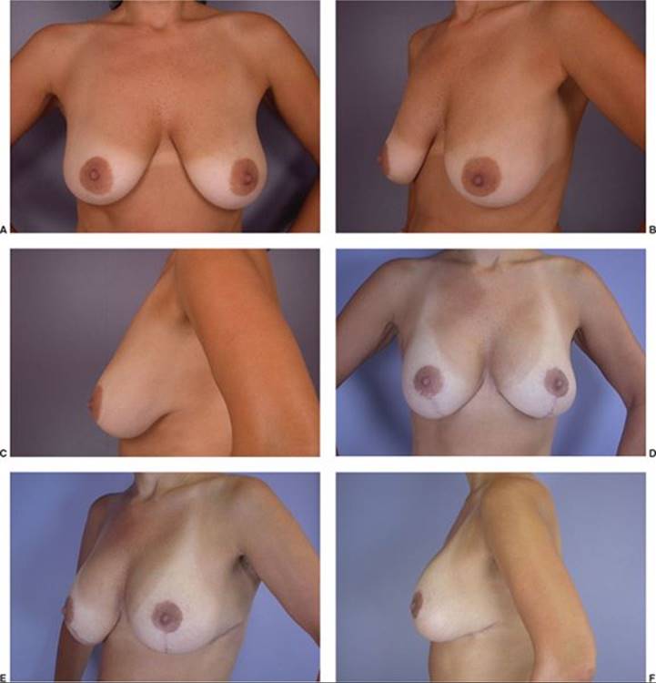
FIGURE 54.6. T mastopexy with augmentation. A–C. Before T mastopexy with augmentation. D–F. After T mastopexy and placement of 200 cc cohesive gel implants.
A “snoopy deformity” is caused by descent of the nipple and breast tissue over an underlying implant. In this scenario, mastopexy may be performed with or without adjustment of the implant or capsule. Most often, patients select re-augmentation in conjunction with mastopexy, where the implant is exchanged for a different size42 (Figure 54.8). The least aggressive mastopexy pattern to achieve the desired result is preferred to avoid nipple ischemia. Initial skin excision should be conservative—additional tissue may be resected after the implant is inserted. This reduces the risk of inadvertent skin shortage and diminishes the likelihood of excessive wound tension.31,42
Ptosis of the implant itself in a previously augmented patient can create a “ball in sock” deformity, particularly with subglandular implants. Occasionally, the inframammary fold itself has descended, either because of overdissection at the time of implant placement or disruption during transumbilical augmentation, causing the implant to sit too low on the chest wall. This may exacerbate the “double-bubble” effect or give the appearance of pseudoptosis or bottoming out. Reconstructive options include pocket revision with capsulorrhaphy, submuscular implant placement (in the setting of existing subglandular implants), use of acellular dermal matrix, or creation of a neosubpectoral pocket.43 The implant capsule can also be used to create an internal autologous splint to reposition the implant and inframammary fold.44

FIGURE 54.7. Correction of previous circumareolar mastopexy. A–C. Patient with widened areola after previous 400 cc textured silicone implants and circumareolar mastopexy. D–F. After correction with a circumareolar mastopexy and replacement with 354 cc textured silicone implants.
MASTOPEXY–AUGMENTATION COMPLICATIONS
Although a single-stage procedure is technically difficult, complication, and revision rates compare favorably with rates seen in either procedure alone.30,36,40,42 Complications are separated into either tissue-related or implanted-related categories. Implant-related complications outnumber tissue-related complications and include deflation, capsular contracture, implant palpability, implant malposition, and a desire to change size. More serious complications involve infection, wound-healing problems, and implant exposure or extrusion. Combined secondary mastopexy–augmentation carries an increased risk of infection because of more tissue rearrangement around the implant and risk of exposure.42 Risk for loss of nipple sensation and ischemia or necrosis is increased because of more extensive undermining and soft tissue manipulation around the nipple. Skin flaps and incisions are at risk for the same reasons, particularly with added tension from an underlying implant. Poor scarring and areolar widening may also be exacerbated by tension. Nipple malposition can occur if the surgeon misjudges its elevation following implant placement. Risks are diminished by proper patient selection and judicious planning and technique. This is especially true for secondary mastopexy–augmentations.13,36,40,42
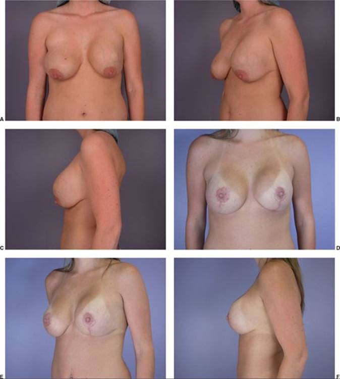
FIGURE 54.8. Secondary mastopexy with augmentation. A–C. Patient with “snoopy” deformity after a breast augmentation 5 years prior. D–F. After secondary mastopexy with placement of 300 cc cohesive gel implants.
References
1. ASAPS. 2009 National Statistics Data. http://www.surgery.org/sites/default/files/2009stats.pdf
2. Rohrich RJ, Thornton JF, Jakubietz RG, Jakubietz MG, Grunert JG. The limited scar mastopexy: current concepts and approaches to correct breast ptosis. Plast Reconstr Surg. November 2004;114(6): 1622-1630.
3. Bostwick JI. Aesthetic and Reconstructive Breast Surgery. St Louis, MO: CV Mosby; 1983.
4. Brink RR. Management of true ptosis of the breast. Plast Reconstr Surg. April 1993;91(4):657-662.
5. Grotting JC, Marx AP, Chen SM. Mastopexy. In: Mathes SJ, ed. Plastic Surgery. Vol 6. 2nd ed. Philadelphia, PA: Saunders; 2006:47-86.
6. Tenenbaum MJ, Stevens WG. Breast ptosis. In: Marsh JL, Perlyn CA, eds. Decision Making in Plastic Surgery. 2nd ed. St Louis, MO: QMP; 2010:306-307.
7. Regnault P. Breast ptosis. Definition and treatment. Clin Plast Surg. April 1976;3(2):193-203.
8. Rohrich RJ, Gosman AA, Brown SA, Reisch J. Mastopexy preferences: a survey of board-certified plastic surgeons. Plast Reconstr Surg. December 2006;118(7):1631-1638.
9. Hall-Findlay EJ. Pedicles in vertical breast reduction and mastopexy. Clin Plast Surg. July 2002;29(3):379-391.
10. Lejour M. Vertical mammaplasty and liposuction of the breast. Plast Reconstr Surg. July 1994;94(1):100-114.
11. Lockwood T. Reduction mammaplasty and mastopexy with superficial fascial system suspension. Plast Reconstr Surg. April 1999;103(5):1411-1420.
12. Graf R, Biggs TM. In search of better shape in mastopexy and reduction mammoplasty. Plast Reconstr Surg. July 2002;110(1):309-317; discussion 318-322.
13. Spear SL, Dayan JH, Clemens MW. Augmentation mastopexy. Clin Plast Surg. January 2009;36(1):105-115, vii; discussion 117.
14. Benelli L. A new periareolar mammaplasty: the “round block” technique. Aesthetic Plast Surg. Spring 1990;14(2):93-100.
15. Goes JC. Periareolar mammaplasty: double skin technique with application of polyglactin or mixed mesh. Plast Reconstr Surg. April 1996;97(5):959-968.
16. Benelli LC. Periareolar Benelli mastopexy and reduction: the “round block”. In: Spear SL, ed. Surgery of the Breast: Principles and Art. 2nd ed. Philadelphia, PA: Lippincott Williams & Wilkins; 2006:977-991.
17. Lassus C. A technique for breast reduction. Int Surg. January 1970;53(1):69-72.
18. Lassus C. A 30-year experience with vertical mammaplasty. Plast Reconstr Surg. February 1996;97(2):373-380.
19. Lassus C. Vertical scar breast reduction and mastopexy without undermining. In: Spear SL, ed. Surgery of the Breast: Principles and Art. 2nd ed. Philadelphia, PA: Lippincott Williams & Wilkins; 2006:1021-1043.
20. Hall-Findlay EJ. A simplified vertical reduction mammaplasty: shortening the learning curve. Plast Reconstr Surg. September 1999;104(3):748-759; discussion 760-743.
21. Hammond DC. The SPAIR mammaplasty. Clin Plast Surg. July 2002;29(3):411-421.
22. Hammond DC. Reduction mammaplasty and mastopexy: general considerations. In: Spear SL, ed. Surgery of the Breast: Principles and Art. 2nd ed. Philadelphia, PA: Lippincott Williams & Wilkins; 2006:972-976.
23. Hammond DC, ed. Atlas of Aesthetic Breast Surgery. 1st ed. Philadelphia, PA: Saunders; 2009.
24. Chiari A Jr. The L short-scar mammaplasty. Clin Plast Surg. July 2002;29(3):401-409, vi.
25. Marchac D, de Olarte G. Reduction mammaplasty and correction of ptosis with a short inframammary scar. Plast Reconstr Surg. January 1982;69(1):45-55.
26. Marchac DA. Vertical mammaplasty with a short horizontal scar. In: Spear SL, ed. Surgery of the Breast: Principles and Art. 2nd ed. Philadelphia, PA: Lippincott Williams & Wilkins; 2006:1049-1059.
27. Chiari A Jr, Grotting JC, Seidel SP. The L short-scar mammaplasty. In: Spear SL, ed. Surgery of the Breast: Principles and Art. 2nd ed. Philadelphia, PA: Lippincott Williams & Wilkins; 2006:1155-1172.
28. Loustau HD, Mayer HF, Sarrabayrouse M. The owl technique combined with the inferior pedicle in mastopexy. Aesthetic Plast Surg. January 2008;32(1):11-15; discussion 16-17.
29. Ramirez OM. Reduction mammaplasty with the “owl” incision and no undermining. Plast Reconstr Surg. February 2002;109(2):512-522; discussion 523-514.
30. Stevens WG, Stoker DA, Freeman ME, Quardt SM, Hirsch EM. Mastopexy revisited: a review of 150 consecutive cases for complication and revision rates. Aesthet Surg J. March-April 2007;27(2):150-154.
31. Handel N. Secondary mastopexy in the augmented patient: a recipe for disaster. Plast Reconstr Surg. Dec 2006;118(7 Suppl):152S-163S; discussion 164S-165S, 166S-167S.
32. Spear SL. Augmentation/mastopexy: “surgeon, beware”. Plast Reconstr Surg. December 2006;118(7 Suppl):133S-134S; discussion 135S.
33. Gonzalez-Ulloa M. Correction of hypotrophy of the breast by means of exogenous material. Plast Reconstr Surg Transplant Bull. January 1960;25:15-26.
34. Regnault P. The hypoplastic and ptotic breast: a combined operation with prosthetic augmentation. Plast Reconstr Surg. January 1966;37(1):31-37.
35. Heden P. Mastopexy augmentation with form stable breast implants. Clin Plast Surg. January 2009;36(1):91-104, vii.
36. Stevens WG, Freeman ME, Stoker DA, Quardt SM, Cohen R, Hirsch EM. One-stage mastopexy with breast augmentation: a review of 321 patients. Plast Reconstr Surg. November 2007;120(6):1674-1679.
37. Nahai F, Fisher J, Maxwell PG, Mills DC 2nd. Augmentation mastopexy: to stage or not. Aesthet Surg J. May-June 2007;27(3):297-305.
38. Spear SL, Boehmler JH, Clemens MW. Augmentation/mastopexy: a 3-year review of a single surgeon’s practice. Plast Reconstr Surg. December 2006;118(7 Suppl):136S-147S; discussion 148S-149S, 150S-151S.
39. Spear SL, Pelletiere CV, Menon N. One-stage augmentation combined with mastopexy: aesthetic results and patient satisfaction. Aesthetic Plast Surg. September-October 2004;28(5):259-267.
40. Stevens WG, Stoker DA, Freeman ME, Quardt SM, Hirsch EM, Cohen R. Is one-stage breast augmentation with mastopexy safe and effective? A review of 186 primary cases. Aesthet Surg J. November-December 2006;26(6):674-681.
41. Spear SL, Low M, Ducic I. Revision augmentation mastopexy: indications, operations, and outcomes. Ann Plast Surg. December 2003;51(6):540-546.
42. Stevens WG, Spring M, Stoker DA, et al. A review of 100 consecutive secondary augmentation/mastopexies. Aesthet Surg J. September-October 2007;27(5):485-492.
43. Maxwell GP, Gabriel A. The neopectoral pocket in revisionary breast surgery. Aesthet Surg J. July-August 2008;28(4):463-467.
44. Rowe NM, Freund R. The autologous internal breast splint: a novel technique for the treatment of postaugmentation ptosis. Aesthet Surg J. November-December 2005;25(6):587-592.