
AL S. ALY AND EMIL J. KOHAN
INTRODUCTION
Body contouring of the lower trunk region is an integral part of the plastic surgeon’s armamentarium. The lower trunk is a circumferential structure that begins at the inferior border of the breasts and ends at the pelvic rim. Although this is a convenient unit, it is difficult to separate from surrounding structures such as the thighs and the thorax. Deformities in the lower truncal region are variable in nature and require different approaches for their treatment. Recent advances in bariatric surgery have resulted in a large population of weight loss patients, which has led to an emphasis on the evaluation and treatment of lower truncal contour deformities. This chapter will focus on excisional procedures, with or without liposuction, in the treatment of lower truncal deformities. Problems that can be ameliorated by liposuction techniques alone are covered in Chapter 65.
PATIENT PRESENTATION
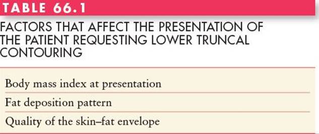
Patients with lower truncal complaints demonstrate a variety of deformities on a continuum from minimal excess fat to circumferential fat and skin excess accompanied by abdominal laxity of the fascia1 (Table 66.1).
Weight is the first important factor that affects the presentation of patients with lower truncal deformities. Because absolute weights can be misleading, body mass index (BMI), which relates weight to height, is the most commonly used parameter. It is calculated in the following manner:
Body mass index = weight in kilograms/(height in meters)2
Body mass index = weight in pounds/(height in inches)2 × 703
Patients who present for lower truncal contouring span the range of BMI from normal to obese.
The upper limit of normal BMI is 25; 26 to 30 is considered overweight; and 30 and above is considered obese. A variety of surgical approaches are required to treat patients in different BMI ranges.
A second factor that affects the presentation of patients is the fat deposition pattern, which is genetically controlled. Women typically deposit fat in the infraumbilical abdomen, lateral thighs, hips, and medial thighs. Men tend to deposit fat in the flanks, the infraumbilical abdomen, and intra-abdominally.2 Although these patterns are common, dramatically different patterns of fat deposition are often present even within the same gender.
The quality of the skin–fat envelope is a third factor to evaluate. Some women who have had one or more pregnancies may present with abdominal skin laxity and stretch marks. The skin in those patients is stretched beyond its ability to rebound back to its original elasticity. A similar process occurs with massive weight gain and subsequent weight loss in which the skin is overexpanded, leading to a skin–fat envelope that is loose and inelastic.
HISTORY OF BODY CONTOURING
Body contouring procedures early in the twentieth century consisted of dermatolipectomies of hanging abdominal panniculi. In these procedures, excess skin and underlying fat were removed to rid the patient of hanging tissues with minimal attention to aesthetic principles. In the second half of the century, advances in abdominoplasty techniques led to improved scar placement, abdominal wall plication, and umbilical transposition. In the 1980s, liposuction was introduced, and it became a tremendous tool in the armamentarium of the plastic surgeon for affecting body contour, replacing a number of excisional procedures. Currently, plastic surgeons routinely use both excisional and liposuction techniques, alone and in combination, to improve abdominal contour.
RELEVANT ANATOMY
Fat in the lower trunk is organized into superficial and deep layers separated by the superficial fascial system, which pervades the entire body. Anteriorly the superficial fascial system is referred to as Scarpa’s fascia (Figure 66.1).
The blood supply of the abdominal skin and fat is important to understand. The skin overlying the rectus muscles is primarily supplied by arteries that originate from the superior and inferior epigastric vessels that run within the rectus muscles. Branches from these vessels perforate the overlying rectus fascia and traverse through the two layers of abdominal fat, finally reaching the skin. This direct blood supply of abdominal skin is interrupted during the elevation of the abdominal flap in a traditional abdominoplasty. A secondary blood supply is derived from lateral intercostal, subcostal, and lumbar vessels that course anteriorly in the fat superficial to Scarpa’s fascia (Figure 66.2). These vessels are the only remaining blood supply of central abdominal skin after traditional flap elevation. Interruption of these vessels by scars, such as cholecystectomy, or chevron scars, can lead to necrosis of tissues inferomedial to the scar. The superficial epigastric vessels supply blood to the skin of the lower abdomen but are also divided during abdominoplasty procedures.
The lower trunk has fascial attachments between the skin and the underlying muscle fascia that act as anchoring points or zones of adherence3 (Figure 66.3). These zones of adherence restrict the overlying skin from moving during the processes of aging and/or weight fluctuations. Posteriorly, the midline has a zone of adherence that overlies the spine. The anterior midline of the abdomen has a less well-defined zone of adherence. Three horizontal zones of adherence are located in the inferior aspects of the lower trunk; one is located at the inguinal region bilaterally and extends toward the anterior superior iliac spine (ASIS). Another is located just above the mons pubis and is variable in its adherence properties. The third is located bilaterally between the hip and lateral thigh fat deposits. Truncal tissues become lax due to aging, pregnancy, and/or massive weight loss. They descend the greatest distance laterally, caused by a combination of tissue laxity and central tethering of the midline zones of adherence. As tissues descend around the pelvis they also migrate centrally (see Figure 66.3).
The inguinal and mons pubis zones of adherence are responsible for holding the final position of abdominoplasty scars in the lower truncal region. Without their effect, the scars would migrate cephalad, possibly above natural underwear lines.
PATIENT SELECTION
Patients who have minimal to moderate subcutaneous fat excess and no abdominal wall laxity are good candidates for liposuction alone. Patients who present with abdominal wall laxity and minimal abdominal skin excess limited to the infraumbilical region are good candidates for mini-abdominoplasty. Patients who present with abdominal wall laxity of both the infra- and supraumbilical regions and generalized skin excess limited to the anterior aspects of the lower trunk are good candidates for a full abdominoplasty. As the deformities increase in magnitude and involve the lateral and posterior aspects of the lower trunk, circumferential truncal liposuction and/or dermatolipectomies become necessary. The indications, goals, and a general description of each procedure are given below.

FIGURE 66.1. Organization of fat and fascia in the anterior abdomen.

FIGURE 66.2. The abdominal wall vasculature. a, artery.

FIGURE 66.3. Fascial zones of adherence. The zones of adherence control the movement of tissue associated with aging and/or massive weight loss. These fascial attachments result in lateral descent of truncal tissues, which rotate toward the midline.
Lower truncal body contouring procedures are often long and extensive in nature. Medical problems such as heart disease, diabetes, and lung disease must be under control before surgery is contemplated. Cigarette smoking also has a deleterious effect on blood supply and, when combined with the already compromised vascular supply of the abdominal skin, can lead to significant tissue necrosis. Many plastic surgeons avoid performing abdominoplasty on active smokers.
MINI-ABDOMINOPLASTY
Women who present with abdominal wall laxity restricted to the infraumbilical region that is associated with minimal infraumbilical skin and fat excess are candidates for a mini-abdominoplasty. Physical examination of the abdomen in the supine position will demonstrate infraumbilical rectus diastasis, which can be confirmed by the “diver’s test” (Figure 66.4).

FIGURE 66.4. The classic “diver’s test” demonstrates how a bend at the waist will reveal the true extent of abdominal wall laxity.
These patients are usually young women who have had one or two pregnancies, have good skin elasticity, and are not overweight. They may or may not have localized fat deposits in other areas of the trunk and lower extremity such as the hips and lateral thighs. The goal of surgery in this patient population is to eliminate the infraumbilical abdominal wall laxity and the minimal skin and fat excess.
Technique (Mini-Abdominoplasty)
An incision is marked in the patient’s natural suprapubic crease and angled toward the ASIS. Often the incision can be limited to the width of the pubic hair or just beyond its lateral edges. Intraoperatively, the proposed incision is made and the dissection extended to the muscle fascia. An abdominal flap is elevated superiorly to the level of the umbilicus. The infraumbilical rectus muscle diastasis is identified, and rectus fascia plication is performed. Some surgeons prefer a single layer, whereas others favor a two-layer plication (Figure 66.5). The abdominal flap is advanced inferiorly and tailored to remove the excess skin and underlying fat. This advancement will usually pull the umbilicus down 1 to 3 cm.
The closure of this incision, as in all subsequent incisions discussed in this chapter, is performed in multiple layers, with the most important layer being the reapproximation of the superficial fascial system, or Scarpa’s fascia.4Permanent or long-lasting sutures are used in this layer in an attempt to limit widening of the scar in the long run. The authors prefer to use interrupted monofilament absorbable sutures in the subcuticular layer to perfectly approximate the skin with an overlying layer of medical-grade skin glue. Drains are inserted and a compression garment is used in the postoperative period by most surgeons.
A variation of this technique can be used in patients who have minimal lower abdominal skin excess, no upper abdominal skin excess, and both infra- and supraumbilical rectus diastasis. To allow access to the supraumbilical rectus diastasis, the base of the umbilicus can be amputated. The abdominal flap is then elevated on either side of the midline in the supraumbilical region, and a supraumbilical rectus plication and an infraumbilical plication are performed. The umbilical stalk is then resutured to the plication at the appropriate level, and the lower aspect of the abdominal flap is tailored appropriately. It is also possible to use a minimal-incision approach to the supraumbilical plication by making an incision in the superior aspect of the umbilicus and using an endoscope to perform a dissection superior to the umbilicus that is wide enough to allow for the desired supraumbilical plication. In any of the mini-abdominoplasty techniques discussed, liposuction can be used to decrease the thickness of any part of the abdominal flap that has not been elevated.

FIGURE 66.5. The abdominal flap elevation and rectus fascia placation in a mini-abdominoplasty.
One of the most difficult aspects of mini-abdominoplasty is avoiding dog-ears because of the short incision.
ABDOMINOPLASTY
Generally, abdominoplasty is indicated in patients whose laxity involves the supra- and infraumbilical regions, limited to the anterior aspects of the lower trunk. The goals of abdominoplasty depend on the presenting deformities. They include creating a flat abdominal contour, eliminating abdominal wall laxity, enhancing waist definition in some patients, and eradicating mons pubis ptosis if present.
Stretch marks are common and may be limited to the infraumbilical region or may include both the infra- and supraumbilical skin. Rectus diastasis of the entire vertical extent of the abdomen is present in these patients, with the infraumbilical diastasis usually more extensive because of the position of the uterus during pregnancy. Preoperatively abdominal wall laxity can again be detected by the “diver’s test” and physical examination. Massive-weight-loss patients who reach a near-normal BMI may also present with lower truncal excess limited to the anterior abdomen. However, most often they present with circumferential deformities that require more extensive circumferential excisions.
Patients who present with excess intra-abdominal fat that would prevent flattening of the abdominal wall by plication are not good candidates for abdominoplasty. The outer skin/fat envelope of the belly always conforms to the shape of an inner balloon whose anterior wall is made up of the abdominal muscle wall. If that wall is rendered convex in profile by virtue of overly abundant intra-abdominal contents, then the final profile of the belly will also be convex. Because abdominal contour flattening is one of the major goals of surgery, these patients are better served by weight loss prior to contemplating abdominoplasty-type procedures.
By the nature of an abdominoplasty, where an ellipse of tissue is removed from the lower abdomen, dog-ears can be created at the edges of the ellipse, especially in patients who already have lateral excess. Patients who present with deformities that extend beyond the anterior aspects of the lower trunk may require 1) extending the abdominoplasty excision laterally, 2) liposuction of the lateral and posterior trunk, and/or 3) circumferential dermatolipectomy to attain the best possible contour.
Some authors advocate the use of fleur-de-lis or “T”-type excisions in which an anterior vertical wedge of tissue is resected, as discussed later in this chapter. Generally, as circumferential lower truncal dermatolipectomy has become more mainstream in plastic surgery because of the massive-weight-loss population, the indications for isolated abdominoplasty have narrowed.
Technique (Abdominoplasty)
The markings for an abdominoplasty are performed prior to surgery. The proposed excision is marked in the lower abdomen. Centrally, the inferior incision line is often marked in the natural suprapubic crease and then carried laterally. Some surgeons utilize a “French bikini/thong pattern” in which the lateral aspects of the proposed inferior incision are angled toward the ASIS, while others prefer a flatter pattern, with many variations described in the literature.5 An attempt is made to avoid the incision beyond the ASIS, but it is more important to avoid dog-ears. With the inferior mark in place the patient is slightly flexed at the waist, and the pinch technique is used to approximate the superior extent of the excision. Ideally, the patient should have enough excess abdominal skin to allow excision of the skin from just above the umbilicus to the suprapubic crease centrally.
In the operating room, a circumumbilical incision is made and the umbilical stalk is dissected to the deep fascia. The inferior mark of the proposed abdominal skin excision is incised. An abdominal flap is elevated superiorly, around the umbilicus, and up to the xiphoid and costal margins (Figure 66.6). The flap is classically elevated at the level of the underlying muscle fascia but many plastic surgeons prefer to elevate the flap at Scarpa’s fascia level. It is felt that this may reduce the rate of seroma formation. Two theories have been invoked as to the etiology of this reduction. The most popular is that the remaining subscarpal fat contains intact lymphatic vessels, which help absorb fluid in the wound. The other possibility is that the fat-to-fat interface leads to better adhesion between the abdominal flap and the underlying tissues. Neither theory has been tested experimentally; thus, it is not currently known why this type of elevation seems to reduce seroma formation. Wide undermining allows the greatest amount of abdominal flap advancement at the time of flap tailoring, but it also leads to the division of the greatest number of superior epigastric muscle perforator vessels, leaving only the lateral intercostal, subcostal, and lumbar vessels as the only viable blood supply of the flap. Some surgeons prefer a more limited dissection above the umbilicus, just to the medial edges of the rectus muscle fascia, to allow for supraumbilical rectus fascia plication up to the xiphoid. The benefit of the limited dissection is the increased number of the perforator vessels left intact to support the blood supply of the tailored abdominal flap. In some patients, however, the limited dissection will not allow the appropriate advancement of the abdominal flap and may reduce the amount of tissue that may need to be resected to create the best contour. As a general rule, flap elevation should be restricted to just what will allow appropriate rectus fascia plication and appropriate flap advancement. Often it is best to limit the initial elevation and then release the tissues incrementally to allow for appropriate contour.
After flap elevation, rectus fascia plication is performed. Many patterns have been proposed for plication, but a vertical plication, in one or two layers, is most common. The patient is then flexed at the waist, and the abdominal flap is advanced inferiorly to facilitate the process of flap tailoring. As the abdominal flap is advanced, the surgeon can control where the greatest tension will be at closure—centrally or laterally. Creating the greatest amount of tension centrally is advantageous in limiting the lateral extent of the final scar but may lead to excessive mons pubis elevation and less waist definition. Lockwood, in his “high-lateral-tension” approach to abdominoplasty, espoused placing the greatest tension on the lateral aspects of the abdominal closure.6 This is based on the fact that the greatest laxity in the lower trunk occurs laterally. However, the increased lateral tension often necessitates extensions of the scar laterally to eliminate dog-ears. This approach can lead to better waist definition and an improvement in anterior thigh contour, but often necessitates leaving behind the original umbilical defect as a closed vertical scar. Thus, placement of tension, centrally or laterally, in abdominoplasty surgery should depend on the patients’ differing needs and desires.

FIGURE 66.6. The extent of abdominal flap elevation and fascial plication in a traditional abdominoplasty.
With the tailored abdominal flap approximated to the lower incision, the position of the umbilicus is noted on the flap and a neo-umbilicus is created. The stalk of the umbilicus is brought through the abdominal flap using one of various incisions advocated by different authors. The authors prefer a simple vertical incision, no defating of the underlying soft tissues, and “three-point fixation” sutures at 3, 6, and 9 o’clock. Whatever method the surgeon chooses, the umbilicus should be fairly small, vertically oriented, superiorly hooded, have a slight hollow around it, and contain the scar on the inside of the cylindrical structure.
The closure of the abdominal incision is accomplished in multiple layers, with the most important layer being the superficial fascial system, or the Scarpa fascia.4This is accomplished with permanent or long-lasting suture, which, it is hoped, reduces the significant tension that can be generated at closure, prevents acute wound dehiscence, and reduces scar widening in the long run. The authors prefer to use interrupted monofilament absorbable suture in the subcuticular layer to perfectly approximate skin. An overlying layer of medical-grade skin glue is then applied. Drains are placed. A compression garment is used in the postoperative period by most surgeons. Figure 66.7 shows an example of abdominoplasty in an ideal patient. Many surgeons, including the authors, use quilting sutures in an attempt to decrease the rate of seromas by reducing dead space between the flap and the underlying muscle wall.7

FIGURE 66.7. Traditional abdominoplasty. A young woman who presented after two pregnancies desiring an improvement in her abdominal contour. She complained of loose abdominal skin and protrusion of her belly, despite a regular exercise program. Her abdominal wall laxity was more prominent in the infraumbilical region, consistent with her pregnancies. She underwent an abdominoplasty using a French bikini pattern of excision and is shown 6 months after surgery. Because her deformities were limited to the anterior abdomen and she was within the normal weight range, there was no need to combine other procedures with her traditional abdominoplasty.
Combining liposuction with traditional abdominoplasty techniques is controversial and is often left to the surgeon’s experience and philosophy.8 Liposuction of the abdominal flap in nonundermined areas is generally considered safe. Liposuction of undermined regions of the flap can potentially lead to flap necrosis because of the compounding effect of liposuction on the already compromised blood supply. Certainly, liposuction of areas outside the bounds of the undermined abdominal flap, such as the hip region and the lateral thighs, can be performed without concern for flap compromise. Studies have demonstrated that combining liposuction with abdominoplasty, whether the liposuction is performed on the elevated flap or the non-flap contiguous areas, may lead to increased risk of seroma formation.9
CIRCUMFERENTIAL LOWER TRUNCAL DERMATOLIPECTOMY OR CIRUMFERENTIAL LIPECTOMY
As a basic principle of plastic surgery, it is always better to reconstruct an anatomic unit in its entirety when possible. Abdominoplasty treats deformities limited to the anterior lower trunk. When deformities involve more than the anterior abdomen, other procedures are required to adequately treat all the deformities. If the surrounding areas such as the thighs, buttocks, hips, and lower back regions contain excess fat without ptosis, liposuction can be added to abdominoplasty to create a better overall lower truncal contour. However, for patients who present with generalized laxity and/or ptosis of those areas, circumferential lipectomies are required. Massive-weight-loss patients make up the largest group of such patients who require circumferential excisional procedures. They will continue to grow in numbers, given that obesity has been recognized as a major health-care issue in the United States and the world. In addition, women who gain moderate weight, 30 to 40 lb, usually with childbirth and/or aging, and are not able to lose the weight through normal means of exercise and nutritional changes may be candidates. They often present with a desire to eliminate anterior abdominal excess, but careful examination will demonstrate circumferential excess that is best treated with a circumferential lipectomy. Finally, normal-weight-range patients who desire remarkable improvements in their lower truncal contour may be candidates as well.
There is a variety of names used to describe circumferential lower truncal dermatolipectomies: extended or circumferential abdominoplasty, central body lift, torsoplasty, and body lift. The authors prefer to divide these different variations into two general categories based on what they treat and what they accomplish. The first category is made up of centrally based procedures that mainly treat the lower truncal unit, which will be referred to as belt lipectomies, as espoused by Aly and Cram.10,11 The second category includes procedures that treat the lower trunk and thighs as a unit, which will be referred to as lower body lifts, as espoused by Lockwood.12,13Each procedure has its benefits and drawbacks. Both should be in the armamentarium of the plastic surgeon who performs body contouring surgery. The choice of procedure is based on the patient’s desires and presenting deformities.
Both procedures eliminate a circumferential wedge of tissue from the lower trunk. A belt lipectomy removes a wedge that is more superiorly located than that removed in a lower body lift (Figure 66.8). The final scar after belt lipectomy is located above the widest aspect of the bony pelvic rim, at the junction between the lower back and buttocks, which may be visible outside of brief undergarments. Because this allows cinching at waist level, more waist definition can be created by this technique. This is often desired in women but may be less desirable in men. Pelvic rim zones of adherence help to prevent the descent of lower truncal tissues as well as inhibit elevation of lower extremity tissues (see Figure 66.4). These fascial attachments are interrupted but not completely eliminated during a belt lipectomy, and they prevent extensive lifting of lower thigh tissues. Thus, overall, a belt lipectomy is capable of creating excellent lower truncal contour by accentuating waist definition, delineating the buttocks from the lower back, and lifting the lateral thighs, but it has limited capability in lifting the distal thighs.
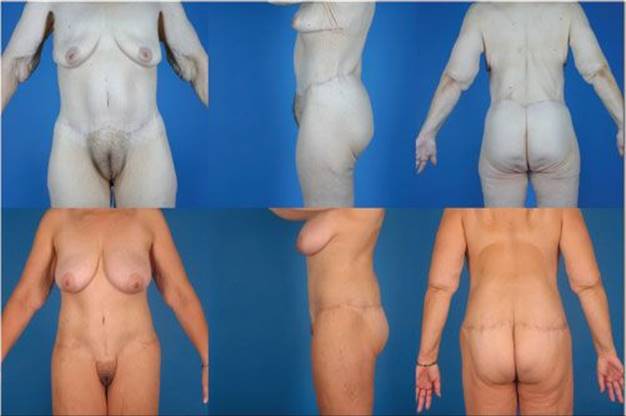
FIGURE 66.8. Belt lipectomy and lower body lift. Two patients who underwent a belt lipectomy above and a lower body lift below. In a belt lipectomy the scar is placed at the junction between the buttocks and lower back, which helps to frame the natural buttocks contour and accentuate waist narrowing. In a lower body lift the scar is onto the buttocks proper and is overall more inferiorly placed, especially in its lateral and posterior aspects. The combination of eliminating the pelvic zones of adherence and the lower position of the excised wedge allows the lower body lift to elevate the thighs more effectively than in a belt lipectomy.
A lower body lift treats the lower trunk and thighs as a unit. The pelvic rim zones of adherence are intentionally interrupted as completely as possible, which allows inferior thigh tissues, down to knee level, to be lifted. This leads to a significant reduction in anterior and lateral thigh laxity. The final scar is located within the buttocks proper, which can blunt waist definition (see Figure 66.8). This is often desirable in men but may be less so in women. The resultant scar is easily covered by normal undergarments, and the thighs are dramatically improved down to the knee level. However, a lower body lift is less efficient in creating waist definition and can result in less than ideal buttocks definition because the scar does not respect the natural junction between the lower back and buttocks.
The majority of patients undergoing a circumferential lipectomy are massive-weight-loss patients. Often they present with a hanging panniculus, mons pubis ptosis, an ill-defined waist, lower back rolls, hip-fat excess, lateral thigh ptosis, and varying types of buttocks deformity. The goals of surgery include elimination of the hanging panniculus, mons pubis elevation, creation of waist definition (especially in women), decrease or elimination of lower back rolls, lifting of the outer thighs, and increase in buttocks definition.
Techniques (Truncal Dermatolipectomy)
Although a circumferential lipectomy is a combination of an abdominoplasty, a lateral thigh lift, and a buttocks lift, the procedure is more complex than simply combining them. The lower trunk of patients who present with circumferential excess has the shape of an inverted cone (Figure 66.9). A wedge of tissue is marked for proposed excision around the lower trunk. The wedge brings a narrower part of the cone down to the level of a wider part of the cone located at, or near, the pelvic rim (see Figure 66.9). As previously noted, the wedge to be excised is generally located in a more superior position in belt lipectomy when compared with the wedge to be excised in a lower body lift. In either method, the anterior aspect of the wedge is wider (in vertical distance) than the lateral or posterior aspects. The lateral resection is the next widest aspect so as to reverse the lateral truncal descent (Figure 66.10).

FIGURE 66.9. Truncal deformity in weight loss patients. In the massive-weight-loss patient, the presenting lower truncal deformity is in the shape of an inverted cone. In a circumferential lipectomy a wedge of tissue is removed. The diameter of the wedge at its superior edge is smaller than its diameter at the inferior edge.
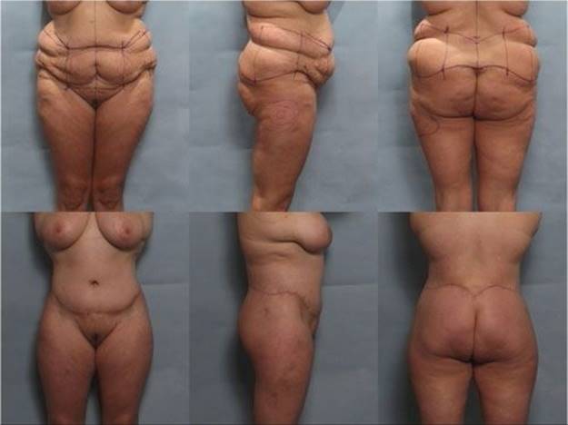
FIGURE 66.10. A 31-year-old woman presented after an 80-lb weight loss to reach a body mass index of 27.31. (Above) Shown with preoperative markings for a circumferential belt lipectomy. Note that the excision laterally is generally aggressive to counteract the lateral descent that occurs with massive weight loss and/or aging. Vertical marks are placed along the circumference of the proposed resection to help alignment at closure. Surrounding areas of the thigh are also marked for liposuction. (Below) The patient 6 months after surgery, demonstrating dramatic waist narrowing, elimination of the panniculus and lower back rolls, and improved buttocks definition.
Because of the circumferential nature of the procedure, more than one position is necessary to accomplish the resection in the operating room. No matter what sequence is preferred by a particular surgeon, the abdominal part of the procedure is performed in the supine position. Surgeons who advocate prone/supine or supine/prone positioning cite the single turn required in the operating room and the ability to control buttock symmetry as their reasons for choosing the “two-position” sequences. The supine/lateral/lateral or lateral/lateral/supine proponents prefer these “three-position” sequences because they allow for easier lateral thigh liposuction and hip abduction in the lateral decubitus position, which facilitates maximal lateral resections. All body positions have potential complications associated with them, especially if the patient is to be maintained in those positions for extended periods of time. The surgeon should be familiar with those complications and how to prevent them.
The extent of anterior flap elevation in the abdominoplasty portion of the circumferential procedure is based on surgeon preference. The lateral elevation is usually more extensive than in an abdominoplasty, which compromises the remaining blood supply to the abdominal flap to a greater extent. Thus, it is important that an effort is made to preserve as many lateral feeding vessels as possible. The plication of the rectus fascia is similar to abdominoplasty plication except that it may sometimes require plication distances that far exceed the usual 5 to 7 cm encountered with routine abdominoplasty. Closure of the circumferential wound should include reapproximation of the superficial fascial system with permanent and/or long-lasting suture.
During the lateral and posterior resection, some surgeons prefer to incise the superior marks first and dissect an inferior skin–fat flap, whereas others prefer the opposite. Some surgeons incise both the superior and inferior extents and excise a predetermined marked amount. The authors prefer to incise the superior side first and tailor the inferior-based flap based on tension and creation of the appropriate contour.
Some surgeons choose to combine extensive liposuction of the surrounding regions, such as the lower back, the upper back, and thighs, whereas others limit their liposuction to the lateral thighs. A major difference between belt lipectomy and a lower body lift is in the treatment of the pelvic rim’s zones of adherence. In belt lipectomy, these attachments are disrupted by liposuction of the lateral thighs, but they are not completely eliminated. In a lower body lift, discontinuous undermining of the anterior and lateral thighs, down to knee level, intentionally destroys the pelvic rim zones of adherence. This allows significant thigh elevation.12
The results attained from circumferential lipectomies depend, to a great extent, on the presentation of the patient and the type of procedure chosen (see Figure 66.10). As a general rule, the lower BMIs at presentation lead to better aesthetic results and lower rates of complications.10,11
COMPLICATIONS
Table 66.2 lists complications that can occur with lower truncal contouring procedures.14 Circumferential procedures are associated with more complications, but they are often performed on patients with higher BMIs. When complications are stratified by BMI, noncircumferential and circumferential procedures have similar rates.
Superficial wound healing problems are the most common complication that occurs with any body contouring excisional procedure because of the high tension created at the wound edges. Conservative wound care will usually allow healing to occur, with the possible need for subsequent scar revisions. Wound dehiscences, defined as separation of the wound at the level of the superficial fascial system, are possible with any of the procedures discussed in this chapter but tend to occur more frequently with circumferential procedures. In procedures limited to anterior resections, mini-abdominoplasty, and abdominoplasty, dehiscences can be prevented by keeping patients flexed at the waist for 5 to 7 days after surgery and educating patients on a slow return to the full upright position over the second week after surgery. Circumferential procedures create competing anterior and posterior tensions, making it difficult to place patients in positions that do not stress at least one aspect of the closure. Avoidance of dehiscences in this patient population entails adjustments of the competing resections to account for opposing tensions, careful ambulation of the patients in the early postoperative period, and education of patients on how to help prevent dehiscences.15
Seromas are common complications with lower truncal contouring procedures. They are due to large dissection surface areas and can develop anywhere in the surgical field but tend to be located posteriorly in circumferential procedures. Patients who present in the high BMI ranges are more likely to develop seromas. Measures that are used to reduce their occurrence include the use of suction drains, compression garments, reduction of activity, and use of quilting sutures. When they do occur, they can most often be treated with serial aspirations. For persistent seromas, sclerosing agents and seroma catheter insertions may be utilized.

Seromas are the most common source of infection after lower truncal procedures. Simple cellulitis is fairly uncommon and is usually treated by appropriate antibiotic coverage and close follow-up. Seroma pockets that become infected usually present with overlying cellulitis, fluid collections that may or may not spontaneously drain, fever, and generalized malaise. A diligent effort should be made to find seromas and treat them whenever suspected. Once seromas become infected, aggressive intravenous therapy and appropriate surgical drainage should be instituted.
Toxic shock syndrome can occur with any body contouring procedure. Postoperatively, patients who appear toxic with fever, chills, generalized malaise, and elevated white blood cell counts should be investigated. Although there is often no evidence of frank pus or large fluid collection in the wounds, aggressive surgical drainage is urgently required in this group of patients.
Vascular compromise can occur with lower truncal body contouring procedures, leading to tissue necrosis. Most commonly the necrosis occurs in the inferomedial aspect of the abdominal flap. A number of factors can contribute to this problem, which include excessive tension on the abdominal closure, aggressive thinning of the abdominal flap, overly aggressive liposuction, and anything that may lead to compromising the lateral feeding vessels of the abdominal flap such as open cholecystectomy incisions. If necrosis occurs, the wound is treated conservatively and eventually allowed to heal by secondary intention. Eventually, a scar revision may be required.
Bleeding after lower truncal contouring procedures can be extensive because of the surface area within which blood can accumulate prior to detection. Although drains do not prevent hematomas, they can often warn the surgeon of a developing hematoma. Small hematomas that are well evacuated by drains in place can be managed expectantly. Large hematomas should be treated by surgical drainage.
Procedures that tighten the abdominal wall are theorized to increase intra-abdominal pressure, leading to a decrease in venous return from the lower extremities. The possible resultant stasis of blood in the deep venous system may cause deep venous thrombosis and/or pulmonary emboli. Measures that are commonly used in the prevention of thrombotic events include early ambulation and sequential compression garments. Some surgeons feel that chemoprophylaxis, low molecular weight heparin (enoxaparin prophylaxis), is indicated in the perioperative period. At the time of the writing of this chapter it is not clear what the proper course of action should be in this arena. The authors prefer to utilize epidural catheter infusions, which help reduce pain, but have been found to reduce the risk of deep vein thrombosis/pulmonary embolism as well, and avoid the use of chemoprophylaxis.16
Patients who undergo large excisional procedures of the lower trunk, especially massive-weight-loss patients, can have psychiatric difficulties in the postoperative period that may interfere with their recovery. Although this can occur with any surgery, the long recovery period that is required after circumferential procedures makes it wise for the plastic surgeon to actively investigate a patient’s psychiatric reserves and consider obtaining psychiatric clearance prior to surgery. The tendency of massive-weight-loss patients to have lifelong psychiatric problems that are not solved by weight loss alone also contributes to the relatively high incidence of these problems.
Although careful marking techniques can help reduce scar and contour asymmetry, it is not possible to eliminate these problems in many patients because of intrinsic skeletal and soft tissue asymmetry. It is best for the surgeon to recognize these natural asymmetries and point them out to patients prior to surgery.
FLEUR-DE-LIS OR T-TYPE PROCEDURES
A fleur-de-lis or T-shaped excision, whether used as an abdominoplasty pattern or in combination with a circumferential lipectomy, is advocated by some authors. The advantage of the vertical wedge is to eliminate horizontal excess, create more waist definition, and decrease lateral fullness. Traditionally, this pattern has not been frequently used because it is difficult to justify a vertical midline incision without a preexisting vertical scar. Recently, however, it has found more use because many massive-weight-loss patients have midline incisions and/or deformities that supersede the created vertical scar in unattractiveness. Even with a preexisting scar, however, there are major disadvantages to the vertical aspect of the T pattern. There is an increased chance of flap necrosis at the T intersection. When used to treat circumferential excess, a fleur-de-lis resection pattern does not eliminate all lateral excess and does not affect lateral thigh descent or buttocks ptosis. When the pattern is used in conjunction with a circumferential lipectomy, it can create a greater mismatch between the upper and lower circumferences of the inverted cone-shaped edges to be reapproximated (see Figure 66.9). Finally, the vertical wedge excised can often lead to epigastric fullness secondary to the dog-ear effect created by the excision. Due to these disadvantages the authors do not utilize this pattern of excision.14
References
1. Aly AS. Approach to the massive weight loss patient. In: Aly AS, ed. Body Contouring After Massive Weight Loss. St. Louis, MO: Quality Medical Publishing; 2006:49.
2. La Trenta GS. Suction-assisted lipectomy. In: Rees TD, La Trenta GS, eds. Aesthetic Plastic Surgery. 2nd ed. Philadelphia, PA: WB Saunders; 1994:1180.
3. Aly AS. Options in lower truncal surgery. In: Aly AS, ed. Body Contouring After Massive Weight Loss. St. Louis, MO: Quality Medical Publishing; 2006:59.
4. Lockwood T. Superficial fascial system (SFS) of the trunk and extremities: a new concept. Plast Reconstr Surg. 1991;87:1009.
5. La Trenta GS. Abdominoplasty. In Rees TD, La Trenta GS eds. Aesthetic Plastic Surgery. 2nd ed. Philadelphia, PA: WB Saunders; 1994: 126.
6. Lockwood T. High-lateral-tension abdominoplasty with superficial fascial system suspension. Plast Reconstr Surg. 1995;96:603.
7. Pollock TA, Pollock H. No-drain abdominoplasty with progressive tension sutures. Clin Plast Surg. July 2010;37(3):515-524. [Epub 2010 May 23].
8. Matarasso A. Abdominoplasty: a system of classification and treatment for combined abdominoplasty and suction-assisted lipectomy. Aesthetic Plast Surg. 1991;15:111.
9. Najera RM, Asheld W, Sayeed SM, Glickman LT. Comparison of seroma formation following abdominoplasty with or without liposuction. Plast Reconstr Surg. January 2011; 127(1):417-422.
10. Aly AS, Cram AF. Body lift: belt lipectomy. In: Nahai F, ed. The Art of Aesthetic Surgery. Principles and Techniques. St. Louis, MO: Quality Medical Publishing; 2005:2302.
11. Aly A, Cram A, Chao M, et al. Belt lipectomy for circumferential truncal excess: the University of Iowa experience. Plast Reconstr Surg. 2003;111:398.
12. Lockwood TE. Thigh and buttock lift. In: Nahai F, ed. The Art of Aesthetic Surgery. Principles and Techniques. St. Louis, MO: Quality Medical Publishing; 2005:2424.
13. Lockwood T. Lower body lift. Oper Tech Plast Reconstr Surg. 1996;3:132.
14. Grazer FM, Goldwyn RM. Abdominoplasty assessed by survey, with emphasis on complication. Plast Reconstr Surg. 1977;59:513.
15. Aly AS. Belt lipectomy. In: Aly AS, ed. Body Contouring After Massive Weight Loss. St. Louis, MO: Quality Medical Publishing; 2006.
16. Rimler JC, Scholz T, Shbeeb A, Chua W, Wirth GA, Paydar K. The incidence of venous thromboembolism in postoperative plastic and reconstructive surgery patients with chronic spinal cord injury. Plast Reconstr Surg. 2010;126:40.