
BENJAMIN CHANG AND SUHAIL K. KANCHWALA
GENERAL PRINCIPLES
Infections of the hand result in pain, disability, as well as lost time and productivity at work. Prompt, accurate diagnosis minimizes disability and facilitates rapid recovery. In addition, infections in the hand are the presenting symptom in a number of systemic illnesses.
Evaluation of a hand infection includes an assessment of the extent of erythema, depth of infection, presence of an abscess, and range of motion of the affected digits. In addition, a medical history is obtained with emphasis on factors that affect immune response (diabetes, human immunodeficiency virus, and immunosuppression).
The anatomy of the hand, with numerous fascial compartments, allows the inflammatory response to infection itself to become pathogenic. For example, excessive swelling in the hand can result in increased pressure on tendons in the fingers and palm, leading to ischemia and tendon necrosis. Potential spaces, such as flexor tendon sheaths and the deep palmar space, can serve as conduits for infection.
Early infections in the hand, regardless of location, are managed initially by rest, elevation, antibiotics, close observation, and splinting in the intrinsic plus position. For those infections that have progressed or whose initial presentation indicates the presence of an abscess (i.e., fluctuance and drainage), the treatment includes surgical drainage and debridement of devitalized tissues. When a delay in operative treatment is unavoidable, the abscess cavity should be aspirated to reduce compartment pressures and the risk of tendon and neurovascular injury.
Antibiotics covering the most likely pathogens for each type of infection are started after wound cultures have been obtained. Antibiotics, however, are not a substitute for adequate surgical drainage and debridement. Infections in the hand are often polymicrobial—a critical consideration when making a selection of empiric antibiotic therapy (Table 72.1).1 All wounds that are a result of exposure to soil, animals, or the oral cavity (i.e., human bite wounds) require tetanus prophylaxis.
Drainage of hand infections can often be performed using regional anesthetic techniques (Chapter 71). However, the infiltration of local anesthesia directly into an area of cellulitis or infection is ill-advised and may spread the infection. For example, fingertip infections can be managed with digital blocks, but deep space infections of the hand should be drained under either axillary block or general anesthesia. A pneumatic tourniquet is used to avoiding excessive bleeding that can impair visualization. When using the tourniquet in the presence of infection, the extremity is exsanguinated by elevation and gravity, rather than compression to avoid the spread of the infection (Figure 72.1).
ACUTE PARONYCHIA
Paronychia or runaround infections of the fingertip are infections of the soft tissue fold surrounding the nail plate, typically with staphylococcal species. Risk factors for paronychial infection include hangnails, nail biting, manicures, and poor hand hygiene. Hallmarks of paronychial infection include pain, swelling, and erythema in the perionychium.
Initial management of paronychial infections includes warm soaks and oral antibiotics. When paronychial infections progress to abscess formation within the eponychial fold or under the nail plate, surgical drainage is necessary. When performing an incision and drainage of a paronychia, it is important to angle the blade away from the nail bed to avoid inadvertent damage to the nail bed and subsequent ridging of the nail. When the abscess extends under the nail plate, the nail is removed (Figure 72.2).
HERPETIC WHITLOW
Commonly confused with paronychial infections, herpetic infections of the hand typically involve the fingertip and soft tissues surrounding the nail plate. While herpetic infections may mimic bacterial infections of the hand, they can usually be distinguished by an adequate history and exam.
Herpetic infections in children and healthcare workers (dentists, respiratory therapists, etc.) are most often the result of viral inoculation from the oropharynx by the herpes simplex type 1 virus (HSV-1). In adults, however, HSV-2 predominates and is most often due to inoculation from genital herpes. Herpetic infections typically have an incubation period of 2 weeks after which patients experience pain and mild swelling in the affected digit. Small 1 to 2 mm vesicles then erupt in the affected digits and coalesce to form large bullae. A Tzanck smear is diagnostic.2
The management of herpetic hand infections does not involve surgery unless there is bacterial superinfection. In fact, surgical intervention in cases of herpetic whitlow can lead to systemic spread.3 Viral infection can lie dormant in the nervous system for many years and then reactivate (Figure 72.3).
CHRONIC PARONYCHIA
Chronic paronychia is a distinct clinical entity from acute paronychia. Chronic inflammation of the soft tissues surrounding the nail plate can lead to repeated episodes of erythema, pain, and drainage from the infected region. Patients who have repeated exposure to water (waiters, dishwashers, etc.) are at highest risk for developing chronic inflammation. Staphylococcus pyogenes, Staphylococcus epidermidis, and Candida are the most common causes of chronic paronychia.
The treatment of chronic paronychia involves the excision of a minimum 3 mm wide crescent of skin and subcutaneous tissue parallel to the eponychial fold running the entire width of the finger. This procedure is referred to as eponychial marsupialization. The wound is then left open for drainage and the patient is placed on a regimen of hand soaks in a variety of solutions such as dilute povidone–iodine solution. The warm soaks are continued until the inflammation/drainage has ceased. Nail irregularities caused by chronic paronychia can be treated by the removal of the entire nail.4 As long as the eponychial fold is appropriately stented, the nail usually regrows without abnormalities.
FELON
A felon is an infection in the soft tissue pulp on the volar aspect of the fingertip. The distal finger pad is an anatomically distinct structure from the rest of the finger. Numerous fibrous septae attach the dermis of the distal finger pad directly to the underlying bone, allowing the fingertip to be used for essential functions such as grasp. If a significant number of these septae are disrupted during drainage of a felon, a mobile, nonfunctional fingertip can result.5


FIGURE 72.1. Subcutaneous abscess of the first web space (A) appearance on presentation and (B) after wide surgical debridement.

FIGURE 72.2. Acute paronychia (A) as seen in the emergency room and (B) incision is made through the most fluctuant region.
Surgical management of the felon requires antibiotics and adequate incision and drainage directly over the point of maximal fluctuance. Incisions should not be carried over the joint flexion crease to prevent postoperative contracture (Figure 72.4).
PYOGENIC TENOSYNOVITIS
Pyogenic tenosynovitis is a closed space infection of the flexor tendon sheath of the fingers or thumb. The most common cause of this infection is penetrating injury to the proximal interphalangeal or distal interphalangeal joint on the volar surface. The flexor tendon sheath can be penetrated by foreign bodies or teeth (as in the case of a human or animal bite). Rarely, pyogenic tenosynovitis is spread to the fingers from a distant source, such as disseminated gonorrheal infection. The most common organisms cultured from patients with pyogenic tenosynovitis are Staphylococcus aureas and β-hemolytic streptococcus species.
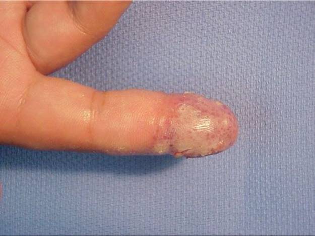
FIGURE 72.3. Herpetic whitlow.
Pyogenic tenosynovitis can be extremely disabling because infections in the tendon sheath impair the normal gliding mechanism of the flexor tendons. Late recognition and treatment of this disorder can result in fibrosis, or tendon necrosis and permanent loss of function.
Hallmarks of this infection include the following clinical signs that were initially described by Kanavel6:
1. Semi-flexed finger position
2. Symmetrical enlargement of the whole digit
3. Excessive tenderness over the course of the flexor tendon sheath
4. Pain on passive extension of the finger
The management of pyogenic tenosynovitis involves adequate drainage and irrigation of the tendon sheath. A variety of drainage procedures have been advocated. There is a consensus that copious irrigation of the tendon sheath with minimized exposure of the tendon itself through carefully placed incisions leads to fewer postoperative complications secondary to adhesion formation within the flexor sheath7 (Figure 72.5).
DEEP SPACE INFECTIONS
Deep space infections of the hand can be broken down into three general regions, the palmar, thenar, and Parona’s spaces. The thenar eminence is the most common region for deep space infections to occur. These occur most commonly as direct puncture wounds to the region or from nearby infection of the tendon sheath. Management includes incision and drainage of the region. Placement of the incision depends on the location of the abscess: (1) palmar—curvilinear from distal palmar crease to hypothenar eminence and (2) thenar—curvilinear along thenar crease (avoid recurrent branch of the median nerve).
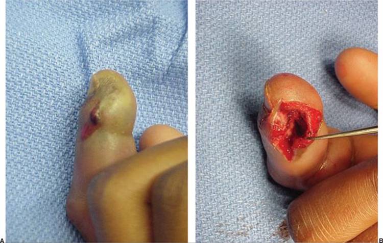
FIGURE 72.4. Felon: (A) Typical appearance and (B) all necrotic tissue is excised.
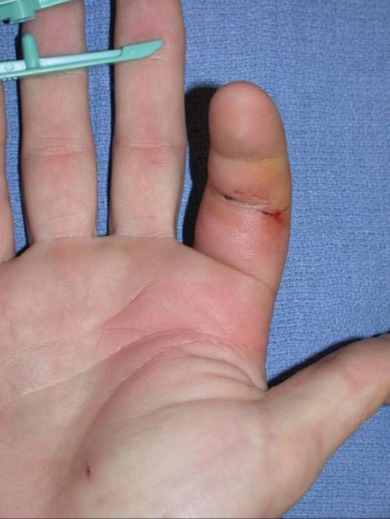
FIGURE 72.5. Pyogenic flexor tenosynovitis.
COLLAR BUTTON ABSCESS
Collar button or web space abscesses form beneath palmar calluses and penetrate through one of the three web spaces and are common in laborers. Since the dorsal skin is more compliant than the palmar skin, swelling and fluctuance from collar button abscesses are often greater dorsally. Complete drainage of these abscesses often requires incisions on both the dorsal and palmar surfaces. The failure to recognize the extent of the infection on the palmar surface can lead to incomplete drainage and spread of the infection to the deep palmar space. It is important to avoid damage to the web itself to avoid contractures postoperatively.
RADIAL, ULNAR BURSA INFECTIONS
Knowledge of the anatomy of the radial and ulnar bursae of the hand allows understanding of bacterial infections in this region. The radial bursa is the proximal extent of the flexor sheath of the thumb, while the ulnar bursa includes the flexor sheath of the little finger and palmar portions of the second, third, and fourth flexor tendon sheaths. Parona’s space (the potential space between the pronator quadratus and the flexor tendons) serves as a bridge between the radial and ulnar bursae and allows the formation of “horseshoe” abscesses. When draining abscesses in Parona’s space it is particularly important to avoid injuring the median nerve and its palmar cutaneous branch.
HUMAN BITES
Human bite injuries lead to some of the most complex of all the common hand infections. Typically, human bite injuries occur through clenched-fist injuries where the patient’s fist strikes an opponent’s tooth, piercing the metacarpophalangeal joint. The initial puncture wound may appear innocuous but leads to a septic joint in a few days. Most of these infections are polymicrobial and include a wide range of possible pathogens due to the high number of bacterial species present in the human mouth.8 Skin flora and Eikenella species are the most common organisms isolated. Additionally, these patients are often noncompliant and there is frequently a significant delay in seeking medical care.9
The management of human bite injuries includes admission, x-rays to evaluate foreign bodies and fractures, culture, and empiric antibiotics. Superficial abrasions and infections are managed with antibiotics and close observation. If the extensor mechanism has been penetrated, or the depth of penetration cannot be determined, the wound is explored in the operating room.
The management of animal bites (i.e., dog and cat) is quite similar to that of human bites. Tetanus prophylaxis is required. Dog and cat bites are more likely unimicrobial with cat bites having a high likelihood of Pasteurella multocida infection. Of the two, cat bites are more likely to become infected because the puncture wounds are small and seal quickly.
SEPTIC ARTHRITIS
Finger joint infections are typically the result of infection from adjacent tissues and less commonly the result of hematogenous spread. Symptoms of a septic joint include swelling, fluctuance, and warmth. The finger is usually held in slight flexion pain on even slight passive movement. Joint aspiration is an important diagnostic tool and will typically produce purulent/cloudy fluid that contains (1) >50,000 white blood cells, (2) >75% polymorphonuclear neutrophils, and (3) glucose <40 mg.
Once pus is identified in the joint, rapid and adequate irrigation and debridement is necessary to minimize cartilage and joint destruction. Cultures are obtained prior to starting antibiotics, which are chosen according to the gram stain results.
OSTEOMYELITIS
Osteomyelitis of the hand is typically the result of penetrating trauma or open fractures. The degree of damage to the soft tissues overlying the affected bone plays a significant role in the pathogenesis of osteomyelitis. Direct spread from a soft tissue infection such as pyogenic tenosynovitis is a rare cause of osteomyelitis. The diagnosis can be made by identifying risk factors as well as plain radiographs, nuclear medicine imaging (bone scan and tagged white blood cell scan), and magnetic resonance.
The management of osteomyelitis depends on the severity of the presenting complaint/disability and the duration of infection. Early infections with minimal complaints may be cautiously managed with intravenous antibiotics alone. However, surgical debridement is necessary in most cases to achieve adequate resolution of the infection.10 When a sequestrum is present, curettage of all necrotic bone is essential and the wound should be packed open. Should a bone defect be present after debridement, reconstruction is only considered after definitive clearance of infection (Figure 72.6).

FIGURE 72.6. Osteomyelitis from untreated paronychia: (A) Clinical appearance and (B) radiograph showing resorbed distal phalanx epiphysis.
NECROTIZING FASCIITIS
Necrotizing fasciitis is a limb- and potentially life-threatening infection that is often caused by minor trauma. Hallmarks of necrotizing fasciitis in the upper extremity include bright shiny skin, nonpitting edema, poorly demarcated redness, violaceous discoloration, and skin necrosis. Patients who are diabetic or immunocompromised are at much higher risk. A single organism is found as the causative agent in nearly 50% of cases, most often group A β-hemolytic streptococcus (occasionally Staph. aureas).11
Because the mortality of necrotizing fasciitis is as high as 40%, early and aggressive surgical debridement of all infected tissues is mandatory. Early empiric treatment with broad-spectrum antibiotics, even before cultures have been obtained, can significantly decrease morbidity. Depending on the degree of infection and soft tissue damage, serial debridements and even amputation may be necessary.
INTRAVENOUS DRUG ABUSE
The direct inoculation of bacteria into the subcutaneous tissues by intravenous drug use can lead to the rapid formation of abscesses. In addition to the introduction of bacteria, the injected material itself can cause local tissue necrosis. The most common causative agents are staphylococcal and streptococcal species. These infections are polymicrobial and present many challenges to treatment.
Appropriate management involves rapid and adequate debridement and antibiotic therapy. Repeated debridements may be necessary. Despite adequate drainage, recurrent abscesses are common. Large abscesses can be effectively drained through the area of maximal fluctuance and multiple small incisions along the periphery of the cavity. Penrose drains are threaded through these incisions, across the cavity, to maintain drainage until purulence resolves (2 to 3 days) (Figure 72.7).11
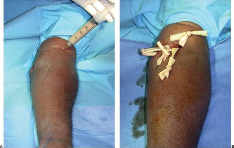
FIGURE 72.7. Subcutaneous abscess from intravenous drug abuse: (A) Clinical presentation and (B) the use of multiple small incisions and penrose drains to manage a large loculated abscess.
NOSOCOMIAL INFECTIONS
Postoperative infections in the hand are quite rare. The routine use of prophylactic antibiotics in clean, routine hand cases is not recommended. Perioperative antibiotics are recommended for operations involving implants, bone or joint spaces, or exceeding 2 hours in length. The most common causative organism for hand infections after surgery is Staph. aureus.
TREATMENT OF RESISTANT ORGANISM INFECTION
There has been a dramatic increase in the number of inpatients with nosocomial methicillin-resistant Staph. aureus (MRSA) wound infections in the last decade. This trend has been seen in the outpatient setting as well. Recently, Karanas et al.12 reported a case series of four patients with community-acquired MRSA hand infections who had no previous risk factors for MRSA infection. The increase in resistant infections underscores the importance of routine wound culture prior to initiating antibiotic therapy, whenever possible, to guide antibiotic therapy. Many institutions publish hospital-specific guidelines for empiric antibiotic coverage, which account for individual drug resistance patterns.13
CONCLUSION
Successful treatment of hand infections requires early diagnosis, appropriate antibiotic therapy, and prompt adequate drainage of abscesses. When possible, cultures are obtained prior to initiating antibiotic therapy. Abscesses should be widely drained, all necrotic tissues debrided, and wounds left open for drainage.
References
1. Cannon DL, Beatty JH. Hand infections. In: Canale ST, Beatty JH, eds. Campbell’s Operative Orthopedics. 12th ed. Philadelphia, PA: Elsevier; 2013:3694.
2. Louis, DS, Silva J Jr. Herpetic whitlow: herpetic infections of the digits. J Hand Surg (Am). 1979;4(1):90-94.
3. Clark DC. Common acute hand infections. Am Fam Physician. December 2003;68(11):2167-2176.
4. Grover C, Bansal S, Nanda S, Reddy BS, Kumar V. En bloc excision of proximal nail fold for treatment of chronic paronychia. Dermatol Surg. March 2006;32(3):393-398; discussion 398-399.
5. Connolly B, Johnstone F, Gerlinger T, Puttler E. Methicillin-resistant Staphylococcus aureus in a finger felon. J Hand Surgery (Am). January 2000;25(1):173-175.
6. Kanavel, AB: Infections of the Hand—A guide to the Surgical Treatment of Acute and Chronic Suppurative Processes in the Fingers, Hand and Forearm. 7th ed. Philadelphia, PA: Lea & Febiger; 1939:241-242.
7. Boles SD, Schmidt CC. Pyogenic flexor tenosynovitis. Hand Clin. 1998;14:567-578.
8. Chuinard RG, D’ambrosia RD. Human bite infections of the hand. J Bone Joint Surg. 1977;59a(3):416-418.
9. Zubowicz VN, Gravier M. Management of early human bites of the hand: a prospective randomized study. Plast Reconstr Surg. 1991;88:111-114.
10. Dormans JP. Hand Infections in Pediatric Orthopaedics and Sports Medicine. St Louis, MO: Mosby;2004:101.
11. Bisno AL, Stevens DL. Streptococcal infections of skin and soft tissues. N Engl J Med. January 1996;334:240-245.
12. Karanas YL, Bogdan MA, Chang J. Community acquired methicillin-resistant Staphylococcus aureus hand infections: case reports and clinical implications. J Hand Surg (Am). July 2000;25(4):760-763.
13. Moran GJ, Krishnadasan A, Gorwitz RJ, et al. Methicillin-resistant S. aureus infections among patients in the emergency department. N Engl J Med. August 2006;355:666-674.