
PART II
SKIN AND SOFT TISSUE
CHAPTER 16 PRINCIPLES OF BURN RECONSTRUCTION
MATTHIAS B. DONELAN AND ERIC C. LIAO
Reconstructive surgery following burn injury involves almost every aspect of plastic surgery. The patient population includes children and adults. All areas of the body can be involved. Deep structures can be injured either acutely or secondarily. Satisfactory outcomes require correction of both functional and aesthetic deformities. Yet, at the same time, the reconstruction of burn deformities requires a unique perspective and an emphasis on certain fundamentals and techniques that make it a specialized area of reconstructive surgery. The surgeon must thoroughly understand the processes of wound healing and contraction. The effect of time on the maturation of scars is of pivotal importance and requires patience and judgment on the part of the surgeon and the patient. Correct timing of surgery is essential. Multiple operations are the rule and frequently take place over a period of many years. Donor sites are frequently limited or compromised. Successful surgical outcomes require a well-functioning support system, including nurses, therapists, psychosocial practitioners, and, hopefully, a supportive family. All of these factors affect the outcome of surgery.
Burn injuries vary greatly in severity and extent, yet virtually all postburn deformities have similar components that must be addressed. This chapter provides a strategic approach to burn reconstruction based on surgical principles particularly relevant to this field that will help in the analysis, management, and surgical treatment of this large and challenging group of patients.
GENERAL CONCEPTS
Over the past 50 years, primary excision and grafting of deep second-degree and full-thickness burns has become the standard of care in the United States and in most developed countries (Chapter 16).1,2Early excision and grafting has decreased the mortality and morbidity of acute burn injuries.3 The duration of acute hospitalization has been greatly reduced. Early excision and grafting has also decreased the frequency and severity of contractures and hypertrophic scarring. Occasionally, however, one still encounters patients who were treated “expectantly” with late grafting and disastrous results (Figure 16.1).
All burns of the second and third degree result in open wounds. Open wounds heal by contraction and epithelialization. Contraction may be decreased by early excision and grafting, but is always present to some degree. Contraction leads to tension, and tension is one of the principal causes of hypertrophic scarring and unfavorable scarring in general. Understanding the role of tension in the evolution of postburn deformities is essential.
Burn reconstruction is fundamentally about the release of contractures and the correction of contour abnormalities. It should not be focused on the excision of burn scars. Scar excision is an oxymoron. A scar can only be traded for another scar of a different variety. When the fundamental problem is that of inadequate skin and soft tissue, further excision of “scars” can add to the clinical problem. Well-healed burn scars, if given enough time to mature, are often an excellent example of nature’s camouflage. The subtle and gradual transition from unburned skin to scar helps the deformity blend into its surroundings. A burn scar that is conspicuous at 1 year because of hypertrophy, contracture, and erythema can become inconspicuous with further maturation. Healed second-degree burn deformities under tension with resulting hypertrophy are unsightly. With time and relief of tension, they will greatly improve. Premature early excision of such scars with primary closure frequently results in a wide iatrogenic scar, which then becomes a more obvious permanent deformity. Lacking camouflage, the surgical scar may be more noticeable than the burn scar, and increased tension from the excision can create contour deformities. Excision and primary closure of burn scars should be reserved for small scars in conspicuous locations that will allow a favorably oriented closure.
Although counterintuitive, it is helpful to learn to love burn scars. After all, without scarring, healing cannot occur, so scars are our friends. For successful burn reconstruction, one must learn to appreciate scars and understand their behavior. Scar rehabilitation is usually a better alternative for the patient than scar excision. Scars under tension are angry and respond with erythema, hypertrophy, pruritus, pain, and tenderness. Relaxed scars are happy scars. They respond by flattening, softening, and becoming pale and asymptomatic. Directing reconstructive surgery toward relieving tension is practical and achievable and often results in great improvement. Advances in laser therapy have greatly facilitated scar rehabilitation, further decreasing the indications for scar excision. Ill-advised attempts to excise scars can be simplistic and are potentially harmful. Burn reconstruction strives to make the patient clearly better, not just different from normal in a different way.
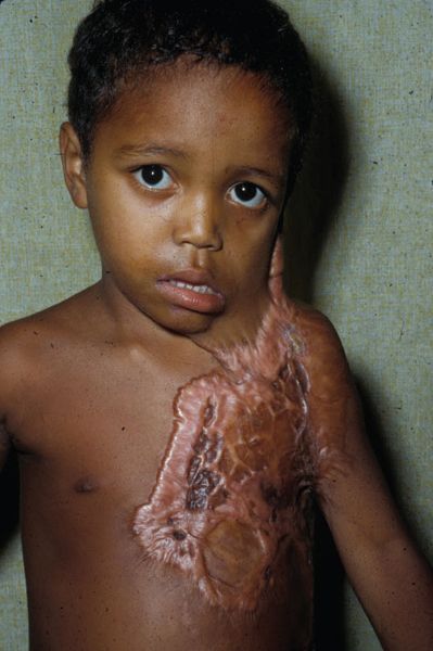
FIGURE 16.1. Contracture due to late grafting. A 4-year-old boy from Central America treated with months of dressings and late grafting, resulting in severe contractures.
Contracture releases can be accomplished with local tissue rearrangement such as Z-plasties or transposition flaps or by releases and skin grafting of the resulting defects. Releases can be performed by either incising or excising scars. Release by incision takes advantage of the healing that has already occurred and because of the relief of tension, it will usually improve the appearance and quality of the tissue that is retained. Mature scars and grafts are a known commodity and will not contract significantly after release. New grafts are less predictable. Incisional releases also create a smaller defect and, therefore, conserve donor sites. When the contracted tissue is of unacceptable quality, or too irregular, excision of scars is required for the best result (Figure 16.2). In most cases, however, it is better to work with the grafts and scars that are already present than to excise them. Grafts can be either split thickness or full thickness. Defects resulting from scar release can also be closed with flaps transferred with either traditional or microsurgical techniques. The choice of the appropriate intervention and the timing of intervention are both essential ingredients that determine success or failure after burn reconstruction.

FIGURE 16.2. Excisional release in the developing breast. A. A 15-year-old girl with bilateral lower-pole breast contractures. B. Excisional release of the lower half of the breasts with split-thickness skin grafting allowed the compressed breast tissue to expand and assume its normal shape. C. Breast augmentation and nipple areola complex reconstruction.
TIMING OF RECONSTRUCTIVE SURGERY
Patients with postburn deformities present to the plastic surgeon in one of three ways. In the ideal circumstance, the plastic surgeon is involved in the patient’s care from the time of the acute injury. The involvement may be as the treating physician or as a consultant with occasional participation in the patient’s acute care. It is a truism that the reconstruction of burn deformities begins with the acute care. Plastic surgical consultation can help prevent secondary deformities by initiating appropriate acute intervention and can also enhance outcomes by assisting with aesthetic decisions such as skin graft donor site conservation. The second group of patients are those with recent burns who received their acute burn care at another facility and come to the plastic surgeon for another opinion. They have immature scars. The third group of patients are those who present with mature scars and grafts and established burn deformities.
The timing of burn reconstruction falls into three distinct phases: acute, intermediate, and late. As a general rule, burn reconstruction is best delayed until all wounds are closed, inflammation has subsided, and scars and grafts are mature and soft. Acute reconstructive intervention is required during the early months following burn injury when urgent procedures are necessary to facilitate patient care, to close complex wounds such as open joints, or to prevent acute contractures from causing irreversible secondary damage. Examples of indications for acute surgical intervention are eyelid contractures with exposure keratitis, cervical contractures causing airway issues, and “fourth-degree burns,” such as in electrical injuries, where acute flap coverage is required.
The intermediate phase of burn reconstruction is best described as scar manipulation designed to favorably influence the healing process. After a patient’s wounds have closed, physical and occupational therapy must continue to correct or prevent contractures, as well as enhance scar maturation with the use of pressure garments, silicone gels, and massage. The efficacy of such treatments has been demonstrated over many years.4,5 Enthusiastic support of these ancillary measures by the plastic surgeon and the entire burn team can be very helpful in maximizing patient compliance. The length of time required to reach the end point of burn scar maturation is considerably longer than is generally appreciated. Scars that are thick, raised, and erythematous after 1 year or longer will often improve dramatically if given more time, often several years. When tension is present, scars never heal well. Judicious surgical intervention to relieve tension during this intermediate period can positively influence scar maturation. A longitudinal scar across the antecubital space subjected to constant tension and relaxation will remain contracted and hypertrophic despite pressure, silicone, massage, and splinting and may result in ulceration or “spontaneous release.” Relieving tension by either carrying out Z-plasties within the scarred tissue or performing a release and graft can help the entire scar to improve after the tension has been eliminated (Figure 16.3). Hypertrophic scars are common in healed second-degree burns under tension. When the tension is relieved, the subsequent improvement in appearance and elasticity is often remarkable.
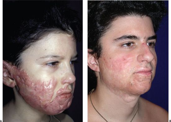
FIGURE 16.3. Multimodal scar manipulation without scar excision. A. An 8-year-old boy 6 months following flame burn injury with diffuse facial hypertrophic scarring and contractures. B. Ten years later following pressure, massage, steroid injections, and multiple Z-plasties within the scar tissue, the hypertrophy has resolved. The depth of the burn is indicated by the absence of beard growth. No scars were excised.
Steroids are effective in diminishing and softening hypertrophic scars. Topical steroids are helpful. Steroid injections are powerful. The latter must be used carefully because of potential problems with atrophy of the scar and the underlying subcutaneous tissue.6 Their use should be limited to situations where time, pressure, silicone therapy, and massage are ineffective and surgery is not an option. For example, isolated hypertrophy without tension such as on the face or shoulders is a good indication. A solution of triamcinolone (10 mg/mL mixed half and half with 1% Xylocaine with epinephrine) administered by intralesional injection with a glass tuberculin syringe, never more frequently than once a month, is efficacious in decreasing hypertrophy and preventing undesirable side effects. Ablative fractional laser therapy provides a new, and potentially more efficacious, way of delivering corticosteroids into the dense collagen of hypertrophic scars.
Intermediate-phase scar manipulation is of particular benefit in the management of facial burn deformities. This is an area where treatment is evolving and there is considerable potential for improvement. Computer-generated clear face masks with silicone lining are expensive but efficacious and well tolerated by patients. Relief of tension on facial scars by eliminating extrinsic contractures from the neck, as well as from the inconspicuous periphery of the face by release and grafting or Z-plasties, can be exceedingly beneficial to the healing of facial burns. The pulsed dye laser (PDL) is effective in decreasing facial erythema when used in this intermediate phase and seems to result in more favorable long-term scar maturation. Z-plasties within the hypertrophic scar to decrease tension and more favorably align scars can achieve dramatic results over time (Figure 16.4).7
Late-phase reconstructive surgery includes all postburn deformities that are stable and consist of mature scars and grafts. It is not uncommon in this group of patients for hypertrophic scars to present with areas of open ulceration. This is almost always caused by chronic tension. The resulting ischemia in the scar causes unstable epidermal coverage. Operations directed at relief of the tension will usually cure the chronic open wounds.
The transition from acute burn injury to the late phase of reconstructive surgery can be prolonged and is unique for each patient. The experience, judgment, and expertise of the plastic surgeon are extremely important during this period. After the acute phase of a burn injury, the patients and the patients’ families desire expeditious reconstructive surgery. Patients would like their scars to be “removed” and they want to “get on with their lives.” Most of the time, this is not in the patient’s best interest. As mentioned above, the amount of time that is required for burn scars to reach their final state of maturation is not generally appreciated. If the prolonged process of scar maturation is allowed to occur, particularly when aided by appropriate help from the surgeon and therapists, hypertrophic, contracted, and conspicuous scars that are problematic at 1 year or longer can improve greatly with more time. Because of the gradual and subtle transition from unburned skin to burn scar, mature scars are usually less conspicuous than would be surgical scars resulting from excision and primary closure. Education of the patient and the patient’s family is essential in order to help guide them to the best possible outcome. The desire for “excision” can lead to iatrogenic deformities such as shown in Figure 16.5. This unfortunate result could have been avoided with more time and Z-plasties performed within the maturing hypertrophic scar tissue.
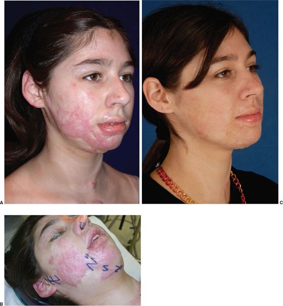
FIGURE 16.4. Pulsed dye laser and tension relief with Z-plasties. A. An 11-year-old girl, 2 years after burns with hypertrophic scarring of right cheek and lower lip ectropion. B. Z-plasties relieve tension and separate broad areas of scarring. After softening of scars, the lower lip was elevated with Z-plasties. C. Six years after burn and 12 laser procedures. The scars are flat, soft, and pale. The lower lip ectropion is corrected. No scars were excised.
Reconstructive Plan
A prospective plan for reconstructive surgery is developed with the patient and the patient’s family during the intermediate phase or at the time of consultation with a patient who has established postburn deformities. Planning the reconstructive sequence is helpful to the patient, the family, and the surgeon. Because the patient’s priorities may be different from the surgeon’s, education, careful consultation, and mutual agreement are of extreme importance. Operations to improve essential function are the initial priority, but appearance, particularly of the face and hands, is always a consideration. The goal of reconstructive surgery is to return patients as much as possible to their pre-burn condition. Therefore, all reconstructive procedures aim to improve both the function and the appearance of the operated area. The planning process gives the patients perspective and helps them develop a positive attitude as they look forward to significant improvement in the future. Enthusiasm and optimism on the part of the surgeon and the entire reconstructive team is essential. Including the patient’s family in these discussions is important. A strong support system is necessary for what is often a long and arduous process.

FIGURE 16.5. Iatrogenic deformity of the lower extremity. A. A 13-year-old girl with hypertrophic, contracted, medial popliteal scar 1 year following burn injury. B. Tissue expander in place prior to scar excision and flap rotation. C and D. Postoperative result of scar excision shows a conspicuous surgical scar, and abnormal leg contour with compression of the calf. The flap fills and deforms the medial popliteal concavity.
Fundamentals
Several basic concepts and techniques are worth reviewing in the context of burn reconstruction.
Contractures. Burns cause tissue loss, wounds heal with contraction, and contractures result. Contractures can be either intrinsic or extrinsic. Intrinsic contractures result from injury or loss of tissue in the affected area, causing distortion and deformity of the part. Extrinsic contractures occur when tissue loss at a distance from an affected area creates tension that distorts the structure. Eyelid ectropion, for example, can result from either intrinsic or extrinsic contractures. Although this concept is obvious and well known, the frequency with which it is ignored in burn reconstruction is astounding. Contracture deformities must be carefully evaluated and an accurate diagnosis made. Corrective measures can then be directed at the cause. There is rarely any indication for release and graft or Z-plasty in unburned skin because of a deformity resulting from an extrinsic contracture.
Tension. For scars to mature as well as possible, tension must be eliminated. Tension deforms normal body contours, and the resulting abnormal shape draws attention to the injured area. Relief of tension and restoration of normal contour by either release and grafting or Z-plasties is perhaps the most basic fundamental of all burn reconstruction. The amount of tension in the skin following a burn injury is often not obvious, particularly to inexperienced surgeons. When releases are carried out and defects are created, the amount of tissue required to close the open defects can be surprising.
Donor Sites. Donor site availability is often problematic in burn reconstruction. Severe burns are usually extensive, and successful reconstruction requires careful allocation of donor site material. Split-thickness grafts from the buttocks, thighs, and postaxial trunk are best used for contracture releases of the trunk and extremities. Full-thickness skin grafts from the retroauricular area, cervicopectoral area, and the upper inner arms are best reserved for head and neck reconstruction. The lower abdomen and groin are excellent donor sites for full-thickness grafts, usually allowing primary closure of the donor site. Full-thickness skin grafts from these areas tend to have a yellowish hue in fair-skinned patients, which is a disadvantage for facial reconstruction.
Release and Grafting. Nothing could be simpler than the concept of a surgical release and graft. Attention to detail is important, however, to obtain the best result. Burn contractures are usually limited to the superficial scars or grafts and a thin layer of fibrous connective tissue just beneath the skin surface. The underlying structures, such as subcutaneous fat, breast gland, and orbicularis muscle, are merely compressed and displaced. Releasing incisions or excisions should be limited whenever possible to the superficial scarred tissues alone. When this is done, normal contour is restored as the deep tissues unfurl, expand, and return to their normal shape (Figure 16.2). Failure to limit the release to the superficial scar causes iatrogenic contour deformities that are often impossible to correct (Figure 16.6). Overcorrection of the contracture is always attempted and grafts are sutured with a bolster dressing. Placing fishtail dart at the ends of the releasing incisions adds additional skin and helps to prevent recurrent contracture by creating W-plasties at the ends of the graft. Postoperative management of grafts with pressure and conformers is essential to minimize graft contracture and wrinkling. The raised edges of the grafts that result from overcorrection and pressure from the tie-over dressing will virtually always flatten. If not, they can easily be excised or revised.
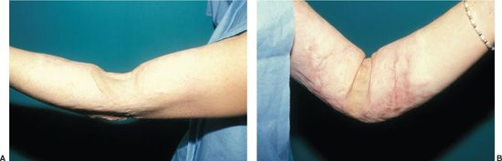
FIGURE 16.6. Failure to limit release to superficial tissue. A. Incisional release in the antecubital space violated the subcutaneous fat creating a severe contour abnormality. B. With elbow flexion, the depression and skin prolapse is a conspicuous iatrogenic deformity.
Z-plasty. The Z-plasty operation is an essential and powerful tool in the surgeon’s armamentarium for burn reconstruction. For more than 150 years, the Z-plasty has been used for its ability to lengthen linear scars by recruiting relatively lax adjacent lateral tissue. The Z-plasty, however, is much more than a simple geometrical exercise in lengthening a linear scar. When executed properly, it causes a profound beneficial effect on the physiology of scar tissue. Burn scar contractures are frequently diffuse, and excision is neither practical nor desirable. When a Z-plasty is performed properly, recruiting lateral tissue, two goals are accomplished. The central limb is lengthened, decreasing longitudinal tension on the scar, and the width of the scarred area is decreased by the medial transposition of the lateral flaps (Figure 16.7). The narrowing of scars by Z-plasty revision can be very effective. A 60° Z-plasty lengthens a scar by 75% while narrowing it by approximately 30%. The Z-plasty also adds to scar camouflage by making the borders more irregular. For a Z-plasty to lengthen a burn scar and restore elasticity, the lateral limbs must extend beyond the margins of the scar. After a successful Z-plasty, the hypertrophic scar resolves and becomes more elastic, and it also has been narrowed by the procedure. The physiology of this phenomenon is related to the immediate and ongoing remodeling of collagen that occurs in hypertrophic scars following the relief of tension.8 Hypertrophic scar remodeling also takes place when tension is relieved by release and graft, but the use of the Z-plasty is simple, elegant, and powerful. As John Stage Davis said, “It is difficult to realize how much permanent relaxation can be secured by the use of scar infiltrated tissue in this type of incision until one is familiar with the procedure and its possibilities. In addition, the improvements in the appearance of scars following Z-plasty revision is often dramatic”.9 Z-plasties can also be used to flatten hypertrophic scars and elevate depressed scars. This occurs because the lateral limbs of the Z-plasty are extended into normal tissue. When the flaps are transposed, the transverse limb goes straight across from normal to normal with a resulting leveling effect. This benefit is obtained immediately in the operating room. When the Z-plasty flaps are incised, the tips should be cut perpendicular to the central limb for a short distance as shown in Figure 16.7. This adds additional tissue to the flap tips and improves blood supply.
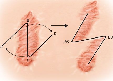
FIGURE 16.7. Z-plasty. Transposing the flaps of a Z-plasty lengthens the central limb and also narrows the involved scar by the medial transposition of the flaps. The flap tips should be incised perpendicular to the central limb for a short distance to supply more tissue and enhance the blood supply. Following transposition, the more irregular borders help to camouflage the scar.
Wherever burn scar crosses a concave surface, there is a tendency for the scar to contract, hypertrophy, and “bowstring.” Z-plasty helps alleviate this common problem. The Z-plasty can also be used at the same time to enhance contour by appropriately designing the flaps. A Z-plasty release is designed such that, following transposition of flaps, the tight transverse limb is located where a normal concavity would occur. For example, the Z-plasties shown in Figure 16.8 release contractures and are used to emphasize jawline definition. The axilla, antecubital space, and popliteal space are frequent sites of hypertrophic scar contracture with bowstringing and are often suitable for treatment with Z-plasty. The medial popliteal scar in Figure 16.5 could easily have been corrected with one or two Z-plasties within the scar, releasing the contracture, improving the appearance, and restoring the normal concave contour. Linear hypertrophic scar contractures are seen less frequently across extensor surfaces. The two exceptions are the wrist and anterior ankle because of their ability to dorsiflex.
Laser Therapy. Hypertrophic scarring is a frequent complication after partial-thickness burn injuries that take longer than 3 weeks to completely epithelialize. Despite conservative management and close monitoring, hypertrophic scars can become severe during the first 2 years after the burn and persist for years afterward. The PDL has emerged as a successful treatment modality during this period of scar proliferation and is an effective alternative to scar excision, particularly in patients with hypertrophic facial burn scars.10 Multiple studies have demonstrated its beneficial effect on scar erythema and hypertrophy. The PDL also rapidly decreases pruritis and pain and provides an additional, low-morbidity, therapeutic intervention for patients and their families during the often prolonged period of scar maturation. When combined with tension-relieving Z-plasties, the improvement can be profound (Figure 16.4). Restoration of hypertrophic facial scars to their previous state of a flat, epithelialized surface is a superior outcome compared with surgical excision with its concomitant increase in facial tension. Similar benefits can be obtained in other anatomic areas as well. The development of fractional ablative and non-ablative laser therapy using various types of lasers including CO2 and erbium offers new options for the management of burn scars in the future. Scar relaxation and improvement in texture and pigmentation have been reported following fractional laser therapy.11-13 These promising interventions are further enhancing our ability to rehabilitate burn scars.

FIGURE 16.8. Z-plasty to increase jawline definition. A. Hypertrophic contracted neck scars create an extrinsic contracture deforming the lower eyelid, oral commissure, and the jawline. B. Z-plasty design incorporates the scarred tissues in the flaps. C. Four years following Z-plasties, the facial deformities are corrected and the scar is flat, soft, and asymptomatic.
Grafts. Skin grafts are pivotal in burn reconstruction. A few generalizations about their characteristics may be helpful. Split-thickness skin grafts contract more than full-thickness skin grafts, have more propensity to wrinkle, and always remain shiny with a “glossy finish.” Thick split-thickness skin grafts contract less and provide a more durable skin coverage, but do not possess elastic properties. Meshed split-thickness grafts are rarely indicated in burn reconstruction surgery. The meshed pattern is permanently retained and has an unattractive “reptilian” appearance. Hyperpigmentation of grafts is a frequent problem in dark-skinned patients, particularly those of African descent.
Full-thickness skin grafts are reliable workhorses in facial burn reconstruction. The use of full-thickness grafts in other areas should be carefully considered. Full-thickness grafts are elastic, contract less, have a “matte finish” like normal skin, and create a durable, resilient, skin surface. Full-thickness grafts, however, require a well-vascularized bed and primary closure or grafting of the donor site and are best reserved for reconstruction of the head and neck or the hand. Composite grafts from the ear are useful for complex facial burn reconstruction, but should only be used when there is adequate blood supply in the recipient bed.
Flaps. Flaps, with or without tissue expansion, are useful for burn reconstruction. They are mandatory for complex defects such as open joints or exposed vessels or to provide tissue coverage that allows for later complex reconstruction, such as tendon or nerve grafting in the hand. Large flaps involve a considerable trade-off because of their donor site morbidity. Their elasticity and minimal contracture, as well as excellent color and texture match, make them an excellent option when available for the correction of cervical contractures (Figure 16.9). Flaps are frequently recommended in the literature for axillary contractures. The normal axilla is concave and lined with thin skin. This allows the arm to rest comfortably at the side. Transposition of flaps into the axilla can effectively release contractures, but can also create terrible contour deformities. When potential flap tissue is available, either posterior or anterior to the axilla, multiple Z-plasty flaps in series can usually release axillary contractures and preserve or restore normal contour (Figure 16.10). When diffuse axillary scarring is present, release and graft is the best option, even though it requires postoperative splinting and often more than one intervention.
Tissue Expansion. Tissue expanders have transformed the treatment of postburn alopecia. Bald areas of 50% of the scalp or more can be successfully reconstructed, frequently requiring more than one expansion. The scalp is an ideal site for tissue expansion because of its blood supply, convex shape, and the unyielding skull against which to expand (Figure 16.11). Although there are conflicting data in the literature regarding complications, in general the scalp is a privileged site for tissue expansion.14,15 The use of tissue expansion in other areas of burn reconstruction is more problematic. Because the underlying theme of burn deformities is tension and tissue deficiency, stretching adjacent tissue in order to carry out scar excision can result in increased tension and iatrogenic contour abnormalities (Figure 16.5). The complication rate of tissue expansion in burn patients can be high in the extremities, reaching 25% to 50% in some reports.16 In contrast, tissue expansion of the scalp is well tolerated and very successful. After alopecia, the most common use of tissue expansion is probably in the reconstruction of facial burn deformities. Care must be taken when advancing or transposing expanded flaps from the cervicopectoral area to the face. It is easy to create extrinsic contractures with a downward vector resulting in a “sad” facial appearance that is distressing to patients. Contour deformities can also be created in the neck with loss of jaw line definition.

FIGURE 16.9. Correction of cervical contracture using regional flap. A. Recurrent anterior cervical contracture in a 17-year-old boy following split-thickness skin grafting. B and C. Transposition flap from the unburned right cervicopectoral area restores normal function and appearance.
Evaluation and Treatment
Successful burn reconstruction requires perspective, patience, a thorough understanding of the problem, and judicious application of the fundamentals of burn reconstruction. As noted previously, burn reconstruction is primarily about the release of contractures and the correction of contour abnormalities. When contractures have a predominantly linear component and there is a relative excess of vascular, elastic tissue lateral to the contracture, the Z-plasty is simple, is reliable, and has the least morbidity. Z-plasty minimizes the need for most postoperative therapy, including pressure garments, and the benefit of the procedure is prolonged. The relaxed scar tissue will continue to soften, flatten, and loosen for many months to years after the operation is performed.
Z-plasties can also be used on the narrower, linear, components of diffuse areas of hypertrophic scarring to separate islands of scar and restore elasticity. Contour abnormalities can be corrected at the same time. The relief of tension leads to improved maturation. When erythema is present, the PDL is an effective adjunctive therapy. Following the benefit of initial scar revision, repeat surgery can be carried out 1 or 2 years later. Typically, this secondary surgery is directed toward scars that previously were not conspicuous or symptomatic but have become so after the treated scars flatten, soften, and become less noticeable. It is often remarkable how much improvement in appearance, contour, and softness can be accomplished by such “separating” Z-plasties. Patients are almost always pleased with the outcome and frequently ask for subsequent similar procedures, a true indication of successful surgery (Figure 16.12).
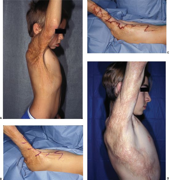
FIGURE 16.10. Multiple Z-plasties for axillary contracture. A. Extensive posterior axillary contracture with hypertrophic scarring. B and C. Multiple Z-plasties and local flaps in series easily release the contracture and flatten the hypertrophic scars. D. Eight years later complete release has been maintained, the scars are flat and soft, and the contours are normal.
When contracted scars or grafts are diffuse and Z-plasty or other local flap rearrangement is not possible, then release and split-thickness skin grafting is usually the best option to correct contractures. Care must be taken to preserve and restore normal tissue contours when releases are carried out to prevent unsightly iatrogenic contour abnormalities (Figure 16.6).
Flaps are excellent for cervical contractures when they are available (Figure 16.9). Otherwise, release and split-thickness skin grafting is a reasonable option, although this requires meticulous postoperative management and often more than one release and graft.17 Microvascular free tissue transfer has been advocated for anterior neck contractures, but its use has been limited because of complexity and morbidity.
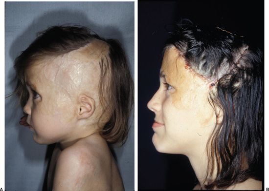
FIGURE 16.11. Tissue expansion for burn alopecia. A. A 7-year-old girl with extensive alopecia involving the vertex, parietal, and occipital areas of the scalp. B. Ten years later following two tissue expansions, alopecia has been eliminated and a normal temporal hairline and sideburn restored.
Tissue expansion is the ideal treatment for postburn alopecia. Even when the area of alopecia is relatively small, scalp expansion should be considered. Excision and direct closure of scalp alopecia usually results in a straight line scar under tension that tends to widen and become conspicuous over time. Tissue expansion allows the closure to be carried out without tension, incorporating interdigitating local flaps and Z-plasties that obscure the scar and prevent widening. Whenever possible, the use of a single large expander is desirable, even if that requires expansion of some areas of alopecia. The larger the expander, the lesser the separation of hair follicles. When expansion is accomplished with a single large expander placed through a single small incision, manipulation of the scalp at the time of alopecia excision is facilitated because the flaps have not been compromised.
Facial burn reconstruction is complicated and can seem overwhelming in severe cases. The importance of time and allowing for maximal scar maturation to occur, along with the use of ancillary techniques such as pressure, silicone gel, steroids, judicious surgical intervention, and the use of the laser for erythema cannot be overemphasized.
It can be helpful to think of patients with facial burn deformities as falling into two fundamentally different categories as described in Table 16.1. Type I deformities consist of essentially normal faces that have focal tissue loss or diffuse burn scarring with or without associated contractures. Type II deformities make up a much smaller number of patients who have “panfacial” burn deformities consisting of what can be referred to as facial burn stigmata. Table 16.2 lists the stigmata of facial burns, which include lower eyelid ectropion, shortening of the nose with ala flaring, a short retruded upper lip, lower lip eversion, inferior displacement of the lower lip, flattening of facial features, and loss of jawline definition. The surgical goals when treating type I deformities should be different from those appropriate for treating type II deformities.
Type I patients have essentially normal faces and surgical intervention should not adversely affect overall facial appearance. The surgeon must not fall into the trap of compromising normal features and contours to “excise scars.” Iatrogenic deformities create an abnormal look and can easily become grotesque. A normal looking face with scars is more attractive than an even slightly grotesque face with fewer scars. Surgery should only be performed when it is reasonably certain that it will make the patient better, not just deformed in a different way. Scar revision with Z-plasties and local flaps is usually the best option for type I patients (Figure 16.13).18 Full-thickness skin grafts from the most appropriate available donor sites are excellent for focal contractures. All human appearance is a mosaic to some degree and mosaic faces with normal movement and expression look much better in real life than they do in images. The PDL is helpful in decreasing erythema.
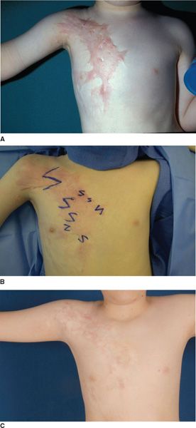
FIGURE 16.12. Effect of Z-plasties on hypertrophic scars. A. Diffuse hypertrophic scarring of the anterior chest and axilla in a 6-year-old boy with contracture and deformity of the normal contours. B. Broad areas of scar were separated with multiple Z-plasties on two separate occasions as noted in the text. C. Seven years later, after two Z-plasty procedures and treatments with the pulsed dye laser, the scars are flat, soft, and elastic. The normal chest and axillary contours have been restored.


Type II patients present a completely different clinical situation. The surgical goals for these patients should be the restoration of normal facial proportion and the return to normal of the position and shape of facial features. When normal facial proportion has been restored and facial features have been returned to their normal location and shape without tension, it is remarkable how much improvement in appearance can be accomplished in even severe facial burns (Figure 16.14).
Cosmetics are effective in covering or minimizing abnormalities of color and texture in all body areas, but their application requires skill and commitment, and their use is usually limited to the face. Many female patients become exceedingly adept at cosmetic camouflage. Male patients are less likely to take advantage of this opportunity to minimize their deformity.
CONCLUSION
Advances in the care of acutely burned patients have created a challenge and an opportunity. More patients survive today with extensive areas of healed burn scar and graft. But this increased challenge provides great opportunity for plastic surgery. Although much gloom and doom surrounds the acute care of burn patients, the injuries are usually superficial. Other than the burn scars and contractures, these patients are usually completely healthy, and successful reconstructive surgery can often restore them to a happy and productive life. Large series have shown excellent long-term outcomes in even extensively injured patients when compared with normal controls.19 Patience, persistence, and determination are essential to accomplish successful reconstruction. The skillful application of basic surgical techniques to the reconstruction of postburn deformities can be gratifying to patients and surgeons alike. Ancillary techniques of scar rehabilitation and photomedicine are providing less morbid and more effective ways to reconstruct burn deformities. The future is bright for further progress. The ultimate principle of burn reconstruction is learning to understand, appreciate, and favorably influence the processes of wound healing and scar maturation.
ACKNOWLEDGMENTS
The authors thank Ms. Aisling Fitzpatrick for assistance with preparation of this manuscript.
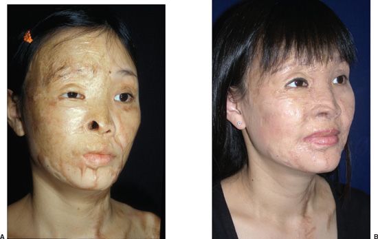
FIGURE 16.13. Type I patient. A. A 24-year-old woman following extensive acid burns to the face. B. Nasal reconstruction was performed with turn-down flaps and split-thickness skin grafting. Four facial scar revisions with Z-plasties and local flaps were carried out over a 3-year period.

FIGURE 16.14. Type II patient. A. A 30-year-old male firefighter following a severe facial burn with facial burn stigmata. B. Eight years later following extensive reconstructive surgery with full-thickness grafts, composite grafts, and multiple scar revisions.
References
1. Cope O, Langohr JL, Moore FD, Webster RC Jr. Expeditious care of full-thickness burn wounds by surgical excision and grafting. Ann Surg. 1947;125:1-22.
2. Janzekovic Z. A new concept in the early excision and immediate grafting of burns. J Trauma. 1970;10:1103-1108.
3. Burke JF, Bondoc CC, Quinby WC. Primary burn excision and immediate grafting: a method shortening illness. J Trauma. 1974;14:389-395.
4. Larson DL, Abston S, Evans EB, Dobrkovsky M, Linares HA. Techniques for decreasing scar formation and contractures in the burned patient. J Trauma. 1971;11:17.
5. Ahn ST, Monafo WW, Mustoe TA. Topical silicone gel: a new treatment for hypertrophic scars. Surgery. 1989;106:7.
6. Ketchum LD, Robinson DW, Masters FW. Follow-up on treatment of hypertrophic scars and keloids with triamcinolone. Plast Reconstr Surg. 1971;48:256-259.
7. Donelan MB, Parrett BM, Sheridan RL. Pulsed dye laser therapy and Z-plasty for facial burn scars: the alternative to excision. Ann Plast Surg. 2008;60:480-486.
8. Longacre JJ, Berry HK, Basom CR, Townsend SF. The effects of Z plasty on hypertrophic scars. Scand J Plast Reconstr Surg. 1976;10:113-128.
9. Davis JS. The relaxation of scar contractures by means of the Z-, or reversed Z-type incision: stressing the use of scar infiltrated tissues. Ann Surg. 1931;94:871-884.
10. Parrett BM, Donelan MB. Pulsed dye laser in burn scars: current concepts and future directions. Burns. 2010;36:443-449.
11. Waibel J, Beer K. Fractional laser resurfacing for thermal burns. J Drugs Dermatol. 2008;7(1):12-14.
12. Haedersdal M, Moreau K, Beyer D, Nymann P, Alsbjorn B. Fractional nonablative 1540 nm laser resurfacing for thermal burn scars: a randomized controlled trial. Lasers Surg Med. 2009;41:189-195.
13. Lee SJ, Kim JH, Lee SE, Chung WS, Oh SH, Cho SB. Hypertrophic scarring after burn scar treatment with a 10,600-nm carbon dioxide fractional laser. Dermatol Surg. 2011;37:1168-1172.
14. Neale HW, High RM, Billmire DA, et al. Complications of controlled tissue expansion in the pediatric burn patient. Plast Reconstr Surg. 1988;82:840-848.
15. Pisarski GP, Mertens D, Warden GD, Neale HW. Tissue expander complications in the pediatric burn patient. Plast Reconstr Surg. 1998;102: 1008-1012.
16. Friedman RM, Ingram AE Jr, Rohrich RJ, et al. Risk factors for complications in pediatric tissue expansion. Plast Reconstr Surg. 1996;98: 1242-1246.
17. Cronin TD. The use of a molded splint to prevent contracture after split skin grafting on the neck. Plast Reconstr Surg Transplant Bull. 1961;27:7-18.
18. Taylor HO, Carty M, Driscoll D, Lewis M, Donelan MB. Nasal reconstruction after severe facial burns using a local turn-down flap. Ann Plast Surg. 2009;62(2):175-179.
19. Sheridan RL, Hinson MI, Liang MH, et al. Long-term outcome of children surviving massive burns. JAMA. 2000;283:69-73.