
PART IV
HEAD AND NECK
CHAPTER 29 SOFT-TISSUE AND SKELETAL INJURIES OF THE FACE
LARRY H. HOLLIER JR., PATRICK KELLEY, AND JOHN C. KOSHY
The treatment of the facial trauma patient continues to evolve with progress in imaging, bone fixation technology, and the application of microsurgical reconstructive techniques. Many of the principles of access, reduction, and fixation remain constant, but the application of these principles has been greatly facilitated with improvements in instrumentation and osteosynthesis technology. Facial trauma continues to be treated by a variety of specialists, including plastic surgeons, otolaryngologists, and oral surgeons. Plastic surgeons, however, are uniquely trained to handle the full range of issues present in the trauma patient.
INITIAL MANAGEMENT
Facial injuries themselves are rarely life threatening, but are indicators of the energy of injury. Initial care of all trauma patients focuses on the algorithmic protocol of ATLS (Advanced Trauma Life Support). Facial injuries should alert the examiner to the possibility of airway compromise, cervical spine injuries, or central nervous system injuries.
Airway
Airway compromise is the result of excessive bleeding from an upper airway source, foreign bodies (including aspirated teeth and bone fragments), or direct laryngeal injury. Often, upright positioning with cervical spine protection will improve airway function compromised by excessive bleeding or foreign bodies. Foreign bodies, when present, can also be mechanically removed by the finger-sweep technique. Airway compromise can also occur when the floor of the mouth and tongue lose support from a comminuted mandible fracture and can be alleviated by simple anterior traction on the mandibular symphysis.
The trauma team should have a low threshold for definitive airway protection via endotracheal intubation. The use of blind nasal intubation should be carried out with caution, as the procedure can exacerbate nasal and nasopharyngeal bleeding. Additionally, the tube may be inadvertently placed intracranially in the obtunded patient with a skull base fracture. Endoscopic nasal or oral intubation improves safety by avoiding cervical spine manipulation and further provides immediate confirmation of tracheal intubation.
Emergent tracheotomy is considered in the unusual circumstance of laryngeal fracture or inability to secure an upper airway route to intubation. Tracheotomy performed in the controlled environment of the operating room is far superior to either emergent tracheotomy or cricothyrotomy (also called cricothyroidotomy); however, certain situations will require this emergent procedure, and there should be a low threshold for performing an emergent tracheotomy or cricothyrotomy. There should also be a low threshold for a controlled, temporary tracheotomy procedure, in the patient with significant soft-tissue trauma to the floor of the mouth and tongue, especially the base of tongue, even if the airway appears stable initially. These injuries are more commonly penetrating in nature and initially misleading as there may be minimal early signs of distress. The swelling that develops over the next 24 to 48 hours, however, may be sufficient to compromise the airway, which may result in a tracheotomy under less than favorable circumstances.
Hemorrhage
The dense vascularity of the head and neck can cause significant blood loss from soft-tissue injuries. Fortunately, most of these injuries allow sufficient access to apply direct pressure and control bleeding. Pressure should be applied accurately and directly, as a number of critical structures can become collateral victims by attempting to clamp sources of bleeding with poor exposure and visualization. Bleeding that cannot be controlled with direct pressure requires packing. Packing in the nasal cavity is usually effective and only rarely requires augmentation with a transnasal balloon catheter in the nasopharynx. These catheters only serve to impede blood from entering the oropharynx where it can more easily enter the lungs. If massive hemorrhage is present, the airway should be managed first by emergent intubation, followed by packing and direct pressure. The source of bleeding is most commonly a branch of the external carotid system, which is most appropriately controlled with angiographic embolization. The radiologist frequently requires the assistance of the surgeon to remove the packing so that the bleeding source can be identified. At this time, and with all cases of significant hemorrhage, type-specific blood should be readily available. Surgical ligation of the external carotid artery is not adequate and will not control bleeding from its injured branches because of the robust collateralization present and should not be attempted (Figure 29.1).
Central Nervous System
Neurologic injury is commonly associated with severe facial trauma. A retrospective study of the National Trauma Data Bank found that the risk of head injury in the setting of isolated facial fractures ranged from 29% to 80%, with increasing rates as the fracture involved more cranial portions of the facial skeleton. In patients with multiple fractures, however, the incidence of head injury becomes more uniform, affecting between 66% and 89% of all cases presenting at major trauma centers. Identifying these injuries is important, as patients with facial trauma rarely die from facial injuries, but can die from associated injuries of the central nervous system. Most patients with facial trauma undergo computed tomography (CT) scanning. The most widely accepted method for expressing the degree of neurologic injury is the Glasgow Coma Score. This evaluates the motor, verbal, and eye-opening responses of the patient on initial evaluation, rating the patient from a lowest score of 3 to a highest score of 15 (Table 29.1).
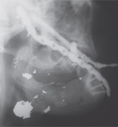
FIGURE 29.1. Hemorrhage treated by embolization. Patient with gunshot wound to mandible requiring embolization of lingual artery (note coil) to prevent exsanguination. Segmental defect is treated with a distraction device.
As a general rule, concomitant head injury is not a contraindication to facial fracture repair, assuming the neurologic injury is stable and not in the process of evolution. In the event of acute brain injury, the surgical repair of facial fractures generally is delayed to avoid the fluid overload associated with surgery and, most importantly, to avoid undetected decline of neurologic function during the period of general anesthesia when clinical neurologic examination cannot be performed. Once the central nervous system injury and concomitant swelling have stabilized, facial fracture repair can generally be undertaken safely.

Facial fractures are also often complicated by cervical spine injuries. In fact, a recent study of the National Trauma Database found that 5% to 8% of isolated facial fractures and 7% to 11% of cases of multiple facial fractures will have an associated c-spine injury.1 Suspicion for injury and vigilant care of the cervical spine are key elements in the care of facial trauma patients. Cervical spine precautions are mandatory until the spine is cleared both clinically and radiographically. For the obtunded patient, the cervical spine is best evaluated with a CT scan, although a negative exam does not rule out unstable ligamentous injury. These patients require additional examination when the sensorium is clear and possibly flexion/extension radiographs or magnetic resonance imaging to definitively evaluate the cervical spine.
FACIAL TRAUMA EVALUATION
History
Use of the AMPLE acronym (allergies, medications, past history, last meal, events surrounding the accident) facilitates a complete trauma history.
Head and Neck Examination
A thorough head and neck examination is performed in a logical and consistent manner to avoid missed injuries. The examination includes the skin, soft tissue, neurovascular structures, and bone. Initially, gross examination identifies skin and soft-tissue defects and any exposed bone. Next, ecchymoses and soft-tissue swelling serve as red flags for potential underlying injury and are used with information regarding the mechanism of injury to develop a level of suspicion about the underlying injuries. Bony structures should be palpated in a systematic fashion to identify tenderness, deformity, or step-offs. In the acutely injured patient with facial trauma, however, the physical exam is greatly impaired by facial swelling, and facial asymmetries secondary to fractures are usually concealed. Additionally, it may be difficult to elicit tenderness because of simultaneous distracting injuries. The examiner must not be misled by more impressive injuries and overlook less obvious but potentially significant problems.
The examiner then carefully assesses the patient for neurologic deficits, including the trigeminal and facial nerves. Sensory disturbances in the forehead, cheek, and lower lip should be well documented, as should any deficits in facial nerve function. Nerve injuries that are not documented preoperatively may be attributed postoperatively to surgical intervention. Lacerations, contusions, and abrasions of the skin may focus the exam by indicating which nerves are at risk.
Much of the long-term morbidity of facial trauma is associated with ocular and orbital injury. Although there should be a low threshold to involve the ophthalmologist, the physician treating facial trauma should be well versed in the ocular examination. A complete ocular examination includes the evaluation of ocular history, acuity, light and red light perception, ocular motility, pupillary exam, and examination of the conjunctiva and eyelids. Each eye requires assessment individually.
Examination of the oral cavity is essential, especially in the obtunded patient who may have loose teeth, bone fragments, or foreign bodies. Identification and removal of prosthetics (e.g., dentures) is essential. The occlusion and intercuspation are carefully evaluated, as both mandibular and maxillary fractures can result in malocclusion. Patients are capable of sensing the slightest change in their occlusion. Even in patients with unusual bites, careful analysis of the wear facets may enable the surgeon to determine if an underlying malocclusion is present.
Proper record keeping of facial injuries includes rough sketch drawings in the medical chart and photographs to document injuries. These photographs may prove invaluable in the treatment of secondary deformities and can also be beneficial in medicolegal disputes. As such, photographic consents should routinely be obtained as part of the treatment consent upon entrance to the emergency department.
Imaging
In almost all patients with facial trauma, CT scanning is performed and is acceptable for the diagnosis of essentially all facial fractures. The scan is performed with axial cuts no greater than 3 mm apart, from the top of the cranium through the bottom of the mandible. Additionally, in cases of complex facial trauma, it is helpful to have a three-dimensional reconstruction of the facial skeleton formatted so as to provide for a better overall orientation.
The one area where the CT scan may not be entirely sufficient is the mandible. Although the CT is essentially 100% sensitive and specific for the fractures, it does not give detailed information about dental structures. This is most critical in the region of the mandibular angle with respect to the condition of the second and third molars. Information regarding root damage and tooth position relative to the fracture affects the planning and treatment of angle fractures and is necessary to achieve optimal outcomes. For more detailed information regarding these variables, a panoramic radiograph is extremely beneficial. These radiographs evaluate the entire mandible, from condyle to condyle, in a single image and provide excellent detail of the condyles and dentition. Several downsides exist, though. First, the more commonly used panoramic devices require upright positioning and cervical spine clearance to be used, although certain less common devices allow the patient to be imaged while supine. Additionally, care must be taken when interpreting fractures based solely on a panoramic radiograph of the mandible, especially the symphysis and parasymphysis, as distortion of these regions can be misleading. In these cases, a supplemental posteroanterior film of the mandible complements the panoramic image by providing additional detail of the region. Lateral radiographs and/or CT scans can provide additional information about the regions posterior to the parasymphysis (Figure 29.2).
TREATMENT OF SOFT-TISSUE INJURIES
Preparation and Anesthesia
Facial injuries frequently involve contaminated wounds. The most important initial responsibility of the surgeon is to convert the contaminated wound to a clean one and then perform wound closure. Wounds are closed as soon as possible. Although facial wounds can usually tolerate up to a 24-hour delay in repair, the longer the wound is open, the greater the chance of infectious complications. Cleansing of wounds is best performed with a mild surgical soap with the light use of a scrub brush. More extensive wounds or those with a great deal of contamination should be irrigated with a pulsed lavage system. All foreign debris are removed from the wound prior to closure. Adequate cleansing usually requires total anesthesia of the region of concern and a cooperative patient. These are the primary factors that dictate the method of anesthesia necessary.
Although general anesthesia may be necessary for some wounds, many facial wounds can be repaired under a regional nerve block. If regional blockade is impractical, a field block may be necessary to avoid direct infiltration of excessive amounts of local anesthetic. Excess local anesthetic may cause distortion of anatomic landmarks that are useful in restoring the tissues to their anatomic positions.
Only 1 to 2 mL of anesthetic at the site of the nerve trunk is needed to provide complete anesthesia for the respective region. Often multiple regions require blockade. We recommend 1% lidocaine with 1:100,000 epinephrine solution mixed at a 9:1 ratio with 8.4% sodium bicarbonate solution (e.g., 9 mL lidocaine with epinephrine and 1 mL of bicarbonate solution). The bicarbonate solution neutralizes the pH of the lidocaine, which has two important benefits. First, it minimizes the pain of the injection. Second, the lidocaine more effectively crosses the neural membrane to affect its sodium ion channel in the neutral state and, therefore, has a quicker onset of action. Topical cocaine (4%) is an excellent choice for anesthesia in the nasal cavity as it also stimulates vasoconstriction of the nasal mucosa providing for a painless and bloodless field.
Traumatic Wounds
Traumatic wounds can be variously described as lacerations, punctures, contusions, abrasions, and crush injuries. The wound configuration, whether linear or stellate, is much less important to the final result than the degree of crush, contusion, and vascular compromise of the tissues. The importance of wound cleansing prior to closure cannot be overemphasized. Removal of all foreign bodies is essential as they are the source of a prolonged inflammatory response and possibly infection. Abrasions with residual foreign bodies will form a traumatic tattoo when not properly debrided. When tissue laxity allows removal of crushed tissue margins, a judicious sharp debridement of the wound margins is undertaken. Clearly devitalized tissue is excised. Freshening the wound margins contributes to rapid healing and improves the final result.

FIGURE 29.2. Fracture missed by Panorex. A. The mandible appears normal on panoramic radiograph. B. Computed tomography scan of same patient revealing complete fracture. Although panoramic radiographs provide valuable information about the mandible and dentition, distortion in the image can conceal fractures.
Perhaps the most important step in the repair of skin lacerations is excellent approximation of the deep dermal layer. By placing the tension of the closure deep to the skin, the resulting scar is improved. A good choice of the suture material for this deep layer is poliglecaprone 25 (Monocryl; Ethicon, Somerville, NJ). Because of the monofilament nature of this suture, it may have a lower likelihood of suture contamination and extrusion. It also maintains tensile strength for a sufficient period of time to allow for uncomplicated wound healing.
The choice of suture for the skin depends on the patient. Assuming a good deep dermal layer has been placed, the skin suture serves only to more accurately approximate and evert the skin edges. In children, it is beneficial to avoid a permanent suture to obviate the need for suture removal. An excellent choice in this case is 5-0 or 6-0 fast-absorbing gut suture. This suture type dissolves so rapidly that suture marks are not left on the face. It provides very little tensile strength, however, requiring the use of adhesive strips. If a skin adhesive is chosen in the pediatric population, one must take care not to place any within the wound itself, which may cause a profound inflammatory response, resulting in breakdown of the closure.
A subcuticular skin suture is also an option in the face. Monocryl is again a good choice. Should one desire to remove the suture, polypropylene may be the best choice as it slides out of the skin easily. Skin edges under a greater degree of tension are usually best closed using interrupted nylon or Prolene. In heavily contaminated wounds, interrupted sutures or running sutures in short segments can be used. This allows for removal of focal areas of suture in the case of infection, avoiding a complete wound dehiscence. As a general rule, sutures in the face can be removed by 5 to 7 days when they are load bearing. When a layer of reliable deep dermal sutures is in place, superficial skin sutures can be removed as soon as 3 days to avoid suture marks.
Injuries to Special Facial Regions
Eyelids. The most important aspect of evaluating trauma to the eyelids is ensuring that injury to the globe has not occurred. A thorough ocular examination is an essential element. It is important to remember that the Bell phenomenon results in an upward and lateral rotation of the globe. As such, one may find penetrating injuries to the globe in locations that do not intuitively correspond to the eyelid injury.
Often a general anesthetic is required to provide sufficient anesthesia to explore eyelid injuries and allow for adequate exploration of the globe. General anesthesia is particularly recommended in the pediatric population where additional damage can be caused by working with sharp needles and instruments around the orbit in an uncooperative child. Direct injuries to the globe warrant urgent ophthalmologic consultation.
The most critical step in eyelid repair is placement of an everting suture along the lid margin. This facilitates proper alignment and makes notching of the lid margin less likely. The suture in the lid margin can be left long and taped down to the cheek to prevent the suture ends from irritating the eye. In general, all layers of the eyelid (inner, middle, and outer lamellas) should be repaired. Although the conjunctiva will heal well without sutures, injuries associated with significant deformity should be sutured with plain gut suture, burying the knots to avoid irritation of the globe. The middle lamella, including the tarsus, is repaired with resorbable suture. The skin of the eyelid is then repaired. Sutures are removed within 5 days. Depending on the magnitude of the injury, it may be helpful to place a Frost suture to support the lid position during healing, especially in injuries to the lower lids (Chapter 32).
Ears. Ear lacerations can usually be sutured in one layer, addressing the skin only. It is typically unnecessary to place a separate layer of sutures within the cartilage. The firm adherence of the skin to the underlying cartilage framework of the ear ensures that skin approximation accurately aligns the cartilage.
The two most prominent concerns in ear injuries are hematoma and chondritis. Collections of blood in proximity to the cartilage can result in cartilage resorption or a reactive chondrogenesis, which ultimately leads to cauliflower ear deformity. Hematomas are evacuated as quickly as possible to avert this adverse sequela. Hematomas are drained through incisions in the overlying skin, making an effort to conceal incisions if possible. Because of the robust perfusion to the auricular skin, a bolster is often required to prevent reaccumulation of the hematoma. Alternatively, a small suction drain or Penrose-type drain may be used. A compression dressing is employed regardless of the type of drainage technique employed. Following treatment of significant lacerations, the convolutions of the ear are lined with antibiotic-impregnated gauze and the ear bandaged in a light head wrap, providing gentle compression of the ear.
As a general rule, ear trauma is not terribly painful. The development of pain in the posttreatment period may indicate hematoma or infection. Delayed onset of pain, therefore, warrants immediate inspection. Infection involving the cartilage (chondritis) is a serious complication. Cartilage has poor blood supply, making it difficult to treat chondritis with oral antibiotics. These patients typically require admission for intravenous antibiotics and possibly debridement. It is rare to develop a significant chondritis without concomitant pain. Chondritis that is overlooked or not treated promptly may result in loss of a significant portion of the auricular cartilage.
Nose. Soft-tissue injuries of the nose are somewhat different from auricular trauma. When lacerations involve the underlying cartilaginous support system of the nose, all layers should be repaired after appropriate anatomic reduction. Simple reapproximation of the overlying skin does not necessarily align the underlying cartilage. As such, any lacerations or transections of the upper or lower lateral cartilages should be separately addressed. Because of the difficulty in achieving adequate anesthesia and control of bleeding with the use of local anesthetic alone, general anesthesia is warranted to maximize patient comfort and control.
Lips. The most important consideration in repairing soft-tissue injuries involving the lips involves accurate reapproximation of the injured structures, especially the vermilion. A discrepancy in alignment of the vermilion border as little as 1 mm is noticeable at conversational distance. As such, prior to infiltration of any local anesthetic, the location of the vermilion border on either side of a laceration should be tattooed using a needle with methylene blue. The vermillion should be accurately reapproximated using a 6-0 nylon or similar suture.
Great care must be taken to separately reapproximate the underlying orbicularis oris muscle. Failure to do so will result in bunching of the muscle on either side of the laceration with attempted animation and typically results in a shortened scar with an exaggerated notching of the lip. Mucosal lacerations are repaired using a resorbable suture such as chromic or Vicryl (Ethicon, Somerville, NJ).
A careful examination is performed to rule out underlying damage to the dentition. Any loose or damaged teeth are documented. Particularly unstable teeth may benefit from a bridle wire securing them to adjacent stable teeth. Panoramic radiographs or periapical images may help to better delineate the underlying dental trauma.
Facial Nerve. Soft-tissue injuries to the face involving the facial nerve are particularly devastating. In examining the patient with facial soft-tissue injury, particularly penetrating wounds, facial motion is examined carefully. One should specifically test elevation of brow, forced closure of the eyes, voluntary smile, and eversion of the lower lip. Eversion of the lower lip is not very well tested by asking a patient to purse the lips; rather, it is best seen in attempted full-denture smile. Deficits in the presence of a penetrating injury likely represent transection of a facial nerve branch. As a general rule, all such injuries should be explored operatively. The exception may be suspected injuries to the buccal branches medial to the lateral canthus of the eye, as a consequence of the extensive arborization of the nerve at this level. Most such injuries will undergo spontaneous reinnervation over a 3- to 6-month period. Injuries lateral to this and any deficit in brow elevation, eye closure, or lower lip depression should be explored.
Timing is of importance in these situations. The ability to identify the distal transected nerve end is facilitated by stimulating with a facial nerve stimulator and detecting the facial motion. After approximately 48 to 72 hours, the distal nerve end can no longer be stimulated, greatly complicating accurate identification because of the small size of the nerve and the inflammatory response in the surrounding tissues. These injuries should be repaired using microscopic magnification and 9-0 or 10-0 nylon epineural sutures. The time to recovery for a repaired nerve can be approximated by measuring the distance between the site of injury and the target muscle. Nerve regeneration typically occurs at a rate of 1 mm/d after a 1-month lag (Chapter 9).
Parotid Gland/Duct Injuries. The most significant concern in parotid injuries is the possibility of facial nerve injury. The facial nerve separates the parotid into a superficial and deep lobe, and lacerations in this region frequently injure both the parotid gland and facial nerve. A parotid gland injury does not require intervention unless the underlying parotid duct is involved. Involvement of the Stensen duct may result in a parotid fistula unless corrected. These injuries may be difficult to identify and may only be seen following repair of skin lacerations with subsequent accumulation of saliva. Identification and access to the distal segment of the Stensen duct can also be facilitated by cannulating the papilla opposite the maxillary second molar with a blunt parotid or lacrimal probe. The ends of the duct are freshened and repaired over a stent. We often employ a 5 French pediatric feeding tube brought through the papilla and secured intraorally to prevent inadvertent displacement during the healing period.
FACIAL FRACTURES
Orbital Fractures
Orbital Examination. In patients with trauma involving the orbit, a thorough examination of the globe and associated structures is performed. If the patient has had previous iatrogenic globe penetration, such as cataract surgery or radial keratotomy, the risk of globe rupture following trauma is substantially increased. A visual examination is then performed, including an exam of the visual fields. Any damage to the optic nerve may manifest first as a limitation in the visual field rather than a significant change in gross acuity.
Additionally, one should test for color desaturation. The first indication of optic nerve compression may be red color desaturation. The easiest way to test this in the emergency department is to dim the lights and hold a penlight up to the finger. The light through the skin appears red. The patient should be asked with alternate eyes closed whether there is any difference in the intensity of the red color between the two eyes.
Direct and consensual pupillary responses are elicited to determine the function of the second and third cranial nerves. Anisocoria may be an indication of second or third nerve damage, or direct trauma to the iris. An afferent pupillary defect is indicative of optic nerve injury and can be elicited by a swinging flashlight test. Range-of-motion testing of each eye will determine the function of the third, fourth, and sixth cranial nerves. Restrictions in the range of motion of the globe should be confirmed with a forced duction test to determine if the restriction is caused by mechanical entrapment or by injury to the nerves or muscles. These emergency department maneuvers, although potentially quite informative, are no substitute for a thorough dilated exam by an ophthalmologist. Ophthalmologic consultation should be considered in every case of orbital trauma.
Indications for Surgery: Orbital Floor. Indications for the repair of orbital fractures are an area of controversy. Mechanical entrapment of an extraocular muscle may be demonstrated on forced duction testing or on imaging studies and is an indication for surgical repair. A second surgical indication is evidence of enophthalmos. With the initial swelling present secondary to the trauma, any enophthalmos that is manifest indicates a significant deformity as it would be expected to worsen with the resolution of swelling. Deferring surgery will complicate the eventual repair that is required.
Defect size is the most controversial parameter in determining the indication for surgery. Various authors have used different guidelines.2 Many believe that any defect greater than 1 cm2 benefits from surgical repair because of the likelihood of subsequent enophthalmos. Other authors have tried to quantitate, via CT imaging, the actual increase in orbital volume compared with the uninjured side. This volume is then used to assess the risk of postinjury enophthalmos. Currently, there are no firm data confirming the usefulness of this approach.
There is some benefit, regardless of the indications for surgery, in delaying surgery until there is a modest improvement in swelling. As a successful outcome in these injuries is dependent on reestablishing the proper anteroposterior projection of the globe, it is helpful to have most swelling largely resolved. Because the surgical exploration of the floor evokes some degree of swelling, the cumulative effect can make it difficult to gauge the degree of correction necessary. However, whether the surgery is performed early or late, the operated eye is overcorrected so that it projects a little farther than the uninjured eye immediately following surgery. Delays in care greater than 3 weeks may result in greater difficulty with the surgery as the periorbita becomes increasingly scarred within the defect.
Incisions/Technique. The most common complication from incisions used to access the orbital floor is eyelid malposition. The subciliary approach, according to the literature, has the highest risk of associated lid retraction. The transconjunctival approach decreases the risk of lid malposition by avoiding incisions through the skin and orbicularis muscle. Although some authors believe that a lateral canthotomy is unnecessary, there is no question that detaching the lateral canthus improves exposure. If a lateral canthotomy is performed, a canthopexy must be performed at the conclusion of the case in order to reestablish the appropriate position of the lateral canthal ligament. The subtarsal incision can also be employed, especially in older patients with prominent lower-lid rhytids within which the incision is concealed. This incision provides the most direct access to the orbital floor and is unlikely to cause lower-lid retraction. If a lateral canthotomy is not performed, the surgeon should be careful that the assistant does not retract with excessive force on the lower eyelid, which can result in inadvertent tears in the eyelid and may also predispose to entropion.
Dissection of the lower lid is directed toward the periosteum of the infraorbital rim, which is incised on the outer margin of the rim. A periosteal elevator is used to dissect posteriorly. When the infraorbital nerve is encountered within the orbital floor, it is protected and kept down with the bone. The goal of the dissection is to identify the portion of the floor that is still intact and contiguous with the orbital apex. Whatever material is used to reconstruct the floor must be suspended on this posterior ledge to ensure accurate anatomic reconstruction. At times, the posterior ledge can be difficult to identify. In these cases, it can be safely identified by placing the elevator straight posteriorly into the maxillary sinus and gently moving the elevator superiorly (Figure 29.3). The posterior floor is then encountered. Once the margins of the defect are defined, reconstruction can proceed.
Floor Implants. Tessier recommended bone for reconstruction of the orbital floor. Although reliable, the use of bone does entail a longer operative time, potential donor site morbidity, and some degree of resorption of the graft postoperatively. Alloplastic materials enjoy several advantages over bone grafts, including immediate availability and no risk of resorption. The disadvantages are the potential risks of implant infection and extrusion, but these complications are rare. Among the implants available are titanium mesh, high-density porous polyethylene, combination polyethylene–titanium implants (Figure 29.4), and resorbable materials. Very large orbital defects, particularly those involving the medial wall, are best reconstructed using implants containing titanium mesh because of the support provided and the ease of contouring. Resorbable implants may be useful in smaller defects, particularly in children, where there is some reluctance to use permanent implants because of growth concerns. Silastic, although used commonly in the past, is best avoided as a floor implant because of a relatively higher risk of late extrusion and infection.
Complications. The most common complications following surgery for orbital fractures are lower-eyelid retraction and enophthalmos. As previously discussed, lower-eyelid retraction is minimized by appropriate choice of incision. It may also be helpful postoperatively to place a Frost suture maintaining the lower eyelid in an elevated position for 24 hours following the procedure. If retraction is noted in the early postoperative period, the patient should begin aggressive lower-eyelid massage and forced eye-closure exercises. This resolves the problem in the majority of cases. Early operative intervention for lid retractions should be avoided unless the patient is experiencing problems with corneal exposure and constant irritation of the eye. In the absence of symptoms, operative intervention for lid retraction should be delayed and then considered after 4 to 6 months if conservative measures fail. Regardless of the initial incision, patients with postoperative lid retraction should be approached through a transconjunctival incision. Scarring of the middle lamella is released and a spacer such as hard palate mucosa placed.
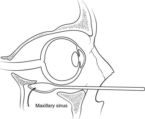
FIGURE 29.3. Identification of posterior ledge in orbital floor fractures. Technique of safely identifying the posterior limit of an orbital floor defect. The elevator is placed into maxillary sinus and used to identify the posterior ledge by sweeping upward and forward.
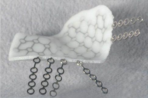
FIGURE 29.4. Combination polyethylene–titanium implants can be used to provide both support and ease of contouring. These function well in managing large orbital defects.
Enophthalmos is particularly resistant to secondary correction and is most frequently caused by inaccurate reconstruction of the orbital floor and excessive orbital volume. As a result of the incline of the floor, placement of the floor implant must be directed superiorly to anatomically reconstruct the defect. Dissecting straight back rather than cephalically results in the implant being placed within the maxillary sinus (Figure 29.5), maintaining the expansion in the orbital volume and resulting in enophthalmos. One must also remember that appropriate reconstruction of the orbital floor results in the operated eye being slightly proptotic in the early postoperative period when compared with the uninjured side. Failure to achieve this at the time of surgery is an almost certain guarantee of enophthalmos when the swelling resolves. Although fat atrophy may also increase orbital volume somewhat postoperatively, the contribution of this mechanism to enophthalmos is likely not significant. The most direct approach for correction of enophthalmos is accurate and anatomic reconstruction of the orbital floor. Secondary reconstruction is more difficult than repair at the initial procedure. However, established postoperative enophthalmos never improves and, if bothersome to the patient, will require reoperation.
Persistent diplopia may also be seen following repair of orbital fractures. This is most troublesome if it is in the primary field of gaze or in downgaze, as walking may become problematic. Performing a forced duction test intraoperatively to ensure that implant placement has not resulted in a mechanical interference with globe movement makes postoperative diplopia less common. In the event that the patient complains of new-onset diplopia in the immediate postoperative period, a CT scan is performed. If no mechanical impedance is appreciated, the patient is followed conservatively with ophthalmologic consultation to assist with care. Although this problem is usually a result of low-grade neurapraxia, muscle contusion, or swelling, which resolves spontaneously within several weeks, rebalancing of the extraocular muscles may be required and is not uncommon. Permanent diplopia is more likely after secondary reconstruction than after skeletal reconstruction at the time of the injury.
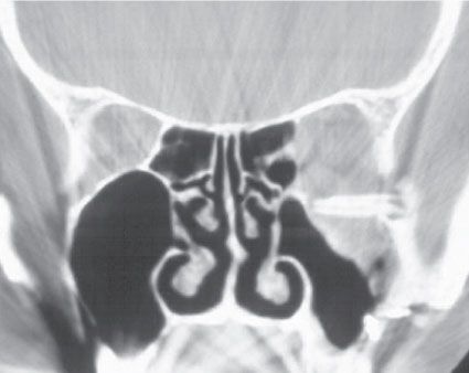
FIGURE 29.5. Incorrectly placed orbital floor prosthesis. Orbital floor implant misplaced directly posteriorly into the maxillary sinus. Note the flat lie of the implant, which should incline superiorly.
Orbitozygomatic Fractures
Diagnosis/Examination. The physical examination in orbitozygomatic fractures is important, but often misleading. Swelling from the injury frequently conceals the malar recession and any evidence of enophthalmos. Anesthesia in the distribution of the infraorbital nerve is common and should be documented on initial examination. Additionally, severely displaced fractures may cause trismus secondary to impingement of the coronoid process by the medial displacement of the zygomatic arch. The orbital aspect of these injuries necessitates careful ophthalmologic examination and is perhaps the most important aspect of the preoperative workup.
The decision to operatively reduce an orbitozygomatic fracture is largely dependent on the CT scan data, as swelling often precludes accurate determination of the degree of deformity. When evaluating the CT scan, it is most useful to look at the lateral orbital wall on the axial cuts, which represents the articulation of the zygoma with the greater wing of the sphenoid. This broad articulation will manifest any displacement present and determine the degree of deformity. One must also assess four other articulations for displacement, including the infraorbital rim, the zygomaticomaxillary buttress, the zygomaticofrontal suture, and the zygomatic arch.
Nondisplaced fractures may be safely managed with a soft diet and close clinical follow-up. A nonchew diet will prevent the activation of the masseter from displacing the fracture segment. Displacement of the fracture is a definitive indication for operative reduction and fixation, as the results of early operative repair far exceed those of delayed repair. Repositioning of a displaced fracture in a delayed setting requires osteotomy of all buttresses and extensive craniofacial exposure. It is best to reduce and fixate fractures anatomically in the acute setting.
Operative Techniques. The operative treatment of orbitozygomatic fractures depends largely on the degree of displacement and comminution. The majority of patients can be accurately reduced and fixated using incisions in the upper gingivobuccal sulcus, the lower eyelid (transconjunctival), and the lateral extent of the supratarsal fold of the upper eyelid. The coronal incision is necessary only when the zygomatic arch must be exposed and reduced as a guide to appropriate alignment of the zygoma. This is typically only necessary when there is extensive comminution of the infraorbital rim and zygomaticomaxillary buttress, so that these cannot be used as an accurate guide to reduction. In practice, only three of the four buttresses require reduction as long as the zygoma is a single, large fracture segment.
As a general rule, the intraoral incision is typically performed first. In lower-energy fractures, it is occasionally possible to elevate the displaced zygoma using an instrument placed behind the zygomatic arch. If the fracture reduces with this maneuver, a plate may be placed across the zygomaticomaxillary buttress and the operation terminated. Even if this does not allow anatomic reduction of the fracture, it is still beneficial in that it moves the zygoma to a more anatomic location, facilitating the lower-eyelid dissection of the orbital rim. As with orbital fractures, the subciliary incision is typically best avoided. Generally, either the transconjunctival or subtarsal incision is best. If a coronal incision has not been used, the zygomaticofrontal suture requires exposure through the lateral extent of the supratarsal fold. Although eyebrow incisions have been used in the past, these may leave prominent scars and provide less direct access to the suture.
Once all of the articulations of the zygoma have been visualized, the fracture segments should be reduced and then stabilized. It is very useful to first align the zygomaticofrontal suture using either a wire or a 1.0-mm plate. This sets the vertical height of the zygoma while still allowing rotation and further alignment at the level of the rim and the zygomaticomaxillary buttress. Once this suture is stabilized, the surgeon should anatomically reduce the other two buttresses. Reduction may be facilitated by using a Carol-Gerard screw placed through the lower-eyelid incision into the body of the zygoma (Figure 29.6). It acts like a joystick and allows easy control of the fracture fragment in three dimensions.
A 1.5-mm plate is typically placed along the superior aspect of the infraorbital rim rather than anteriorly to avoid either visibility or palpability by the patient postoperatively. The buttress that provides the most stability to the reduced fracture is the zygomaticomaxillary. A 2.0- or 1.5-mm L-plate is most commonly used at this location. In the most severe fractures, when the arch has to be exposed to align the zygoma, this is best plated first with a 2.0-mm plate to reestablish the width and projection of the zygoma.
It is not necessary to provide three or four points of fixation for every zygoma fracture. Although multiple plates are commonly used, it is usually a result of the sequence of steps used in aligning the fracture. Although three or four plates may be necessary in high-energy injuries, many fractures can be adequately fixated using a single 2.0-mm L-plate along the zygomaticomaxillary buttress.
When the zygomatic arch is significantly displaced and comminuted, it requires full exposure and reconstruction. There is a tendency for surgeons to view the arch as an arch when in reality it is much more of a straight anteroposterior structure. If reconstructed as an arch, it will result in excessive facial width and prominence to the region relative to the contralateral side. In addition, the malar eminence will not be projected as far forward as it should. This is an especially important factor when there is significant destruction to the malar and arch complexes bilaterally and the surgeon must reestablish facial width in proportion to facial height without the benefit of other reference points (Figure 29.7).
Arch Fractures. Unlike true orbitozygomatic injuries, isolated arch fractures frequently do not require operative reduction. Operative treatment is indicated for severe depression of the arch causing either a cosmetically significant contour depression or impingement on the coronoid process and trismus.
The fracture may be approached intraorally through an upper buccal sulcus incision or an incision in the temporal scalp. The intraoral approach, although more difficult, avoids an external scar. The scalp incision, commonly referred to as the Gillies approach, allows more direct access to the fracture site. The incision is placed horizontally within the temporal hairline. This better camouflages the scar as hair can drape over the area, as opposed to a vertical incision, which allows the hair to part on either side. Dissection is carried directly through the temporoparietal fascia and deep temporal fascia into the muscle. The elevator is then placed in plane between the deep temporal fascia and the temporalis muscle and advanced to a position deep to the arch. Any resistance to sliding the arch inferiorly usually indicates that the plane of dissection is too superficial. Outward pressure on the arch forces the fracture into an anatomically reduced position.
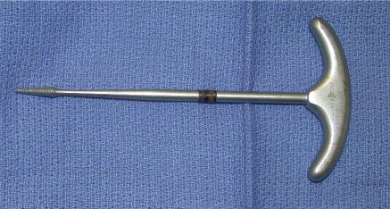
FIGURE 29.6. Carol-Gerard screw. This instrument can be used to gain control of the zygoma in orbitozygomatic fractures. It can be placed either through an intraoral incision or percutaneously.

FIGURE 29.7. Incorrect reduction of the zygomatic arch. Computed tomography scan of a zygomatic arch that was inappropriately reconstructed as an arch. The normal arch lies in an almost straight anteroposterior direction, as can be seen on the normal side.
Occasionally, arch fractures essentially snap back into position and are stable. In the majority of cases, however, the fracture is unstable in the reduced position. In such situations, it is helpful to splint the reduced arch using permanent sutures placed transcutaneously and encircling the arch. The suture is tied over a metal eye shield to provide constant upward and outward traction. Typically two sutures are required to hold the arch in position. Excessive tension on the sutures may lead to skin necrosis at the margins of the eye shield.
Complications. After lower-lid malposition, the most common complication following repair of orbitozygomatic fractures is enophthalmos resulting from malreduction of the fracture. As in orbital floor injuries, displaced orbitozygomatic fractures almost always expand orbital volume, as the zygoma constitutes a large portion of the orbital floor and lateral wall. Both the increased orbital volume and the malar recession must be addressed in the treatment of the malreduced fracture. To address this, the zygoma is best osteotomized, repositioned in its appropriate anatomic location, and secured with plates. Occasionally, the malar recession is not significant and treatment can be directed at correction of the orbital volume alone. As in an orbital floor fracture, additional alloplastic material may be placed in the floor or along the wall of the orbit to diminish volume and correct the enophthalmos. Maxillary sinusitis and persistent numbness in the infraorbital nerve distribution may complicate orbitozygomatic fractures less frequently.
Nasal Fractures
Diagnosis/Examination. The diagnosis of a nasal fracture is clinical. Patients presenting with a history of acute trauma with evidence of a deviated nose have an underlying nasal fracture. Radiographs, although frequently obtained, are superfluous.
When examining a patient with a suspected nasal fracture, an intranasal inspection is mandatory to identify a septal hematoma. Untreated septal hematomas may lead to resorption of the cartilaginous septum and result in a saddle nasal deformity. Septal hematomas should be expeditiously evacuated. Reaccumulation can be prevented with the use of either septal quilting sutures or intranasal splints.
Treatment. Treatment of nasal fractures can be divided into early and late phases. If one sees a patient with nasal fracture immediately following the injury, it is beneficial to perform a closed reduction. This may be performed in the emergency department using local anesthesia and an intranasal vasoconstrictor. Although closed reduction can be performed any time in the first 2 to 3 weeks, in the first few days following the injury, the swelling may be so severe as to camouflage the magnitude of the deformity and complicate accurate reduction. Swelling usually improves significantly 5 to 10 days after injury facilitating closed reduction.
The late phase is defined as a period where sufficient bony union has taken place such that osteotomy is required to correct the deformity. Correction in the late phase should be viewed as a complete rhinoplasty. The contribution of the septum to the deviation should be appreciated and addressed. Simply performing osteotomies is unlikely to correct the deformity in the face of septal involvement, particularly in high dorsal deviations. Extensive septal mobilization, resection, and scoring may be necessary. Airway compromise should also be a focus of the rhinoplasty.
Maxillary Fractures
Diagnosis. Fractures of the maxilla have been classically described as Le Fort I, II, and III patterns. These patterns, by definition, are fractures that detach the maxilla from the skull base. The maxilla is mobile and may result in malocclusion. Although it is not uncommon that the anterior wall of the maxillary sinus, and even the zygomaticomaxillary and nasomaxillary buttresses, are fractured in facial trauma, injury to these structures alone do not constitute a true Le Fort injury. The fracture must extend through the pterygoid plates to create a complete Le Fort fracture (Figure 29.8).
The Le Fort I injury classically passes through the maxilla transversely, somewhere between the tooth roots and the infraorbital rims, with preservation of the integrity of the infraorbital rims. The Le Fort II fracture extends through the infraorbital rim and nose and is sometimes referred to as a pyramidal fracture. The Le Fort III fracture involves the zygomatic arch, lateral orbital wall, and the nasofrontal region. Le Fort fractures rarely exist in pure form. More commonly, patients will present with a combination of injuries such as a Le Fort I on one side and a Le Fort III on the contralateral side.
Preoperative evaluation should be focused on the occlusal relationship. Any sign of malocclusion is evaluated carefully. Correction of the occlusion to the preinjury state will guide the appropriate anatomic reduction.
Operative Technique. Le Fort I injuries can be adequately exposed through an upper gingivobuccal sulcus incision and maxillary degloving. Le Fort II injuries often require a lower-lid incision. As in orbital fractures, a transconjunctival incision is preferred over a subciliary incision. In older patients, a subtarsal incision can be concealed in a lower-lid crease. Le Fort III injuries may be approached through a combination of buccal sulcus and lower-lid incisions in low- to moderate-impact injuries. More severe injuries require a coronal approach for exposure of the nasofrontal and medial orbital regions and the zygomatic arch. Following mobilization of all of these fractures, the patient should be placed in maxillomandibular fixation, the fractures reduced, and the zygomaticomaxillary and nasomaxillary buttresses stabilized with plates.

FIGURE 29.8. Incomplete Le Fort I fracture. A. Three-dimensional computed tomography demonstrating a midface fracture that appears to be present at the Le Fort I level. B. Axial CT scan of the same fracture revealing intact pterygoid plates. This fracture was completely stable on examination under anesthesia and thus does not require stabilization.
Nasoorbitoethmoid Fractures
Diagnosis and Examination. Fractures of the nasoorbitoethmoid (NOE) region involve the medial orbit, nasal bones, septum, and nasofrontal junction. On examination, patients with NOE injuries may exhibit substantial loss of dorsal nasal support. Frontal sinus fractures (discussed later) and cerebrospinal fluid leaks also commonly accompany NOE fractures. Additionally, telecanthus may be present secondary to lateral displacement of the bone fragments bearing the medial canthal tendon. These patients demonstrate an increased distance between the medial canthi and rounding of the medial canthal angle.
The CT scan is the most helpful radiologic tool in determining the location of the injury and the degree of comminution—the two factors critical to determining the appropriate treatment. Analysis of the axial cuts should focus on the lacrimal fossa and the origin of the nasolacrimal duct. This region identifies the level of insertion of the medial canthal tendon. Comminution at this level may necessitate transnasal fixation of the bone fragments bearing the medial canthal ligaments. Bilaterally mobile medial canthal tendons represent a significant challenge to reconstruction as there is no normal frame of reference with which to judge preoperative location of the medial canthi.
On occasion, it is difficult to determine if the bone bearing the medial canthal insertion is truly mobile. Definitive diagnosis can be made under anesthesia by placing a hemostat in the nose to the level of the medial canthus. With a finger palpating externally over the medial canthus and outward pressure on the internally positioned hemostat, one can determine the degree of mobility and lateral displacement, and consequently the need for operative reduction and stabilization.
Operative Technique. The majority of NOE injuries should be exposed through both coronal and lower-eyelid incisions. To improve visualization, it may be helpful to score the periosteum in the glabellar region to allow expansion of the soft-tissue envelope. As the fracture is dissected, one must take great care not to strip the insertion of the medial canthal tendon off the fracture fragments. Markowitz et al.3 have devised a classification scheme based on the relation of the insertion of the medial canthal tendon to the fracture. Type 1 fractures involve a large bone fragment to which the medial canthal tendon is inserted. These injuries may be treated essentially by fixation of the bone fragment to adjacent surrounding bone. Type 2 fractures involve more extensive comminution; however, the medial canthal tendon is still attached to a bone fragment that can be stabilized directly. When the fragment is too small for fixation, transnasal wiring can be used to position the fragment and medial canthal tendon appropriately. Additional bone grafting may be required. Type 3 injuries involve avulsion of the medial canthal tendon from its skeletal insertion. In addition to appropriate reduction of bone fragments and bone grafting, these patients require transnasal fixation of the medial canthal tendon.
When transnasal medial canthoplasty is necessary, it must be performed so that the direction of pull on the canthal tendon is posterior and superior. A common mistake is to reattach the canthal tendon too anteriorly, resulting in persistent telecanthus. The procedure is performed by drilling from the contralateral side through the ethmoid bones, with the exit point planned at approximately the level of the superior aspect of the lacrimal fossa. This is an appropriate vector for pull on the tendon. The medial canthal tendon is either grasped from the underside of the coronal incision or looped from an anterior incision medial to the medial canthal angle. The wire or permanent suture is then placed through the drill hole using a wire-passing drill bit toward the contralateral side. The suture or wire can be affixed to a screw placed on the contralateral side.
Many of these injuries involve substantial loss of nasal support that cannot be restored by direct plating. In these situations, onlay bone grafting of the nasal dorsum will reestablish the appropriate projection and width of the nasal radix. Multiple techniques have been described to stabilize bone grafts in this region including miniplates, lag screws, and K-wires.
A soft-tissue bolster over the medial canthal valley is also often necessary to restore the normal contour to this region. The medial canthal valley is a unique region of the face where the skin is intimately adherent to the underlying skeleton. The soft-tissue bolster prevents the subcutaneous accumulation of blood and fluid that interferes with the healing of the skin directly to the underlying bone. These bolsters are usually left in place for 7 to 10 days. The skin underlying the bolster should be watched carefully for necrosis and the bolster should be tied in such a way to allow for adjustment of tension postoperatively.
The most common complication following NOE fractures is telecanthus. This is difficult and sometimes impossible to correct secondarily. Once it is established, the area must be approached in a similar fashion, the scar contractures completely released, bone grafts placed to reestablish contour, and transnasal canthoplasties performed. The results of secondary repairs are always disappointing when compared with accurate acute repair.
Frontal Sinus
Diagnosis and Examination. Patients with frontal sinus fractures may present with obvious contour deformities of the forehead, but often the swelling associated with the injury blunts the degree of deformity. Injury to the frontal sinus is commonly associated with injury to the central nervous system, and early evaluation should focus on this possibility. Axial cuts of the CT scan are useful in determining the degree of injury and involvement of the anterior table, posterior table, and the nasofrontal duct. These three structures are used in the classification of frontal sinus fractures as well as subsequent treatment.
Isolated anterior table fractures may be treated simply by reduction and plate fixation via a coronal incision or through existing cuts in the forehead. If extensive comminution exists, the anterior wall is replaced with split calvarial bone graft. The function of the nasofrontal duct should be kept in mind at all times. In many cases, involvement of the nasofrontal duct is obvious from the CT scan (Figure 29.9). This is particularly true for fractures located inferior and medially in the sinus, where the meatus typically originates. During operative exploration, direct instillation of dye (fluorescein) into the region of the nasofrontal duct within the sinus cavity has been advocated to test the function of the duct. The presence of dye on pledgets placed intranasally indicates a functional duct system. Generally however, this should not be necessary. Based upon the fracture pattern seen on the CT scan and intraoperatively, the surgeon should be able to make the decision regarding compromise of the nasofrontal duct. Any significant concern should prompt the surgeon to obliterate the sinus.
The first step in frontal sinus obliteration is removing the anterior table entirely. The limits of the sinus may not be obvious based on direct examination, particularly in small fractures. Placing one arm of a bayonet forceps into the sinus until the limits of the sinus are reached can help delineate the margins of the sinus. Additionally, the operating room lights may be turned down and a light source placed within the sinus, defining the margins of the sinus (Figure 29.10).

FIGURE 29.9. Axial computed tomography scan of the frontoethmoidal region revealing complete obliteration of the nasofrontal duct by bone fragments. The frontal sinus requires obliteration in the case of a nonfunctioning duct to avoid development of a mucocele.

FIGURE 29.10. After removing the anterior table of the frontal sinus, one must delineate the limits of the sinus. This may not always be visible directly, but it helps to turn down the operating room lights and place a light source within the sinus.
Once the anterior table is removed, all mucosa is removed from the sinus. Because of small mucosal invaginations into the bone, termed the vascular crypts of Breschet, a burr should be used to ensure complete mucosal obliteration. Once this has been accomplished, the nasofrontal drainage system is plugged to prevent ingrowth of the mucosa from the ethmoid sinus and nose below. This may be accomplished with a graft of muscle, bone, fat, or a pericranial or galeal flap. At this point, many surgeons fill the sinus cavity with graft material, most frequently fat. The need to obliterate the sinus has been challenged by some on the basis of the concept of osteoneogenesis. Rohrich and Mickel4 demonstrated spontaneous obliteration by bone in cat frontal sinuses that were surgically burred out and not filled with graft material. This technique has been used by the authors for years without evidence of any increase in complication rates over those published in the literature. From a conceptual standpoint, one must question the superiority of filling a bone cavity with nonvascularized material over simply not filling it at all. In reality, the pericranial or galeal flap used to obliterate the nasal communication may in itself completely fill smaller sinus cavities (Figure 29.11).
Fractures of the posterior table of the frontal sinus place the patient at risk for acute meningitis and late intracerebral mucocele formation. Fractured fragments of the posterior table may develop trapdoor-type phenomena leaving small bits of mucosa within the cranial cavity. These areas of trapped mucosa are at risk for mucocele formation. Evaluation of the axial cuts through the frontal sinus will reveal displacement of the posterior table within the cranial cavity. Any significant posterior displacement or the presence of a cerebrospinal leak is considered an indication to cranialize the sinus. Cranialization involves performing a frontal craniotomy and removing the entire forehead as a bone flap. The posterior table is then removed and the mucosa of the anterior table burred out. As in obliteration, the nasal communication is eliminated using a pericranial or galeal flap. The bone flap is then plated back into position and the brain is allowed to expand into the space previously occupied by the sinus. The extensive exposure provided by the approach also allows repair of the dura, if necessary.
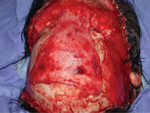
FIGURE 29.11. Pericranial flap. This tissue is often used to interrupt the communication between the ethmoid sinuses and nose with an obliterated frontal sinus.
Complications. The most significant complications related to frontal sinus fractures are infectious in nature. In cases in which the frontal sinus has been preserved, one must give consideration to serial CT scans in the postoperative period to assess adequate drainage of the sinus. Failure of the sinus to clear radiographically may indicate impaired drainage that may lead to infection. Evacuation of a poorly draining sinus should be achieved with aggressive medical therapy, including topical and systemic decongestants and mucolytics, or via an endoscopic or open surgical drainage procedure. Mucocele formation may be a late complication of these fractures (Figure 29.12). Unfortunately, most mucoceles and infections present very late following these injuries. It is not uncommon for this to occur years following the injury or surgery. Such delayed presentation makes accurate data regarding the success of the various treatment options difficult to obtain.
Mandible Fractures
Diagnosis and Examination. A complaint of malocclusion after trauma is a reliable indication of a mandible fracture. Because the goal of treatment is to restore the mandibular arch to its preinjury occlusal state, as much information as possible is obtained regarding the preinjury occlusion, orthodontic treatment, and any history of dental extraction. The wear facets of the dentition are perhaps the most valuable indicators of the preinjury occlusion. Stable mandible fractures with no evidence of malocclusion can occasionally be treated with a nonchew diet for a period of 4 to 6 weeks, depending on the age, state of dentition, and compliance of the patient. For the most part, this is the exception and not the rule, as patients suffering from mandible fractures tend to be young, male patients notorious for their noncompliant behavior.
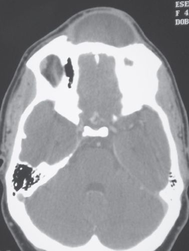
FIGURE 29.12. Frontal sinus mucocele. Axial computed tomography scan of a mucocele several years following a frontal sinus obliteration with bone cement.
Because of the constant motion of the lower jaw in normal daily function, mandible fractures tend to be painful until stabilized. Some of the immediate discomfort is relieved by temporary immobilization of the fracture with a wire joining the teeth adjacent to the fracture site. This must be done carefully to avoid dislodging the teeth, which may be destabilized by the fracture. Documentation of mental nerve function is performed. Mental nerve neurapraxia often indicates involvement of the body of the mandible between the mental foramen and the mandibular foramen. Because of the likelihood of further neurapraxia related to surgical correction, it is wise to discuss the condition of the nerve and its clinical course prior to operative repair.
As mentioned earlier, an excellent single test for mandibular fractures is the panoramic radiograph. This requires that the patient be able to sit upright with a mobile neck, so cervical spine clearance is obligatory. The Panorex provides an excellent evaluation of the condyles and dental anatomy. Because of the nature of the technique, however, there can be significant distortion in the region of the symphysis, which may conceal fractures in this region (Figure 29.2). The symphyseal region is best evaluated with a plain, posteroanterior radiograph of the mandible. A CT scan is frequently obtained in the emergency center to evaluate facial trauma and, as mentioned previously, is quite sensitive and specific for detecting mandibular fractures, but provides less information with regard to dentition.
Fixation. Choice of fixation depends on the specifics of each fracture. One must choose between rigid fixation (load bearing) and what one might term functionally stable fixation (load sharing).5 When internal fixation was first applied to the mandible, it was generally felt that absolutely rigid stabilization was required. Since then it has become appreciated that many fractures may do just as well with lesser degrees of stabilization or load-sharing fixation (Table 29.2).
Rigid fixation typically implies plates that accommodate screws that are 2.4 or 2.7 mm in diameter. Functionally stable fixation most often uses plates accommodating screws 2.0 mm in diameter or smaller. The decision is determined largely by the fracture pattern and its intrinsic stability. Patients with severely displaced fractures or with multiple mandible fractures have lost more of the intrinsic stability of the mandibular arch and may require fixation that is more rigid and bears more of the functional mandibular load. Additionally, patients at high risk for poor healing, such as those with atrophic mandibles or established infection, usually benefit from absolutely rigid fixation. Uncomplicated, isolated mandible fractures heal uneventfully using smaller plates and screws placed along the lines of osteosynthesis as delineated by Champy (Figure 29.13).
Operative Technique
Symphysis/Parasymphysis Fractures. As with most mandible fractures, the operative procedure should begin by reestablishing the patient’s occlusion using maxillomandibular fixation. In severely displaced fractures, it may be beneficial to expose the fracture first to achieve some degree of initial reduction. If the arch bars are applied initially without preliminary reduction, the arch bar itself may lock the arch into a malocclusion.

Maxillomandibular fixation does not usually achieve sufficient immobilization and stabilization of symphyseal fractures because of the lack of intercuspation of the anterior dentition, and therefore displaced symphyseal fractures typically require internal fixation. These fractures may be stabilized with either large or small plates. Placement of a single, large plate (= 2.4 mm) along the inferior border using bicortical screws is sufficient. It should be noted, however, that contouring a 2.4-mm plate to the acute curvature of the symphyseal or parasymphyseal region can be difficult and time consuming. Equally rigid fixation can be achieved by placing a 2.0-mm plate with bicortical screws at the inferior border and a 2.0-mm tension band (smaller plate at superior border) with monocortical screws just below the root apices. A well-placed arch bar can take the place of the tension band and be left in place until bone union. We prefer a tension band as we use it to assist with reduction prior to placing the inferior border plate. After the patient is placed into maxillomandibular fixation, two small holes are drilled into the inferior border on each side of the fracture to accommodate a reduction clamp. With the bone aligned and the patient in occlusion, the monocortical tension band plate is applied. With this in position, the bone reduction clamp may be removed without impacting the reduction and the inferior border plate placed onto a relatively stable platform. Most of these fractures may be treated via an intraoral incision. External incisions are typically reserved for the most severe fractures in this region (multiple fractures and comminuted fractures).
Body Fractures. Mandibular body fractures are treated with either maxillomandibular fixation alone (for 4 to 6 weeks) or internal fixation. Most practitioners prefer to perform an open reduction and internal fixation in these cases. As with symphyseal and parasymphyseal fractures, a single 2.4-mm inferior border plate or two 2.0-mm plates may be used. Care must be taken to avoid the mental nerve when placing the screws. As with symphyseal fractures, all but the most severe fractures are treated using an intraoral incision.
Angle Fractures. Mandibular angle fractures are associated with the highest risk of infection and postoperative complications. The presence of a partially erupted or impacted third molar is the cause of the majority of complications. These teeth predispose the angle region to fractures by weakening the bone in the region. The fracture is considered open because of communication with the third molar socket. Although the decision to remove or retain the third molar is controversial, it is generally believed that any significantly damaged, loose or diseased third molar should be removed, as should any tooth that prevents reduction.
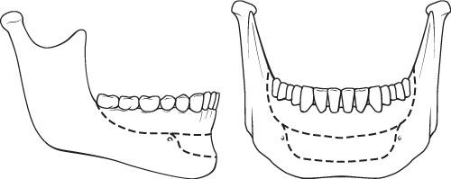
FIGURE 29.13. Champy’s lines of osteosynthesis.
Mandibular angle fractures have been treated with a variety of fixation techniques. Ellis6 advocate using a single miniplate along the external oblique ridge of the mandible along the lines of Champy. When comparing this with all other forms of fixation, the authors found their complication rate to be the lowest. Additionally, the fracture may be stabilized along the buccal cortex using a single 2.4-mm plate at the inferior border. Alternatively, one may use a strut plate (essentially two plates combined into one) (Figure 29.14). This is placed over the inferior alveolar canal (midportion of the mandible) and secured with monocortical screws. Intraoral exposure is preferred in the majority of patients with simple angle fractures. A small incision in the cheek is required for placement of the screws. However, in complicated or comminuted fractures consideration should be given to an external incision. The external incision provides much greater exposure and control over the fracture. The external scar and risk to the marginal mandibular nerve are less important than proper stabilization of a complex mandibular fracture.
Subcondylar Fractures. No fractures of the mandible are as controversial as subcondylar fractures. In addition, the terminology in the literature is confusing. Injury may occur at the level of the condylar head, the condylar neck, or the region below the sigmoid notch, the subcondylar region. Condylar head injuries are intra-articular, are not amenable to internal fixation techniques, and are associated with a high risk of ankylosis. Condylar neck fractures are defined as those that occur between the head and the sigmoid notch.
Historically, most of these injuries have been treated using maxillomandibular fixation for a period of 2 to 6 weeks. Although this results in excellent functional occlusion, it rarely reduces the fracture into anatomic alignment. Rather, the patient develops a functional occlusal adaptation to the malreduction. Patients frequently continue to deviate to the fractured side with maximal opening and lose some of the contour of the mandibular border on the fractured side.
Many authors advocate open reduction and internal fixation. Opponents are concerned with the risk to the facial nerve. Although most published series report a low risk of permanent injury to the facial nerve, even neurapraxia is a distressing complication. In an effort to avoid this, endoscopic instrumentation has been developed to fixate these fractures largely through an intraoral incision with small trochar sites externally.7,8
Zide and Kent9 published a list of absolute and relative indications for open reduction and internal fixation of subcondylar fractures. This list has been modified by the body of literature over the years. As a general rule, internal fixation is considered in cases where maxillomandibular fixation is contraindicated (e.g., poorly controlled seizure disorder), when an acceptable occlusion cannot be reestablished, and with bilateral fractures in the setting of panfacial injury (to reestablish appropriate posterior facial height).
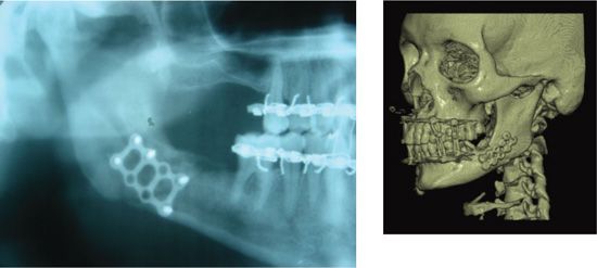
FIGURE 29.14. A strut plate can be used for the treatment of mandibular angle fractures. The combination of two plates functions to provide greater support to these fractures and are placed over the inferior alveolar canal and secured with monocortical screw.
When maxillomandibular fixation is chosen as the definitive treatment, the period of immobilization varies, depending on the age of the patient and the fracture pattern. Patients in whom uncomplicated healing is expected are maintained in maxillomandibular fixation for 2 to 3 weeks, followed by an additional 2 to 3 weeks of nonchew diet. As a general rule, the primary determinant of the period of treatment is the occlusion. Patients who present with no significant occlusal change may not require maxillomandibular fixation at all. Additionally, high condylar fractures may have a greater risk of ankylosis and should be immobilized for shorter periods of time. The same can be said of children in whom maxillomandibular fixation is limited to 2 weeks. Many surgeons advocate the use of elastics rather than wire to allow some motion and guide the occlusion in the postoperative period. The hope is that the motion will prevent the stiffness associated with long-term maxillomandibular fixation. The use of elastics should be restricted, however, to reliable patients.
Complications. The most common complication after mandibular fracture repair is malocclusion, usually secondary to maladaptation of the plate used for fixation. Inappropriately contouring the plate (especially a large plate such as the 2- to 4-mm system) results in the mandible shifting to adapt to the plate. It is less commonly seen when miniplates are used, as the plates tend to adapt to the bone instead of the reverse. This phenomenon is also less likely with locking plates. These are plates designed with threads within the screw hole. As the screw head approaches the plate, it locks into the plate. As such, the screw does not continue to tighten and pull the bone up to the maladapted plate. The screw stops when it reaches the plate.
Once accurate reduction fixation has been applied to a mandibular fracture, maxillomandibular fixation is released and the patient’s occlusion carefully assessed. Malocclusion at this point mandates removal of the fixation and recontouring of the plate. It is a mistake to rely on postoperative maxillomandibular fixation or elastics to correct a malocclusion secondary to a malreduction.
Infection is also a common complication of mandibular fractures, most often as a consequence of mobility at the fracture site or because of loose hardware. During the exploration of an infected fracture that had been previously repaired, the operative site is thoroughly irrigated and the stability of the fixation assessed. In patients in whom the fixation has not failed and continues to provide stabilization of the fracture, the plates and screws are left in place and the wound cultured and closed over drains. Culture-specific antibiotic therapy is instituted after an appropriate operative culture is obtained. If, on the other hand, the fixation is loose, it is removed and a more rigid fixation applied to ensure stable fixation of the fracture site. The orthopedic adage is apt: “An infected union is better than any nonunion.”
SECONDARY DEFORMITIES IN FACIAL TRAUMA
Enophthalmos
Enophthalmos is defined as posterior displacement of the globe within the orbit. Clinically, it is noticeable when the displacement is 2 mm or greater. Inferior displacement of the globe is termed hypoglobus. Often there is some element of both enophthalmos and hypoglobus in the presence of malreduced or unreduced orbital injuries.
Failure to reestablish the correct orbital volume is the most common mistake in the repair of these fractures, resulting in late enophthalmos. This occurs if the orbital floor prosthesis is not appropriately angled superiorly toward the anterior edge of the remaining intact orbital floor but is placed straight back into the maxillary sinus. Some suggest that correct placement of the implant or bone graft should be confirmed with an endoscope placed into the maxillary antrum.
Delayed enophthalmos is evaluated with a maxillofacial CT scan with 1.0-mm cuts through the orbits. Sagittal and coronal reconstructions are useful adjuncts to the orbital analysis and comparison with the normal contralateral side is extremely helpful. Thorough examination of the eye is performed by the ophthalmologist to assess abnormalities in vision or extraocular movement. The most common finding in patients with enophthalmos is diplopia, more often in peripheral than central gaze. Diplopia is most frequently related to disorders of the nerves or scarring of muscles controlling ocular motility, but may be related to malposition of the globe. The central nervous system can accommodate some degree of ocular displacement before diplopia becomes evident.
Correction of enophthalmos should be directed toward correction of the orbital volume. In the case of fractures of the orbital floor or medial wall, placement of bone grafts or an alloplastic implant in the deficient areas will help correct the displacement. Titanium mesh is easier to mold and contour to larger and more complex defects. Porous polyethylene is useful in the reconstruction of isolated floor fractures. The orbit is typically approached through a transconjunctival incision in much the same way as in primary repair. If the problem with globe positioning is strictly one of posterior displacement (enophthalmos) with no vertical component, and there is no well-defined defect to reconstruct, the implant is typically placed posterolaterally in the orbital cone (Figure 29.15). This achieves forward displacement of the globe without changing the vertical dimension. It is also important to perform a thorough subperiosteal dissection of the orbital cone to prevent the globe from being tethered posteriorly by scar. A slight degree of overcorrection is warranted in these cases to compensate for swelling that occurs during the dissection.
Enophthalmos secondary to malunion of an orbitozygomatic fracture requires careful evaluation. When the malar eminence is displaced in the presence of enophthalmos, corrective osteotomies of the entire zygomatic complex are performed at the level of the zygomaticomaxillary buttress, infraorbital rim, arch, zygomaticofrontal buttress, and sphenoid articulation within the orbit. This is accomplished through the coronal, lower lid, and buccal sulcus incision. Bone grafting is often required to compensate for the inevitable loss of bone that occurs from the original injury. Orbitozygomatic osteotomies for posttraumatic malunion with enophthalmos are challenging, morbid and are associated with the need for revisionary procedures even in the most experienced hands.
Malocclusion
Malocclusion is a difficult problem to correct secondarily once bone union has occurred. It is far easier to avoid it in the first place. Although malocclusion can be related to malunion of maxillary, palatal, or mandible fractures, it is most commonly associated with poorly treated mandible fractures.
It cannot be overemphasized that the occlusion seen following plating of the fractures must be meticulously evaluated. Any discrepancy seen in intercuspation and alignment of wear facets is corrected at that time. Removal and replacement of hardware is a minor inconvenience when compared with the inconvenience to both the surgeon and the patient of a postoperative malocclusion. To fully assess the corrected occlusion, it is necessary to release the patient from maxillomandibular fixation. Maxillomandibular fixation can displace the condyles or bone fragments enough to give the appearance of a good bite. When checking the occlusion at the conclusion of the procedure, the surgeon should use only gentle upward pressure on the symphysis to check the occlusion. Because of the relatively lax configuration of the mandibular articulation with the skull, it requires only a mild degree of force to dislocate the condyles and force the patient into an occlusal state that appears normal, but is not centric. Centric occlusion is seen when the condyles are seated within the articular fossae. The challenge is to ensure that the occlusion at the end of the case is equal to centric occlusion.
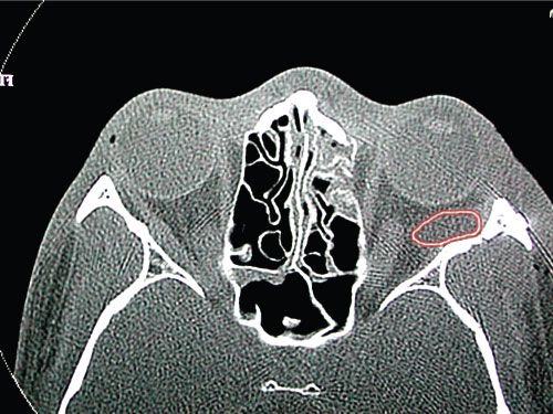
FIGURE 29.15. If enophthalmos is strictly secondary to posterior displacement and there is no well-defined defect to reconstruct, bony orbital volume can be reduced by placing a porous polyethylene implant wedge posterolaterally in the orbital cone.
When malocclusion is discovered in the immediate postoperative period, Panorex and plain radiographs can often determine if the condition is amenable to operative correction. Minor tooth interferences can be addressed by burring down teeth at the points of contact. Orthodontics is a powerful tool to address less-significant degrees of malocclusion. Orthodontics is often more useful in the malaligned alveolar fracture or segmental fractures of this nature. The majority of postoperative malocclusions, however, should be taken back to the operating room for exploration, removal of hardware, appropriate reduction, and stabilization. If internal fixation cannot be achieved for some reason, then the fail-safe maneuver is 4 to 6 weeks of maxillomandibular fixation.
In the case of malunion, where bone fragments are no longer mobile, osteotomies must be made. This approach requires the fabrication of models and mock surgery. The models are cut to correct the malocclusion, and an occlusal or lingual splint is fabricated to guide the repair. Although reproducing the initial fracture often suffices, in late cases in which there has been some degree of dental compensation, sagittal split, and Le Fort I osteotomies may be required.
Temporal Hollowing
Temporal hollowing is caused by injury and subsequent loss of volume within the temporal fat pad. The temporal fat pad lies between the two layers of the deep temporal fascia (the thick layer of fascia immediately superficial to the temporalis muscle) that encompass the fat pad and insert onto the zygomatic arch. In dissection of the temporal region, some surgeons believe it is important to dissect deep to the superficial layer of the deep temporal fascia as one approaches the zygomatic arch in an effort to protect the temporal branch of the facial nerve. This results in some degree of trauma to the fat pad, which may result in devascularization and some loss of volume in this region. Nerve injuries are more likely the result of excessive traction, which is not necessarily prevented by this fascial layer. To minimize the risk of devascularization and temporal hollowing, the authors prefer to dissect just on top of the superficial layer of the deep temporal fascia (or temporoparietal fascia) with a moist sponge using a sweeping motion. The dissection should elevate the superficial temporal fascia off the deep temporal fascia, ensuring that the nerve is raised with the flap, and avoids the fat pad altogether.
Once temporal hollowing has occurred, one effective treatment is placement of a porous polyethylene implant in the subperiosteal plane and secured to the temporal fossa. Because of the deep placement of the implant below the temporalis muscle, the size of the implant is frequently larger than one would anticipate relative to the defect (Figure 29.16). Porous polyethylene offers several advantages, including availability, permanency, and tissue ingrowth. It can also be stabilized with screws to the temporal region to prevent displacement. For smaller volume defects, autologous fat grafts are also an option (Chapter 44).
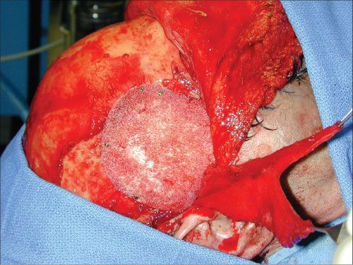
FIGURE 29.16. Porous polyethylene implant used to treat temporal hollowing. The implant is placed in the subperiosteal plane and larger than the anticipated defect.
Telecanthus
Telecanthus is even more difficult to resolve secondarily than it is primarily. The most common findings are tenting of the soft tissues of the medial canthus, lateral displacement of the canthus, and rounding of the medial canthal angle. The goal of revisional surgery is to correct all of the above deformities and ensure that the skeletal contour in the region is correct. Because of the nature of soft tissue in the region, most deformities in bone contour are readily visible. Exposure is obtained most frequently through coronal and lower-lid incisions. The entire soft-tissue envelope is mobilized and scar tissue resected or released. The correct position of the insertion of the medial canthal tendon is at the posterosuperior aspect of the lacrimal fossae. If this area has been distorted, it should be reconstructed to the appropriate contour, using the uninjured side to guide the reconstruction. The same is true for positioning of the medial canthus. The typical error is in reinserting the canthus too far anteriorly. It should be remembered that it is impossible to overcorrect the medial canthus in this deformity.
Redraping the soft tissue of the medial canthal valley is a significant challenge. Transnasal wiring or suturing can be used to set the correct position of the medial canthus, but does little to ensure that the overlying soft tissues adhere down to the bone. It is useful in this regard to employ a soft-tissue bolster to compress the skin to the bone. It is best to use a technique that allows for adjustment of tension on the bolster postoperatively to avoid tissue necrosis. The bolster helps eliminate dead space and evacuate blood so that hematoma formation does not impede soft-tissue adherence.
Gunshot Wounds and Panfacial Fractures
Gunshot wounds cause any combination of soft-tissue injury and fracture within the craniofacial skeleton. With most handgun and rifle injuries to the face, the entrance wound tends to be inconspicuous relative to the degree of skeletal injury. The exit wounds are more variable and depend to a large degree on the caliber (energy) of the weapon. Shotgun wounds, on the other hand, impart a great deal of soft-tissue injury along with a significant degree of underlying skeletal injury (Figure 29.17).
The goal of skeletal reconstruction must first be the restoration of the anteroposterior projection and width of the face. Primary bone grafting has proven a reliable technique in the face of bone loss and severe comminution.10,11 Although the order in which the craniofacial skeleton is addressed is somewhat controversial, the zygomatic arch serves as a useful guide and should be reduced and fixed early in the sequence. Correct positioning of the arch essentially establishes the proper facial width, framing the face. Failure to accurately reconstruct the arch results in the remainder of the reconstruction being set to the incorrect frame.
When a large soft-tissue component is also present, as occurs with shotgun wounds, soft-tissue reconstruction becomes an early priority. Stable soft-tissue coverage is vital to restoration of the skeleton, especially when bone grafting is required.12 Additionally, damaged and devitalized tissue can lead to significant scar contracture that ultimately can limit facial form and function. Although debridement of soft tissues in the facial region should be tempered with the goal of preservation of critical structures, all efforts should be made to achieve complete soft-tissue coverage and wound healing within 1 to 2 weeks in an effort to avert scar contractures. At times, this goal can only be achieved with free tissue transfer (Figure 29.18).
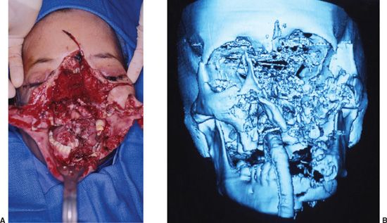
FIGURE 29.17. A and B. Shotgun wounds. Shotguns cause both extensive soft-tissue and skeletal destruction, often resulting in severe comminution of the facial skeleton.
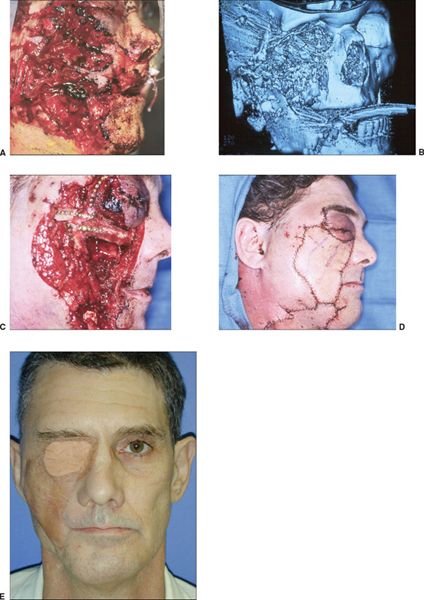
FIGURE 29.18. Complete phases of treatment of facial shotgun wound. A. Initial injury B. Three-dimensional computed tomography scan revealing degree of skeletal destruction. C. Reconstruction of the zygomatic complex with extensive primary bone grafts fixed with miniplates. D. Soft-tissue reconstruction with a combination of local flap advancement from cheek and radial forearm free flap for the central region. Stable soft-tissue reconstruction is critical to bone graft survival. E. Two-year follow-up.