
PART I
PRINCIPLES, TECHNIQUES, AND BASIC SCIENCE
CHAPTER 5 MUSCLE FLAPS AND THEIR BLOOD SUPPLY
JAMIE P. LEVINE
FLAPS
A flap is a unit of tissue that is transferred from a donor to a recipient site with its blood supply. Numerous classification schemes exist. Flaps may be characterized by their component parts (e.g., cutaneous, musculocutaneous, and osteocutaneous), their special relationship to the defect (local, regional, distant, or free), the nature of the blood supply (random vs. axial), and finally by the movement that is required for the flap to fill the desired defect (e.g., advancement, pivot, transposition, and interpolation). This chapter will focus on the blood supply and classification systems for muscle flaps and their associated uses. The blood supply to the muscle is intimately involved with the overlying fascia and skin. In general, muscle flaps are “axial” pattern flaps, with a known blood vessel oriented longitudinally within the flap. These vessels will have perforating branches that then supply contiguous territories including the overlying skin. With the advancement of and enthusiasm for perforator flaps, the knowledge of the muscle blood supply has expanded into blood supply for territories of tissues (Chapter 4). These territories can be harvested as composite flaps incorporating all of the tissue layers or individual components can be taken separately based on the perforator blood supply.
HISTORY
Skin and subcutaneous tissue was initially elevated as “random” pattern flaps either from a site adjacent to the wound or from a distant site. Due to unpredictable circulation, these flaps often went on to partial or complete necrosis. This era of flap surgery required adherence to length and width ratios in hopes of maintaining adequate vascularity for flap survival. “Delay” techniques (Chapter 1) were also utilized to augment the vascular supply. The great advance in this area was the identification of specific vascular pedicles in consistent and reliable locations (dorsalis pedis, groin flap, etc.). Since these flaps could be elevated with a defined vascular pedicle, it became possible to transfer larger flaps.1 These early axial flaps were a vast improvement with regard to size and reliability, but because they remained pedicled, they were limited to specific topographic locations.
Next, the identification of muscle flaps as a source of tissue offered tremendous flexibility and more options for wound coverage and defect reconstruction.2 Muscles are available in almost all topographical areas. As the vascular anatomy to these muscles was elucidated, it became possible to detach the muscle origin, insertion, or both and to transfer the muscle to a new site while maintaining vascular perfusion. The decision of which muscle to utilize for a given defect takes into account multiple factors: the size and location of the defect, damage to regional tissues, and the presence of exposed vital structures. The ability to transfer muscles changed the way we are able to manage complex wounds of any variety.
With increasing interest in muscle circulation, the contribution of muscle flap circulation to the overlying skin was then recognized. This further advanced our ability to close complex, composite defects with improved function, cosmetic appearance, and donor site variety. Each superficial muscle provides vascular connections via musculocutaneous perforating vessels to the overlying skin. With the identification of vascular connections to the skin, it became possible to include a segment of skin with the muscle flap.3 Prior to identification of the muscle skin territory, which allows its design as a musculocutaneous flap, the muscle flap was inset into the wound and exposed portions were skin grafted for coverage. With a composite of muscle and its overlying skin, defect closure can be accomplished with muscle, subcutaneous tissue, and skin. The ability to take an island of skin can also increase the coverage area of a single flap if it is designed appropriately and also allows for improved postoperative monitoring as either a free or pedicled flap (Figure 5.1).
As understanding of cutaneous blood supply increased, fasciocutaneous flaps were also described.4 Finally, Ian Taylor was able, through ink injection analysis, to put the various concepts of skin circulation together in its most coherent form and defined the concept of the angiosome (Chapter 4). These studies helped to define the vascular territories of the body (with an average of over 300 cutaneous perforators)5 and provide a reliable guide to composite flap design based on cutaneous vascular anatomy. These concepts of vascular anatomy have opened a new era of flap transfer by defining perforator anatomy. The separation between pure fasciocutaneous and muscle flaps has also been erased. It was believed that the pedicle to fasciocutaneous flaps was always along the intermuscular septae. With the knowledge of perforator vessel dissection, fasciocutaneous flaps can be dissected from muscle-based vessels and perforators.
The next step in utilizing this knowledge of muscular vascular anatomy has been the creation of specially designed, chimeric flaps that can include segments of as much muscle, skin, and other tissues as needed for the reconstruction. This knowledge of vascular anatomy has also developed the concept of the free form perforator flap.6 These flaps can contain any tissue zone supplied by a regional vascular perforator and allows individualization of tissue thickness and texture to the specific needs of the defect.
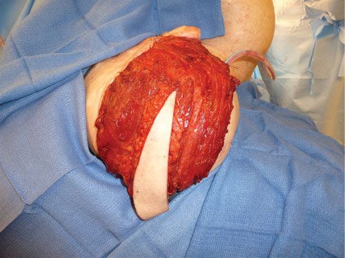
FIGURE 5.1. Myocutaneous latissimus free flap for scalp coverage. Although skin grafting of the muscle is still required, the skin island provides extra coverage and an area for improved monitoring postoperatively.
BLOOD SUPPLY
Blood Supply: Random Flaps
Any flap requires an adequate blood supply after transfer to survive. “Random” cutaneous flaps are based on unnamed smaller vessels. It was observed historically that the ratio of flap length to width was a critical variable for flap survival. These restrictions limit their reliability for use with large defects. When utilized appropriately, however, random flaps can be reliable first choices for coverage of smaller defects throughout the body. The term “random” really means that the surgeon does not know for sure if there is enough longitudinally oriented (axial) vessels to keep the flap alive.
Blood Supply: Axial Flaps
In contrast to random pattern flaps, axial pattern flaps are based on a reliable, anatomically defined vascular territory that is oriented longitudinally within the flap and that extends beyond the base of the flap. Since the description of the first axial flap (the deltopectoral flap) nearly four decades ago, the knowledge of the body’s various cutaneous angiosomes and subsequent exploitation of axially based flaps has grown significantly.7 The advances in anatomic vascular knowledge have increased the type and the reliability of axial pattern flaps and have fostered the development of microsurgical free flap transfer. Because of their significantly greater reliability, axial flaps (flaps with known blood supply) are preferred for coverage of moderate to large defects.
The reliability and volume of tissue that can be placed into a defect is markedly greater than with any type of random pattern flap. Due to the axially oriented circulation, delay procedures are often not necessary even when mobilizing large tissue volumes in one procedure based on this direct circulation. The only limitation, when pedicled, is the limited topographic arc of rotation. These limitations have been essentially overcome by microvascular free tissue transfer techniques (Chapter 8) that are limited only by the availability of recipient blood vessels.
Blood Supply: Delay Phenomenon
In order to extend the somewhat restricted size of random flaps, surgeons rely on the delay phenomenon. This is most commonly achieved by interrupting a portion of the normal blood supply to the flap without transferring the flap from its native position. The associated sublethal ischemia results in (1) opening of “choke” vessels that are normally closed allowing blood flow into the ischemic region of the flap, (2) reorientation of the vessels within the flap to a more longitudinal pattern, and (3) sprouting of new vessels within the flap through angiogenesis, and perhaps via vasculogenesis.8
Vessels within the flap also respond to the stress of delay by increasing in caliber. Most surgeons find it prudent to delay a flap for at least 7 days to 3 weeks prior to final transfer, thereby permitting a maturation of the process of neovascularization.
Incorporating a planned delay can significantly improve the chances of complete survival of a large random pattern cutaneous flap,9 as in patients with an impaired microcirculation (e.g., smokers and diabetics). Furthermore, delay is always considered if a flap demonstrates signs of ischemia or venous congestion after elevation. In such cases the procedure is best performed in a staged manner, following a period of delay. Obviously, a planned surgical delay requires appropriate staging. In these cases intermediate coverage of critical structures may be required to bridge the gap between surgeries. If at all possible, the resection or exposure of any critical structures such as bone, tendon, nerve, or vessels should be delayed till the final coverage is ready. This is not always possible, especially in traumatic wounds. In these cases a delay procedure may not be possible and another flap is chosen.
Vascular delay can also be utilized to extend the size of axial flaps. By pre-incising the skin and subcutaneous tissue in a musculocutaneous or fasciocutaneous flap, or dividing the nondominant or codominant vascular pedicles, the tissue in a pedicled axial flap is maximized. An example is the delay of a pedicled transverse rectus abdominis myocutaneous (TRAM) flap. Since the inferior epigastric vessels are the primary vascular supply to the TRAM, division of this pedicle and attempts to transfer the flap based on the superior pedicle often lead to areas of ischemia in the skin beyond the primary skin territory (zone one). By ligating the deep inferior epigastric vessels with or without incising the skin paddle 1 to 2 weeks prior to breast reconstruction surgery, a much larger and more reliable skin paddle can be transferred.
PATTERNS OF MUSCLE CIRCULATION
The most universally accepted system of muscle flap blood supply was developed by Mathes and Nahai. Every muscle, in part or as a whole, has the potential for use as a muscle flap. Muscle circulation is based on specific pedicles that enter the muscle between its origin and insertion and consist of an artery and single or paired venae comitantes.10 The position, number, and size of the vascular pedicles influence both the likelihood of flap survival and the flap design. The relative importance of each vascular pedicle to muscle circulation has been determined in cadavers by colored latex and barium injections, allowing evaluation of each vascular pedicle with regard to its length, diameter, location, and regional source. Subsequent use of the muscles as flaps has confirmed the relative importance of each pedicle to muscle survival and the potential for various flap modifications. If a pedicle to a muscle is critical to muscle survival based on its size and distribution to the internal vascular architecture of the muscle, it is specified as a dominant (where multiple pedicles are present) or major (where more than one pedicle is dominant) pedicle. Nondominant pedicles are labeled as minor pedicle(s). When a series of segmental smaller vessels are identified that may support muscle flap survival despite ligation of dominant or major pedicles, these minor pedicles are considered secondary pedicles. Variations in major and dominant pedicle anatomy are uncommon, although the location and number of minor pedicles is quite variable.
Five patterns of circulation to the muscle have been identified and are the basis of the classification system (Figure 5.2): Types I to V.
As noted above, Taylor et al. performed injection studies leading to the angiosome concept of the body. They noted that vessels frequently accompany nerves and described a classification system based on these observations.11 These studies revealed that many of the currently used flaps can be considered neurovascular flaps. Muscles were classified into four types according to their extrinsic and intrinsic neurovascular supplies. Type I muscles are supplied by a single unbranched nerve. In Type II muscles, the nerve branches before entering the muscle. Type III muscles receive multiple motor nerves from the same nerve trunk, and Type IV muscles are supplied from multiple nerve trunks. This system provides the clinical information necessary to divide muscles into functional neurovascular units for local and distant transfer.
MATHES AND NAHAI CLASSIFICATION
Type I: Single Vascular Pedicle
A single vascular pedicle enters the muscle and the muscle may be safely elevated on this pedicle.
Muscles identified with this pattern of circulation include the abductor digiti minimi (hand), abductor pollicis brevis, anconeus, first dorsal interosseous, gastrocnemius, genioglossus, hyoglossus, longitudinalis linguae, styloglossus, tensor fascia lata, transversus and verticalis linguae, and vastus lateralis.
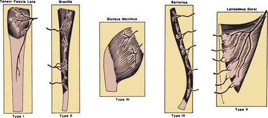
FIGURE 5.2. Patterns of vascular anatomy: Type I, one vascular pedicle; Type II, dominant pedicle(s) and minor pedicle(s); Type III, two dominant pedicles; Type IV, segmental vascular pedicle; Type V, one dominant pedicle and secondary segmental pedicle. (From Mathes SJ, Nahai F. Classification of the vascular anatomy of muscles: experimental and clinical correlation. Plast Reconstr Surg. 1981;67:177, with permission.)
Type II: Dominant Vascular Pedicle(s) and Minor Vascular Pedicle(s)
Use of a Type II flap generally requires division of part or all of the minor pedicles with preservation of the dominant pedicle. The muscle survives when elevated based on the dominant vascular pedicle. Muscles with a Type II vascular pattern include the following: the abductor digiti minimi (foot), abductor hallucis, brachioradialis, coracobrachioradialis, flexor carpi ulnaris, flexor digitorum brevis, gracilis, hamstring (biceps femoris), peroneus brevis, peroneus longus, platysma, rectus femoris, soleus, sternocleidomastoid, trapezius, triceps, and vastus medialis.
Type III: Dominant Pedicles
Type III muscles contain two large vascular pedicles, each of which may support the entire muscle. Muscles with a Type III vascular pattern include the following: gluteus maximus, intercostal, orbicularis oris, pectoralis minor, rectus abdominis, serratus, and temporalis.
Type IV: Segmental Vascular Pedicles
This group of muscles contains a series of segmental pedicles—generally of equal size—that enter the muscle along its course. Each segmental pedicle provides circulation to a portion (segment) of the muscle. Generally, division of two or more pedicles is feasible for transposition of a portion of the muscle as a flap. However, the muscle generally will not survive if an excessive number of the segmental pedicles are divided during flap elevation. Muscles with a Type IV vascular pattern include the following: the extensor digitorum longus, extensor hallucis longus, external oblique, flexor digitorum longus, flexor hallucis longus, sartorius, and tibialis anterior.
Type V: Dominant Vascular Pedicle and Secondary Segmental Vascular Pedicles
In this pattern of circulation, the muscle receives a large vascular pedicle that will reliably provide circulation to the muscle when it is elevated solely based on this particular vascular pedicle. However, the muscle has secondary vascular pedicles, which generally enter the muscle at its opposite end from the site of entry of the dominant vascular pedicle. These secondary pedicles will also support the muscle if the dominant vascular pedicle is divided. Thus, the muscle may be utilized as a flap based on either of the two sources of circulation. Muscles with a Type V pattern include the following: internal oblique, latissimus dorsi, pectoralis major.
ARC OF ROTATION
Each muscle and myocutaneous flap has a limited arc of rotation when transferred as a pedicle flap. The distance from the point where the pedicle enters the flap to the distal end of the flap defines the capability of that flap. A muscle flap that can be based on a dominant vascular pedicle can reach adjacent areas that fall within the radius created by the pedicle and the most distal portion of muscle that is supplied by that circulation. Generally, the muscle is released from either its origin or its insertion. The muscle is then mobilized on the major or dominant pedicle being utilized. In pedicle flap elevation, the pedicle is not usually skeletonized in order to avoid vascular injury and kinking. These rotational limitations should be incorporated into the surgical plan so that defect coverage will be maximized. With progressive mobilization of the pedicle, the arc of rotation of the flap can be increased. Release of the bony attachments overlying the point of entry of the vascular pedicle will also allow the muscle to be elevated as an island flap based only on its vascular pedicle with subsequent increase in its arc of rotation (Figure 5.3A–C).
Specific knowledge of anatomic landmarks including muscle insertion and origin and where the vascular pedicle enters the muscle will allow for better planning. A template can be made of the defect and then the arc of rotation of potential regional muscles can be plotted. Certain defects require two or more regional flaps, but knowledge of the muscular anatomy will allow reliable planning Muscle flap elevation based on the dominant pedicle is designated as the standard flap. If a muscle flap is elevated on its secondary pedicle, which requires division of the dominant pedicle, the flap is classified as a “reverse” flap. An example of this is a pectoralis muscle flap that is normally elevated on its dominant axial pedicle, the thoracoacromial vessels. The flap can also be raised as a turnover flap based on the secondary vessels from the internal mammary circulation, to cover a midline sternal defect.

FIGURE 5.3. Arc of rotation. A. Arc of rotation with flap elevation to point of entrance of vascular pedicle to flap. B. Extended arc of rotation based on flap elevation with dissection of pedicle to regional source. C. Extended arc of rotation based on flap elevation with pedicle dissection and release of proximal fascia and/or muscle origin or insertion. (From Mathes SJ, Nahai F. Reconstructive Surgery Principles, Anatomy and Technique. Vol 1. New York, NY: Churchill Livingstone; 1997:115, with permission.)
In a rotation advancement flap such as a gluteal flap for sacral wound coverage, the arc of rotation is based more on the pivot point of the cutaneous incision and any associated backcut rather than on the vascular pedicle. Clearly, these flaps are limited by distance since a large cutaneous component remains attached.
SKIN TERRITORY
Musculocutaneous flaps are composite axial flaps that consist of muscle and overlying subcutaneous tissue and skin. In most cases, the muscle at the base of the flaps is supplied by a single dominant vessel, which gives off one or more perforating vessels to supply the overlying subcutaneous tissue and skin. Examples of musculocutaneous flaps include the TRAM flap and the latissimus dorsi flap. Topographically, nearly any muscle in the direct subcutaneous location provides perforators to the skin either directly through or adjacent to the muscle. This subcutaneous tissue and overlying skin can be incorporated into a multilayered type of reconstruction. The skin territory of each superficial muscle is defined anatomically as that segment of skin extending between the origin and insertion of the muscle and located between its edges along the course of the muscle and can even be extended beyond this territory. The pedicled musculocutaneous flap may be designed with the skin left intact (rotation flap) at the flap base or a skin island (island flap) may be designed over the flap. Generally, the more narrow muscles (i.e., gracilis) have a greater limitation in skin territory because of the decreased number of perforating vessels to the overlying skin and the increased importance of septocutaneous vessels to the skin territory in proximity to the muscle.
FLAP MODIFICATIONS
The goals of reconstructive surgery include safety along with restoration of form and function. The donor site must also be considered. Repair of a defect in one region by creating an equally problematic defect in the donor site is not a satisfactory trade-off. Knowledge of the vascular territory of the donor muscle based on either dominant or segmental supply helps define which portion of the muscle can successfully be transferred or survive regional mobilization. Limiting the amount of fascial harvest and muscle dissection can offer a functional benefit in certain donor regions. A classic example of this is muscle and fascial harvest in TRAM flaps and the associated risks of abdominal wall laxity and weakness. Although the standard design of the muscle flap often represents the most appropriate method to reach these goals, alterations in flap design may avoid problems at the donor site. Muscle sparing and perforator approaches help to decrease the abdominal wall morbidity associated with this type of flap harvest and minimize the need for alloplastic (mesh) reconstruction of the donor site. Certainly, when harvesting bilateral flaps for breast reconstruction, a perforator dissection will minimize the overall tissue loss on the donor site and allow for an easier primary closure of the abdominal wall.
Segmental Flap
As noted above, transferring a portion of a muscle has potential advantages, including functional preservation, decreased bulk at the recipient site, and potential use of the remaining muscle as a secondary flap.12 Type III muscles, especially the gluteus maximus, are ideally suited for segmental design because these muscles have a dual blood supply. Thus, it is possible to split the muscle, leaving half of it attached to its origin, insertion, and motor nerve. The other half of the muscle can then be elevated as a transposition flap. This type of muscle flap modification may be used for both Type I and Type II muscles because the muscle is divided based on branches of the dominant vascular pedicle. Type V muscles, because of their blood supply, have the ability to be split and taken as smaller flaps based on the main or secondary circulations (Figure 5.4).
A Type IV muscle, in particular, requires elevation as a segmental flap, because the entire flap generally does not survive based on a single segmental vascular pedicle. Only a portion of the muscle can be divided and used as a transposition flap. Use of the superior part of the sartorius muscle for groin vessel coverage is an example of segmental muscle flap design. The sartorius is elevated by ligating one or two (as many as needed) perforators and rotating the proximal muscle medially to cover the femoral vessels. More ligation of distal perforators may compromise the blood supply to the proximal flap, which is required for the vessel coverage.
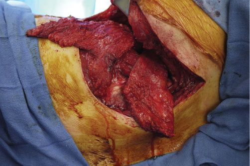
FIGURE 5.4. Split latissimus, along with other muscular flaps being used to obliterate a bronchopleural fistula and empyema cavity. Latissimus was split and used superiorly and inferiorly to help fill the space in this reconstruction.
Distally Based Flaps
Design of a flap on minor pedicles located opposite to the base of the standard flap is classified as a distally based flap.13 Generally, the entire muscle will not survive division of the dominant pedicle and, therefore, only a small part of the muscle is elevated on a specific identified minor pedicle. Delay by ligation of the dominant pedicle prior to flap elevation helps in successful elevation of distally based flaps, including the proximal muscle. The main problem for these distally based flaps can be venous drainage, especially in the lower extremity. Elevation of the extremity to allow for postural drainage and surgical delay, as mentioned above, helps the distally based flap to adapt the venous circulation to its new circuitous pathway. An example of this is the use of the medial hemisoleus as a reversed flap based on the distal posterior tibial perforators for coverage of ankle and distal third defects of the lower extremity.14
Neurotized-Functional Muscle Flap
A muscle flap may be used to provide motor function at the site of reconstruction.15 Flap design requires preservation of both the dominant vascular pedicle and the motor nerve (examples include the latissimus and the gracilis). In order to maintain effective muscle function, the muscle must be inset so that its resting length and tension is the same as it was in the donor site. A muscle may be designed both to provide coverage of a defect and to restore function. An example of this is the use of the latissimus dorsi muscle in the biceps region. It may be used as a pedicled flap on its motor nerve (thoracodorsal nerve) or a neurorrhaphy can be performed to the musculocutaneous nerve. In the forearm region it can be used as a free flap (Figure 5.5A–E).
Sensory Flap
Sensory reinnervation of cutaneous islands after transfer is unpredictable. A musculocutaneous flap may be designed to include a sensory nerve to the cutaneous portion of the flap. If the sensory nerve does not enter the skin territory of the flap adjacent to the dominant or major vascular pedicle, the nerve may require division during pedicled or free flap elevation. If divided, a neurorrhaphy may be performed to another sensory nerve at the recipient site. Examples of this exist with breast reconstruction. Neurorrhaphies can be performed between the 11th intercostal nerve, which is involved in sensation to the rectus flap, or the cutaneous branches of the 7th thoracic nerve, which provides sensibility to the cutaneous component of the latissimus flap, and the lateral cutaneous branch of the 4th intercostal nerve, which provides the major contribution to sensation of the breast. Clinical and research studies have shown more consistent sensory return to the recipient site when a sensory neurorrhaphy is performed.16 The difficulty with this approach is that sensory return is not a functional requirement in all territories of the body. Even in areas such as the plantar aspect of the foot where sensation is important for protection and proprioception, function can be preserved without direct sensory reconstruction. Many patients regain deep sensation from local neural growth into the transplanted tissue. Also, sensory nerves supplying a given cutaneous territory may not be clearly visible or consistent on dissection. The indications for sensory reconstruction in these flaps must be individualized and should be planned to help guide the flap dissection and the patient’s expectations. Division of sensory nerves must be performed appropriately to avoid neuroma formation. Regional dysesthesia is a potential consequence with injury to, or harvest of, sensory nerves supplying a cutaneous area.
Vascularized Bone
Vascular connections between the muscle and bone are generally observed at the muscle–bone interface. If these vascular connections are preserved, it is possible to elevate a segment of vascularized bone with the flap. A segment of the 6th rib with the pectoralis major muscle and a segment of the iliac bone with the internal oblique muscle (deep circumflex iliac artery flap) are examples of muscle flaps that may include bone. In a free fibula flap, the flexor hallucis longus is supplied by the peroneal vessels and interconnected through this vasculature with the fibula bone (Figure 5.6A–C). Although the muscle dissection can be limited during the flap harvest, it is often incorporated to supply extra internal or cutaneous coverage, bulk, and vascular supply.
Tissue Expansion
Although rarely used because of surgical staging difficulties and risk of complications, insertion of a tissue expander beneath a musculocutaneous flap allows for an increase in skin island dimensions and assists in donor site closure (Chapter 10).17 In flap coverage surgery, tissue expansion is more commonly utilized in preparation of fasciocutaneous advancement flaps. Tissue expansion can be utilized to increase the useable skin island in a latissimus musculocutaneous flap and also allow for primary closure of the defect. When used for breast reconstruction, the tissue expander increases the dimensions of both the remaining skin envelope and the associated overlying pectoralis major muscle.
Free Flaps
Free flaps are the natural extension of axially based muscle and musculocutaneous flaps and have further advanced our ability to provide reconstructive options. Pedicled flaps are limited by their arc of rotation. Microvascular free tissue transfer broadens the flaps’ usefulness to all areas of the body. Free tissue transfer should, like all reconstructive techniques, be performed in a well-planned fashion and should not be performed in lieu of appropriate regional options. The reasons for using muscles as free flaps are essentially fourfold. First, to overcome limited regional options such as in distal tibial and foot defects. Second, the volume of the defect is larger than regional tissues can reconstruct. Microvascular transplantation is frequently utilized in the head and neck region where there is a lack of suitable regional muscles to satisfy the reconstructive need for combined facial, oral, and nasal cavity defects. Third, when functional deficits from utilizing the regional muscle supply may limit the outcome, nonessential distant muscle can be utilized to provide a functional outcome. Fourth, for infections or prosthetic coverage when flap re-elevation is likely, even when local, fasciocutaneous coverage can be performed.

FIGURE 5.5. Chimeric flap for thigh reconstruction. A. Massive thigh sarcoma during resection, removing most of the anterior thigh musculature and skin. B. Contralateral thigh chimeric flap including vastus lateralis, tensor fascia lata, and anterolateral thigh tissue along with the accompanying motor and sensory nerves. C. The pedicle and associated nerves are seen. D. Placement of the flap into the defect and neurorrhaphy between the muscular nerve branches and the recipient site nerve branches for eventual neurotization. E. Six months postoperatively with return of nerve function and improving knee extension.
Flap design is essentially the same for both regional transposition and microvascular transplantation of muscle and musculocutaneous flaps. The reconstructive needs are analyzed and treated in a composite fashion. Like tissues are chosen to reconstruct the defect for both functional and aesthetic purposes. The consistent, long vascular pedicle to most Type I, II, and V muscles allows rapid elevation of the muscle with its vascular pedicle for microvascular transplantation (Figure 5.7A–E).
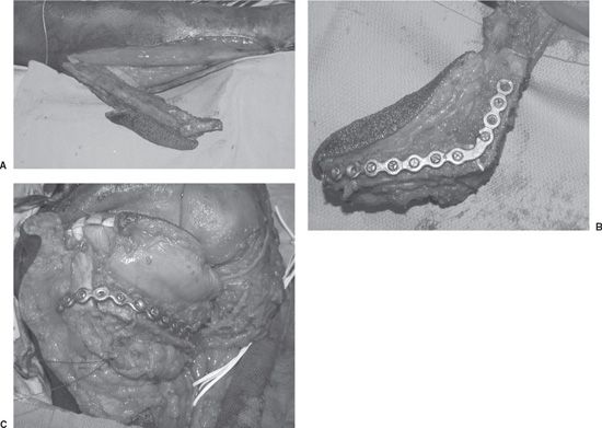
FIGURE 5.6. A. Fibula harvest for mandible reconstruction including a portion of the flexor hallucis longus muscle, which is supplied by the peroneal vasculature. B. Fibula osteotomized and plated while remaining attached to its pedicle in situ. C. Fibula transferred and revascularized. Bone inset has been performed and soft tissue inset is next. Included is a skin island (osteomyocutaneous) for intraoral reconstruction.
Perforator Flaps
A muscle’s axial blood vessel provides perforators, which first supply the muscle and then proceed superficially to supply the overlying skin and subcutaneous tissue. These vessels can be meticulously dissected from the surrounding muscle to produce a direct cutaneous perforator flap (Figure 5.8). These perforator flaps are cutaneous flaps, which are based on the vessels known to traverse various muscle flaps such as the deep inferior epigastric, thoracodorsal, and superior gluteal vessels. These flaps demonstrate that the unnamed cutaneous/perforating vessels arise from larger, named vessels and travel through the muscle or muscular septum to supply a large cutaneous region. The reliability of these flaps is clearly more robust than previously thought. The problem of anatomic variability to these cutaneous perforators is greater when not following known muscle territories. Perforator flaps, while technically challenging, may decrease some of the functional morbidity associated with the harvest of muscles and overlying muscle fascia in myocutaneous flap harvest. These flaps have become widely utilized for breast reconstruction but can be used for any location throughout the body.18
Prefabricated Flaps
Prefabrication represents the future of flap-based reconstruction and is in essence in vivo tissue engineering. The goal of this type of reconstruction is to provide all missing components of a given defect by positioning support, lining, and coverage tissues in preplanned positions and allowing them to vascularize prior to transfer, and minimize donor site morbidity.19,20 Descriptions of prefabrication have been mostly focused on the head and neck region but can be translated to all parts of the body. Defects in the head and neck can be complex involving mucosal loss from the oral, nasal, and pharyngeal cavities; structural loss of either the bony or cartilaginous skeleton; and cutaneous loss. In larger defects there is no one flap that can provide all of these missing layers. By either incorporating multiple flaps (e.g., free osteocutaneous fibula free flap with a pedicled pectoralis myocutaneous flap for a composite mandibular defect) or by precise, planned flap prefabrication, these defects can be reconstructed. The use of thinned flaps with the pre-grafting of autologous or bioengineered structural elements such as bone and cartilage and the creation of new vascular bundles in desired donor sites are well established in the literature. The advancement of in vivo and ex vivo tissue engineering, with, and ultimately without, immune modulation, is one of the next frontiers for reconstructive surgery.
Combined Flaps
Combined flaps are used when either a large volume of tissue is required, more than a single flap can provide, or when multiple tissue types are required in a complex position or orientation. Instead of taking multiple pedicled or free flaps to perform a given reconstruction, conjoined or chimeric flaps can be used. Each of these groups has subcategories and individualized terminology within them but the basic principles remain the same.21 The flaps in these groups are either attached by a common vascular supply or are directly attached, each with its own vascular supply. Conjoined flaps are individual flaps that have their own vascular territory but are attached by a soft tissue bridge to form a larger flap with multiple vascular territories being incorporated. An example of this is the bridging of an extended myocutaneous latissimus flap with a superficial inferior epigastric flap, which was first described by Harii et al., back in 1981.22 These flaps could be rotated on either pedicle, with the other pedicle being attached microsurgically to enhance the blood supply and create a tremendously large flap for cutaneous coverage. These massive flaps can also be taken further as a pure free flap with double microvascular anastomoses. The use of these flaps obviously depends on the defect. A flap can be considered conjoined between individual perforators because each of these territories can be separated and can act as an individual flap unit.
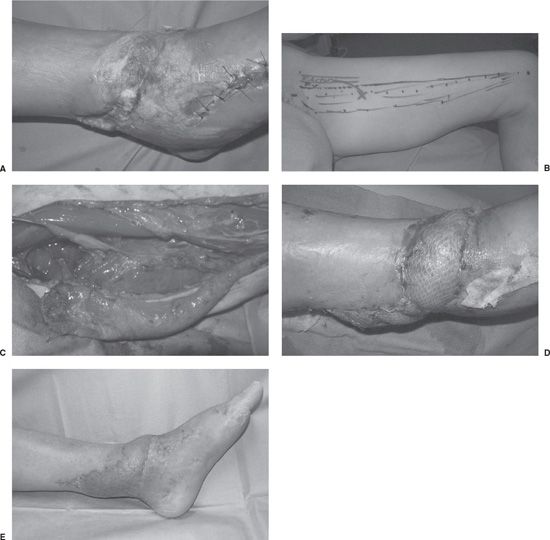
FIGURE 5.7. A. Open ankle defect after orthopedic trauma. Bone and hardware were exposed after debridement. B. Design of a same-leg gracilis flap that approximated the defect size well and with limited donor site morbidity. C. Harvest of the gracilis (Type II) flap. D.Intraoperative coverage of this small defect with well-vascularized muscle. E. Several months postoperative picture showing excellent healing and recontouring.
Chimeric flaps are individual flaps that are fully separated from each other but linked together by a common source vessel. The classic examples come from the subscapular system and from the lateral circumflex femoral system. The subscapular system has a variety of tissue types from bone to skin that can all be taken as separate flaps or in multiple combinations (Figure 5.9A–E). The vascular supply for these flaps is from large independent subfascial vessels that connect to the same source vessel. The anterolateral thigh chimeric flaps are perforator based since islands of tissue can be created on each perforator branch that emanates from the source supply of the lateral circumflex femoral vessels. A final subtype of the chimeric flaps is the fabricated chimeric flap. These are flaps that are attached to each other by microanastomosis either at branch points or at the distal (flow-through) end to create a hybrid flap.
COMPLEX WOUND MANAGEMENT
Muscle and musculocutaneous flaps are ideal for treating difficult soft tissue and bony or prosthetic infections. Although treatment to decrease the bacterial concentration below 105 per gram of tissue is necessary, subsequent coverage with well-vascularized muscle appears to further decrease the bacterial load, protect against recurrence of infection, and maintain wound closure. Planned treatment of complex wounds with staged debridements followed by coverage with well-vascularized tissue and appropriate antibiotic therapy has revolutionized wound management and is the standard of care in most situations. Historically, experimental studies comparing bacterial resistance in musculocutaneous as compared with cutaneous and fasciocutaneous flaps have demonstrated superior resistance to bacterial invasion and subsequent flap necrosis in the muscle and musculocutaneous flaps.23,24 Since muscle flaps appear to provide protection from progressing bacterial injury to the soft tissues and improved tissue vascularity, it has allowed for the management of complex wounds that traditionally did not respond well to local wound care. There have also been studies showing no difference between muscle and fasciocutaneous flaps in infected wounds.25 It remains widely accepted, however, that muscle flaps provide an excellent option for coverage. Also, when comparing the use of muscle and fasciocutaneous free flaps in traumatic wounds, there does not appear to be an increased incidence of long-term postoperative infection when appropriate debridement is performed.26 Muscle flaps are extremely useful in three-dimensional defects, which require the flap to contour to irregular or complex topography.

FIGURE 5.8. Perforator TRAM (transverse rectus abdominis myocutaneous) flap anastomosed to the internal mammary vessels.
Osteomyelitis
Following debridement of the infected bone associated with chronic osteomyelitis, a muscle flap is transposed as a regional flap or transplanted by microvascular technique into the defect. The flap fills the area of bone debridement with well-vascularized tissue and provides stable wound coverage (Figure 5.10A–D). As noted above, short-term culture-specific antibiotic therapy is utilized simultaneously. With this approach, successful management of chronic infection in the site of bone or cartilage injury has been observed.27 Debridements can be performed in a staged fashion depending on the amount of infection and stability of the patient. Coverage with the muscle flap is planned immediately after the final debridement. Sternal wounds are a common and problematic example of osteomyelitis (Figure 5.11A and B). Treatment with serial debridement and antibiotics and coverage with a muscle flap such as the pectoralis major and/or the rectus abdominis are necessary for ultimate wound closure, chest wall stabilization, and patient survival.
Vascular Insufficiency
Nonhealing wounds associated with vascular insufficiency frequently require extremity amputation. Revascularization of the leg may salvage the extremity, but the wound will still necessitate flap coverage. Although revascularization provides macroscopic blood flow to an extremity, the area of a specific wound may still have insufficient microvascular perfusion or be too large to heal on its own. Muscle flap placement provides transplanted microcirculation and tissue bulk to allow these wounds to heal and ultimately provide for limb salvage. Either simultaneous or delayed muscle flap transplantation will allow preservation of a functional extremity despite wound complexity.28 In certain circumstances, a flap can be chosen that has a flow-through ability. With this type of flap design, the vascular supply, more often, from a traumatic injury or atherosclerotic disease can be augmented and flap tissue placed to cover the defect. This type of reconstruction requires precise planning and execution.
Radiation Wounds
Wounds associated with radiation injury do not respond to local wound care and can be some of the most difficult wounds to treat (Chapters 3 and 17). Tissue that has undergone high-dose ionizing radiation therapy has limited resistance to injury and ability to regenerate. The effects of this type of radiation are longstanding. Radiated tissue can remain intact for decades but any form of tissue stress or injury can form a chronic wound with critical structures ultimately being exposed. Treatment of these wounds usually requires wide debridement of necrotic skin, affected soft tissue, and sclerotic or infected bone and results in a complex wound usually associated with exposure of vital structures. If adjacent muscle units have vascular pedicles located distant to the radiation port, regional muscle flaps may be useful for vascularized coverage (Figure 5.12A–C).29 In areas with poor local muscle availability such as the head and neck region, particularly in the skull, microvascular transfer of a muscle flap is generally required for coverage.
Exposed or Infected Prosthesis
When wound coverage overlying the site of a vascular or orthopedic prosthesis fails, early wound debridement, muscle flap coverage, and culture-specific antibiotic therapy frequently allow salvage of the prosthesis and simultaneously provide stable defect coverage.30 Once infection has been established in the prosthesis, however, it is usually necessary to remove the prosthesis. More common areas of exposure for vascular grafts are in the groin and lower extremity. Groin coverage is usually accomplished reliably with a sartorius muscle flap, but larger flaps can also be mobilized if needed. Orthopedic hardware is more commonly exposed in the midline from spine surgery or over joint with limited coverage such as the knee. Spine hardware can usually be well covered with myocutaneous advancement flaps, whereas joint coverage usually requires rotation flaps such as the gastrocnemius muscle. Lastly, hardware exposure can occur in combination with trauma such as in lower extremity injuries or after radiation injury leading to exposure of underlying bony or vascular prostheses. In any of these cases, reconstruction with either a local or free muscle flap is almost always indicated.

FIGURE 5.9. Chimeric flap reconstruction in the head and neck. A. Exposed mandibular plate along with an underlying mandibular defect. B. Chimeric flap design from the subscapular system including bone, muscle, and fasciocutaneous tissue. C. Flap harvested with the separate components visualized, all attached to the main subscapular system. D. Flap being placed into the bony and soft tissue defect prior to microvascular reanastomosis. E. Closure of the cutaneous defect at the end of the procedure.
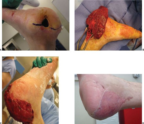
FIGURE 5.10. Reconstruction of osteomyelitis of the heel. A. Chronic draining heel wound with refractory calcaneal osteomyelitis. Prior debridement was performed. B. The heel was opened transversally along its lateral surface to expose the calcaneal wound fully and allow for debridement and subsequent coverage. The incision was carried up to the posterior tibial vessels where a muscle flap was anastomosed and then placed inside of the heel for complete coverage of the debrided osteomyelitic space. C. Muscle in place after inset and prior to skin graft placement. D. Six months postoperatively with a well-healed and contracted wound reconstruction.
CONCLUSIONS
Muscle and musculocutaneous flaps are available in all body regions. With the selection of muscles with a suitable vascular pedicle, the muscle may be safely elevated to provide coverage and simultaneously restore form and function. Thorough knowledge of the muscular anatomy, vascular circulation, and the arc of rotation is required in order to select the optimal muscle unit for specific defects throughout the body. When regional muscle flaps are unavailable or undesirable, the surgeon may elect to transfer distant muscle or musculocutaneous flaps microsurgically. Muscle and musculocutaneous flaps also provide a method to treat complex wounds—such as osteomyelitis, radiation necrosis, traumatic defects, and exposed hardware—that in the past were recalcitrant to wound care. Use of muscle and musculocutaneous flaps has revolutionized reconstructive plastic surgery. Their use has allowed for bolder, more effective oncologic resections, limb salvage in previously irreparable situations, improved functional restoration with motor unit loss, contracture release in secondarily healed or scarred joint and soft tissue contractures, and improved aesthetic outcome for contour defects including breast reconstruction. Nearly any defect can be closed with a careful analysis and a planned approach for reconstruction. Use of muscle and musculocutaneous flaps broadens the options for defect closure in every area of the body. The future of flap reconstruction is also advancing and becoming more refined with the use of perforator flaps, flap prefabrication, and chimeric flaps to more precisely reconstruct the most complex defects. All of these modifications and advancements in flap surgery have positioned the reconstructive surgeon at the forefront of clinical tissue engineering and vascularized composite allotransplantation.
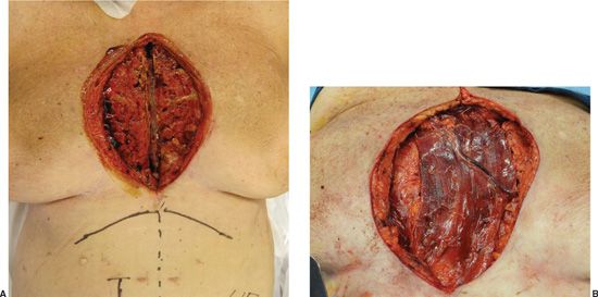
FIGURE 5.11. Reconstruction of sternal osteomyelitis. A. Sternal osteomyelitis after wire removal and an initial debridement. Significant debridement of the pectoralis muscle was also performed. B. Pedicled rectus abdominis flap placement into the defect.
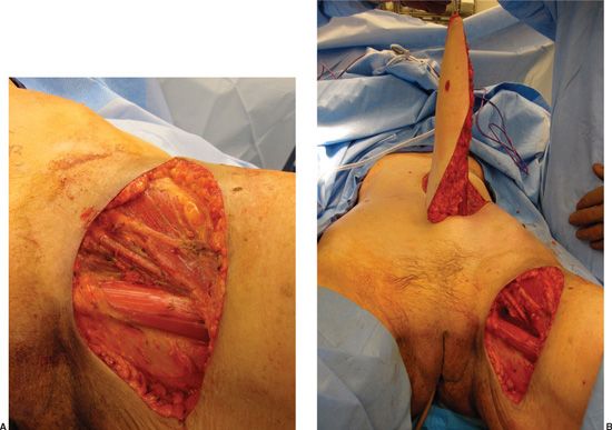
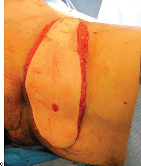
FIGURE 5.12. Reconstruction of a radiated groin wound. A. Excision of a radiated open tumor wound in the groin along with underlying lymph node dissection and removal of surrounding radiated skin. B. Elevation of a vertical rectus abdominis myocutaneous flap. C.Placement of the flap through a subcutaneous tunnel with excellent coverage of the defect including muscle over the entire wound bed and complete cutaneous closure with the overlying skin paddle.
References
1. McGregor IA, Morgan G. Axial and random pattern flaps. Br J Plast Surg. 1973;26(3):202-213.
2. Mathes SJ, Nahai F. Clinical Applications for Muscle and Musculocutaneous Flaps. St. Louis, MO: C.V. Mosby; 1982.
3. McCraw JB, Dibbell DG, Carraway JH. Clinical definition of independent myocutaneous vascular territories. Plast Reconstr Surg. 1977;60(3): 341-352.
4. Tolhurst DE. Surgical indications for fasciocutaneous flaps. Ann Plast Surg. 1984;13(6):495-503.
5. Taylor GI, Palmer JH. The vascular territories (angiosomes) of the body: experimental study and clinical applications. Br J Plast Surg. 1987; 40(2):113-141.
6. Wei FC, Mardini S. Free-style free flaps. Plast Reconstr Surg. 2004; 114(4):910-916.
7. Bakamjian VY, Long M, Rigg B. Experience with the medially based deltopectoral flap in reconstructuve surgery of the head and neck. Br J Plast Surg. 1971;24(2):174-183.
8. Ghali S, Butler PE, Tepper OM, Gurtner GC. Vascular delay revisited. Plast Reconstr Surg. 2007;119(6):1735-1744.
9. Atisha D, Alderman AK, Janiga T, Singal B, Wilkins EG. The efficacy of the surgical delay procedure in pedicle TRAM breast reconstruction. Ann Plast Surg. 2009;63(4):383-388.
10. Mathes SJ, Nahai F. Classification of the vascular anatomy of muscles: experimental and clinical correlation. Plast Reconstr Surg. 1981;67(2):177-187.
11. Taylor GI, Gianoutsos MP, Morris SF. The neurovascular territories of the skin and muscles: anatomic study and clinical implications. Plast Reconstr Surg. 1994;94(1):1-36.
12. Mathes SJ, Vasconez LO. Myocutaneous free-flap transfer. Anatomical and experimental considerations. Plast Reconstr Surg. 1978;62(2):162-166.
13. Mathes SJ, Nahai F. Reconstructive Surgery: Principles, Anatomy and Technique. New York, NY: Churchill Livingstone; 1997.
14. Pu LL. The reversed medial hemisoleus muscle flap and its role in reconstruction of an open tibial wound in the lower third of the leg. Ann Plast Surg. 2006;56(1):59-63; discussion 63-64.
15. Terzis JK, Sweet RC, Dykes RW, Williams HB. Recovery of function in free muscle transplants using microneurovascular anastomoses. J Hand Surg Am. 1978;3(1):37-59.
16. Yap LH, Whiten SC, Forster A, Stevenson HJ. Sensory recovery in the sensate free transverse rectus abdominis myocutaneous flap. Plast Reconstr Surg. 2005;115(5):1280-1288.
17. Radovan C. Tissue expansion in soft-tissue reconstruction. Plast Reconstr Surg. 1984;74(4):482-492.
18. Geddes CR, Morris SF, Neligan PC. Perforator flaps: evolution, classification, and applications. Ann Plast Surg. 2003;50(1):90-99.
19. Garfein ES, Orgill DP, Pribaz JJ. Clinical applications of tissue engineered constructs. Clin Plast Surg. 2003;30(4):485-498.
20. Pribaz JJ, Fine NA. Prefabricated and prelaminated flaps for head and neck reconstruction. Clin Plast Surg. 2001;28(2):261-272, vii.
21. Hallock GG. Further clarification of the nomenclature for compound flaps. Plast Reconstr Surg. 2006;117(7):151e-160e.
22. Harii K, Iwaya T, Kawaguchi N. Combination myocutaneous flap and microvascular free flap. Plast Reconstr Surg. 1981;68(5):700-711.
23. Calderon W, Chang N, Mathes SJ. Comparison of the effect of bacterial inoculation in musculocutaneous and fasciocutaneous flaps. Plast Reconstr Surg. 1986;77(5):785-794.
24. Gosain A, Chang N, Mathes S, Hunt TK, Vasconez L. A study of the relationship between blood flow and bacterial inoculation in musculocutaneous and fasciocutaneous flaps. Plast Reconstr Surg. 1990;86(6):1152-1162; discussion 1163.
25. Salgado CJ, Mardini S, Jamali AA, Ortiz J, Gonzales R, Chen HC. Muscle versus nonmuscle flaps in the reconstruction of chronic osteomyelitis defects. Plast Reconstr Surg. 2006;118(6):1401-1411.
26. Yazar S, Lin CH, Lin YT, Ulusal AE, Wei FC. Outcome comparison between free muscle and free fasciocutaneous flaps for reconstruction of distal third and ankle traumatic open tibial fractures. Plast Reconstr Surg. 2006;117(7):2468-2475; discussion 2476-2477.
27. Mathes SJ, Alpert BS, Chang N. Use of the muscle flap in chronic osteomyelitis: experimental and clinical correlation. Plast Reconstr Surg. 1982;69(5):815-829.
28. McCarthy WJ 3rd, Matsumura JS, Fine NA, Dumanian GA, Pearce WH. Combined arterial reconstruction and free tissue transfer for limb salvage. J Vasc Surg. 1999;29(5):814-818; discussion 818-820.
29. Mathes SJ, Alexander J. Radiation injury. Surg Oncol Clin N Am. 1996; 5(4):809-824.
30. Greenberg B, LaRossa D, Lotke PA, Murphy JB, Noone RB. Salvage of jeopardized total-knee prosthesis: the role of the gastrocnemius muscle flap. Plast Reconstr Surg. 1989;83(1):85-89, 97-99.