
PART VIII
HAND
CHAPTER 83 REPLANTATION STRATEGIES OF THE HAND AND UPPER EXTREMITY
SANDEEP JACOB SEBASTIN AND KEVIN C. CHUNG
REPLANTATION STRATEGIES OF THE HAND AND UPPER EXTREMITY
Replantation is the reattachment of a completely amputated body part by reestablishing arterial inflow and venous outflow. Reattachment of incompletely amputated parts (irrespective of the nature or amount of tissue holding them together) is called revascularization. The term revascularization should ideally be restricted to incomplete amputations that require restoration of both arterial inflow and venous outflow. If the incomplete amputation needs only an arterial repair for restoring circulation, the correct term is critical arterial repair. The digits represent the most commonly amputated body part and replantation offers a result that is superior to any other type of reconstruction or the best available prosthesis. Although survival of the amputated digit depends on the patency of the microvascular repair, replantation cannot really be considered successful, until function is restored. Function is related to the adequacy of bone, tendon, nerve, and skin repairs and the postoperative rehabilitation.
HISTORY
Balfour provided the first scientific report of digital reattachment in the Edinburgh Medical and Surgical Journal in 1814. He reported the successful reattachment of the partial amputation of his son’s index, long, and ring fingers at mid-distal phalanx following a door crush injury. He also reported the successful result in a carpenter with a complete oblique guillotine amputation through the distal interphalangeal joint (DIPJ) of the left index finger. These reattachments were performed without vascular anastomoses and most likely survived as composite grafts. Murphy in 1896 reported the first successful critical arterial repair. He resected a femoral pseudoaneurysm and performed an end-to-end repair in a 29-year-old male 1 month after a gunshot injury. Carrel performed the first extremity replantation with vascular anastomosis in a mid-femoral amputation of a dog hind limb in 1906. He won the Nobel Prize in 1912 for his pioneering work on vascular anastomoses and organ transplantation.
Kleinert performed the first successful extremity revascularization in 1958. A 27-year-old man thrust his arm through a glass window and had a circumferential laceration in his proximal forearm. The radius, ulna, and a 2 cm bridge of skin and soft tissue on the dorsal ulnar aspect remained intact. Repair of the radial and ulnar arteries, and the basilic vein was carried out with 6–0 braided nylon sutures. Kleinert also performed the first microsurgical anastomosis of a digital artery in 1962. The patient was a 52-year-old man with a metal roller press crush injury to the thumb. He had a circumferential skin laceration distal to the metacarpophalangeal joint and the thumb was attached by two dorsal veins and threads of subcutaneous tissue.
Malt performed the first successful extremity replantation in 1962. He reported two cases of above elbow amputations, both following train accidents. One was a 12-year-old boy and the other a 44-year-old man and both regained reasonable function. Wei reported the successful replantation of a below elbow amputation in a factory worker in 1963. Komatsu and Tamai performed the first successful replantation of a completely amputated digit with microsurgical anastomosis in 1965 when they replanted a 28-year-old man’s left thumb that had been amputated by a steel-cutting machine.
CLASSIFICATION OF AMPUTATIONS
Amputations are classified based on the completeness of the amputation, the anatomical level of separation, and the mechanism of injury. It is good practice to describe an amputation based on these three characteristics. Along with an evaluation of the whole patient this will help in the decision-making process.
I. Completeness of amputation: It is important to separate complete (total) amputations from incomplete (subtotal/near-total) amputations, which separates replantations from revascularizations. A revascularization may appear to be an easier operation compared with replantation; however, in practice it is often more difficult. The presence of intact bone in an incomplete amputation means that the surgeon may not be able to shorten the bone significantly, which, in turn, may necessitate the use of vein grafts, nerve grafts, or skin flaps to bridge any defects. It is also not possible to do examine the part on the back table to isolate the neurovascular structures.
II. Anatomical level: Upper extremity amputations are classified into two broad groups.
A. Amputations proximal to the radiocarpal joint: Replantation at this level is termed a major limb replantation because these replantations have a higher risk of systemic complications. Because muscle can withstand ischemia for a short period of time, there is a risk of myoglobinuria and renal failure. The more proximal the amputation, the greater the muscle mass. Systemic complications are directly related to the muscle mass and the ischemia time and therefore the success of a major limb replantation depends on establishing circulation early.
B. Amputations distal to the radiocarpal joint: These amputations are described based on the flexor tendon zones of injury (zones 1–4). Each zone has anatomic characteristics that influence the technique and outcome of replantation.
III. Mechanism of injury: An amputation can occur following a clean-cut, a crush, an avulsion, or any combination of these injury patterns. Survival, as well as late functional outcome, depends to a great extent on the mechanism of injury. Although a clean-cut amputation or a pure avulsion type injury is easily identified, crush injuries vary widely. We have modified the classification of injury mechanism proposed by Yamano to simplify it and eliminate the subjectivity involved in grading crush amputations.
A. Clean cut (sharp cut/guillotine): Results from objects with narrow sharp edges like knives or meat slicers. The wound edges are clean and minimal debridement is required.
B. Blunt cut (dull cut): Results from objects with narrow blunt edges like saws or fan blades. The wound edges are jagged and show crushing, which extends a limited distance proximal and/or distal to the amputation. Moderate debridement is required.
C. Crush: Results from an object with a broad blunt edge, like a punch press or a wooden log. The wound is torn (lacerated) rather than cut (incised) and the wound edges are irregular. There is significant tissue injury that extends proximal and/or distal to the amputation. Extensive debridement is required.
D. Avulsion: Caused by traction, such as an anchor rope or the reins of a horse. Similar to a crush injury, the tissue is torn rather than cut. However, unlike crush where the tissue injury is at the level of the crush, the separation of tissues (vessels, nerve, tendon, bone, and skin) occurs at different levels depending on their tensile strength. A degloving injury is a special type of avulsion amputation usually caused by a tight ring (ring avulsion amputation), which gets caught during motion. The skeletal and tendinous apparatus are preserved, whereas the surrounding soft tissue envelope is pulled off. Peripheral bony parts of varying lengths may be avulsed together with the tissue covering.
E. Combined: Caused by a combination of crush, avulsion, or other mechanisms of injury. An example is an initial incomplete crush amputation by a machine followed by an avulsion when the patient reflexively withdraws the hand resulting in a complete amputation.
PREOPERATIVE MANAGEMENT
I. Transfer to a replantation center: The outcome of replantation depends on (1) patient factors; (2) the nature of the injury; and (3) the skill and experience of the surgical team. The first two cannot be controlled. Therefore, it is important that an experienced microsurgeon ascertains the suitability for replantation. All patients deserve such an evaluation and should be expeditiously referred to appropriate centers, where they can be guided toward decisions that serve their best interests. Before transport, an attempt should be made to contact the center to determine the appropriateness of the transfer and to allow preparation for replantation. A detailed description of the injury, the patient’s age, and general health, and the condition of the injured part are provided to the replant team over the phone. A digital photograph of the injured part helps tremendously.
The amputation stump is covered with a saline-moistened gauze, loosely wrapped, and elevated. Compression bandages may be required to stop bleeding. Once the patient is stabilized, an attempt is made to collect and preserve all amputated parts. The amputated part/s are wrapped in a saline-moistened gauze and then placed in a dry, watertight, plastic bag, that is in turn placed on ice (Figure 83.1). The aim is to keep the part cold to prevent the ill effects of warm ischemia, while avoiding direct tissue contact with ice that can cause frostbite. In cases of incomplete, nonviable amputations, the wound is wrapped in moist gauze, dressed, a simple splint applied to prevent kinking, and ice packs used to surround the distal portion of the amputated part. Radiography and arteriography delay transfer, increase the total ischemia time, and can be expeditiously performed at the replantation center.
II. Management in the emergency department: Once the patient arrives at the replantation center, the patient is examined to rule out associated injuries. This is especially important in major replantations, where the attention is focused on the amputation and there is real risk of missing severe injuries. Resuscitation and stabilization of the patient should take precedence over treatment of the amputated limb. A member of the replantation team obtains a brief history from the patient and the patient’s family that includes the age, hand dominance, occupation, preexisting systemic illness, allergies, and the mechanism of injury. History of smoking, alcohol or drug dependence, and any psychiatric illness should also be obtained. Radiographs of the amputated part and the proximal extremity are obtained. Routine investigations include a chest radiograph, electrocardiogram, complete blood count, serum electrolytes, and blood typing and cross matching.
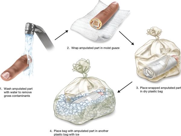
FIGURE 83.1. Preservation of the amputated part for transport.
Active bleeding may be found, especially in incomplete amputations with partial vessel lacerations. Control of bleeding is usually achieved by a pressure dressing. Blind attempts at clamping or ligation of vessels are avoided to prevent further injury to vessels and nerves. Temporary use of a proximal tourniquet is preferable, precisely identifying the bleeder, and clamping or ligating it carefully. Patients are given tetanus prophylaxis if the immunization status is uncertain or if the last shot was received more than 5 years previously. Prophylactic antibiotics with first-generation cephalosporins are indicated in amputation injuries directed toward the most likely organism, Staphylococcus aureus. An aminoglycoside and/or a third-generation cephalosporin may be required in amputations associated with more extensive contamination.
III. Evaluation for replantation (indications/contraindications): In complete amputations, a member of the replant team should take the amputated part to the operating room as soon as possible, to do preliminary bench-work assessing the suitability for replantation and to dissect, isolate, and tag important structures. This is ideally done while the patient is being stabilized in the emergency department. This may also be possible in incomplete amputations in which the amputated part is held by strands of tissue that have no contribution to circulation or innervation. The surgeon can divide the remaining strands of tissue converting an incomplete amputation into a complete amputation. When there is a skin bridge that may be important for venous drainage, however, keeping the skin bridge is important to avoid the most difficult part of the operation, the venous repair.
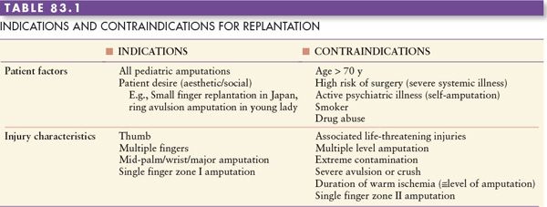
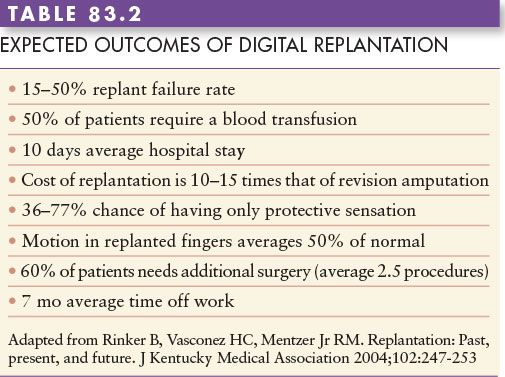
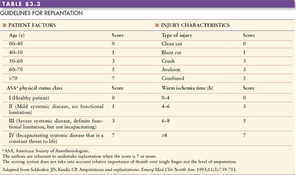
Patients desire reattachment of every amputated part, but they are not aware of the risk, cost, and eventual outcome. Although the indications and contraindications (Table 83.1) for replantation have remained more or less unchanged over many years, the decision to replant or not to replant is individualized. The main concern is “how to make it functional.” Function encompasses all aspects of a person’s performance in society, aesthetic as well as mechanical. Prior to replantation, a frank discussion between the patient (and relatives) and the surgeon is mandatory to discuss the likelihood of a successful result, the duration of postoperative therapy, possible need for multiple secondary surgeries, estimated time away from employment, and to temper patient expectations. The expected outcomes for digital replantation are shown in Table 83.2. The guidelines for replantation suggested by Schlenker and Koulis are useful in decision making, especially in borderline cases (Table 83.3). The long duration of surgery; need for blood transfusions; need for joint fusion; possible use of skin, nerve, and vein grafts; and approximate length of hospitalization are emphasized. Rarely, a flap may be required in the primary setting to cover exposed vessels, and appropriate options are planned and discussed with the patient preoperatively.
OPERATIVE MANAGEMENT
I. Team approach: Once a decision to replant a part has been made, events should progress in an efficient, stepwise manner. Replantation surgery takes a long time, such as 6 to 8 hours for a major limb replantation and 2 to 5 hours for a more distal amputation. In multiple digital amputations, each digit can take up to 3 to 4 hours. A team approach is advised to avoid surgeon fatigue. Realistically, in most centers, one surgeon is on call for replantation and a backup surgeon is often not available. It is important in multiple digit replantations, therefore, to proceed efficiently; fatigue will set in after a few hours of intense activities under the microscope. Ideally, two teams each consisting of a surgeon trained in microsurgery and hand surgery should be available for these procedures. One team can work on the amputated part, while the other prepares the stump. The operating nurses should also have training in the handling of microsurgical instruments and microsutures. We have found it useful to connect a television monitor to the operating microscope. This allows the nurses to see what is happening and improves their participation in what could otherwise be a long and tedious procedure for them.
II. Anesthesia: Regional anesthesia alone or in combination with general anesthesia provides the benefit of sympathetic blockade that results in vasodilatation and facilitates vascular anastomosis. The regional block can also be maintained in the postoperative period.
III. Patient preparation: A urinary catheter is inserted because of the length of the procedure. A padded tourniquet is applied around the upper arm for all amputations except transhumeral. In major limb replantation, a tourniquet is placed on the lower limb which is prepared for the skin, nerve, and/or vein grafts. The operating room and the patient are kept warm and appropriate padding provided at bony pressure points.
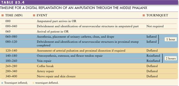
IV. Sequence of steps: The logical sequence is to progress from repair of the deeper structures (bone and tendon) to superficial structures (nerve and vessels) and from repairs requiring gross manipulation (bone and tendon) to those that need an operating microscope (nerve and vessels) for fine precise repairs. The exact order of repair depends on surgeon preference and the level of amputation. There is some disagreement whether arterial or venous repair should be done first. Our preference is to establish venous drainage first. This minimizes blood loss and completes what is technically the most difficult step of replantation early on. Doing the artery first allows selection of veins with good outflow for anastomosis; however, the field is bloody and dissection difficult. Reinflating the tourniquet at this stage may increase the risk of arterial thrombosis as a result of stasis across the anastomosis. The two situations when it is preferable to do the arterial repair first are a distal replant where the arterial inflow helps in identifying the veins and in major limb replantations to decrease the warm ischemia time. A timeline sequence of steps has been depicted in Table 83.4.
A. Bench-work: The amputated part is cleaned with routine bacteriocidal solution such as betadine and placed on a small operating table. In the hand, wrist, and major limb replants, the debridement and dissection of the neurovascular structures are performed with the amputated part on an ice pack (avoiding direct contact with ice) to limit the warm ischemia time. When one digit in a multiple digit injury is being dissected, the other digits are preserved as previously mentioned. Cooling should continue until the arterial anastomosis is complete. Amputated parts or digits unsuitable for replantation should not be discarded, but evaluated for use as a spare part or as a source of nerve, arterial, skin, or bone graft. Irrespective of the level of amputation, the key steps in preparation of the amputated part include meticulous debridement, isolation of the neurovascular structures, and bone shortening.
All grossly contaminated or devitalized tissues are sharply debrided. Ragged bone and the flexor and extensor tendon ends are trimmed. A mid-lateral incision is made on both sides. The digital neurovascular bundles are isolated and tagged. The digital arteries are inspected carefully under loupe magnification or the microscope. Damaged arteries often show separation of the endothelial layer. Stretched or traumatized vessels are frequently speckled due to rupture of the vasa vasorum producing the “measles” or “paprika” sign. A corkscrew appearance of the arteries (ribbon sign) suggests an avulsion force (Figure 83.2A). Vessels should be trimmed until normal appearing vessel ends are present. Bruising of the skin along the course of the digit (red line sign) suggests a severe avulsion injury with disruption of branches of the digital artery at the site of the bruises (Figure 83.2B). Replantation may not be successful in these cases.
The ends of the nerve are examined under the microscope for fascicles protruding from the end of the cut nerve (snail eyes or yeux d’escargot sign) that indicates a repairable nerve. The dorsal dissection for veins can be done in two ways. One is to raise thin dermo-epidermal flaps on the dorsum and identify the veins in the subdermal fat. Our preference is to raise a thicker dorsal skin flap in a plane superficial to the extensor tendon paratenon. The distally based skin flap is retracted with two 5-0 silk sutures at each corner (Figure 83.3). The veins can be identified and dissected such that they lie on the paratenon. Once identified the vessels and the nerves are tagged with small metal clips or 8-0 sutures. Identification of these structures can become difficult and time consuming after bone fixation. Bone shortening is one of the key maneuvers to allow primary repair of vessels and nerves and can often obviate the need for additional soft-tissue coverage. It is preferable to shorten the bone in the amputated part to a greater extent than in the stump. This will maintain a greater length of the stump and facilitate a prosthesis should the replant fail. The extent of bone shortening is also dictated by the proximity to the joint.
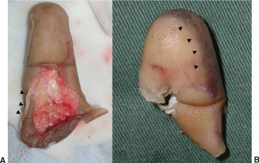
FIGURE 83.2. Clinical signs suggesting injury to the digital arteries (A, B). A. Ribbon sign: avulsion of digital artery. B. Red line sign: disruption of branches of digital artery.
B. Preparation of the stump: Preparation is done under tourniquet control in a similar manner as described for the amputated part. The flexor tendons may have retracted proximally and are retrieved and held out to length, transfixing them with 23G needles. After debridement, identification, and tagging of all structures, the tourniquet is deflated to assess the force of arterial inflow. A positive “spurt” test indicates a good quality vessel. If the outflow is poor, the artery is bathed with 2% lidocaine (Xylocaine) or papaverine to relieve vasospasm. If good flow from either of the arteries is still not evident, it is dissected proximally until good quality vessel is identified. Vein grafts obtained from the volar wrist are employed if the vessel quality does not look suitable under the microscope. Rather than struggling to shorten the bones substantially and to repair the vessels under tension, it is more expedient to harvest the vein graft early in the operation and bypass the crushed vessels. We typically mark out the course of the veins over the volar wrist, before tourniquet inflation, to facilitate vein identification (Figure 83.4).
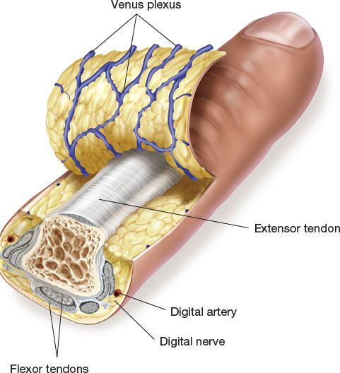
FIGURE 83.3. Exposure of the venous plexus on the dorsum of the finger by raising a thick dorsal skin flap superficial to extensor tendon.
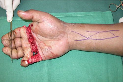
FIGURE 83.4. Surface marking of volar wrist veins prior to tourniquet inflation.
C. Bone fixation: Bony fixation is performed quickly, but not at the expense of stability. The fractures in replantation are usually transverse or nearly transverse. Following bone shortening there is no fracture jigsaw to help with reliable reduction. Our preference is to use parallel Kirschner wires to fix distal phalanx fractures and the Lister technique (single interosseous wire loop with an oblique anti-rotation Kirschner wire) for middle and proximal phalanx fractures (Figure 83.5). Care must be taken to avoid twisting the neurovascular bundle or tethering the tendons by the K-wires. Other options include crossed K-wires or two perpendicular interosseous wire loops (90-90 box wiring). This box construct is quite stable and compresses the fracture allowing early motion and decreasing nonunion. However, it requires precise technique and additional bone exposure. Multiple K-wires are preferred for amputations through the hand with multiple metacarpal fractures. Although mini-plates and screws are an attractive option, alignment of the shortened bones is often imperfect. Additionally, the placement of plates requires more dissection for bone exposure. The main reason plates are not indicated in this scenario, however, is the amount of time required for this type of fixation. If only one finger or just the thumb is replanted, then taking more time for plate fixation may be reasonable. If more than one finger is being replanted, the fatigue factor will set in, making replantation of the additional fingers more tedious and more prone to technical errors or shortcuts. Expedient, meticulous execution of the replantation procedure is a key consideration, because most of these cases occur in the wee hours of the morning when everyone involved is tired.
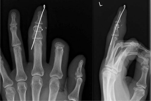
FIGURE 83.5. The Lister technique of osteosynthesis using a single interosseous wire loop and an oblique de-rotation Kirschner wire.
For more proximal amputations through the forearm or humerus, rigid fixation with plates and screws is preferred. A 3.5 mm dynamic compression plate is used for the radius and ulna and a 4.5 mm plate can be used for the humerus (Figure 83.6). A primary arthrodesis is indicated if the amputation passes through or very close to a joint. This is especially indicated in amputations close to the DIPJ, the thumb at the level of the metacarpophalangeal joint, and the hand at the radiocarpal joint. We have occasionally used an intra-articular loop wire technique to preserve the joint in amputations close to the proximal interphalangeal joint (PIPJ) (Figure 83.7). A silicone implant arthroplasty can be considered for amputations of the finger through the PIPJ or the metacarpophalangeal joint.
D. Tendon repair: Any excess tendon resulting from bone shortening is excised. Tendon repairs are meticulous using nonabsorbable sutures to allow early mobilization and decrease adhesions. The extensor tendons are repaired with 4-0 interrupted horizontal mattress sutures and the flexor tendons with a combination of an epitendinous repair (6-0 suture) and a four-strand core suture (4-0 suture). For expediency, most surgeons will omit the epitendinous repair and only repair the profundus tendon to decrease tendon adhesions between the repaired profundus and superficialis tendons.
E. Vein repair: Ideally, the number of venous repairs exceeds the number of arterial repairs by one. For example, at least three veins should be repaired if two arteries are being repaired. Vein repair should not be done under tension. Mobilization of the veins by dividing side branches or dissecting a vein of sufficient length from the dorsum of an adjacent digit will permit tension-free primary repair (Figure 83.8). If not possible, a reversed vein graft or a venous flap from the distal forearm is considered.
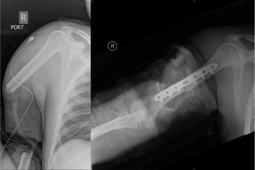
FIGURE 83.6. Rigid bone fixation in major limb replantation.
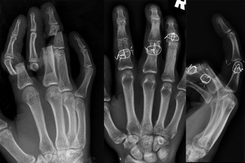
FIGURE 83.7. Intra-articular loop wire fixation in replantation of amputations close to the PIPJ in order to preserve the PIPJ.
F. Arterial repair: Every attempt is made to repair both digital arteries. If a single artery is being repaired, the dominant artery is repaired preferentially. The ulnar digital artery is dominant in the thumb and the rest of the fingers, except the small finger where the radial artery is dominant. We prefer the back wall first technique because it allows visualization of the entire repair. In addition, the vascular clamp does not need to be flipped, a maneuver that may be difficult in limited space and when the vessels do not have laxity. If a primary repair is not possible, the available options include the use of vein graft, cross anastomosis (radial digital artery to ulnar digital artery or vice versa), or transposition of a digital artery from one of the adjacent fingers (Figure 83.9). Occasionally, a cross anastomosis can help with small vessel gaps; however; this makes secondary tenolysis a risky procedure. We reserve transposition of adjacent digital arteries in patients that have an associated skin defect. A vascular island flap is transposed to cover the defect and simultaneously revascularize the digit.
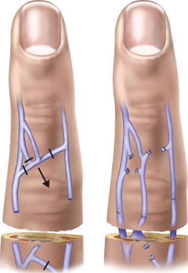
FIGURE 83.8. Dividing venous branches will allow mobilization and primary repair of veins.
G. Nerve repair: Adequate bone shortening usually allows a tension-free nerve repair. If a single 8.0 suture is unable to hold the nerve ends together, a nerve graft is considered. A graft harvested from the distal end of the posterior interosseous nerve is useful in bridging short gaps, whereas a graft from the medial antebrachial cutaneous nerve is more suitable for longer defects. In multiple digital amputations, nerve grafts are harvested from the discarded digits.
H. Skin closure and dressing: Meticulous hemostasis is secured and the skin flaps loosely approximated with a few interrupted sutures. Exposed tendons are covered by transposing local skin flaps as Z-plasties. Other residual raw areas may be left open to heal secondarily or covered with small split-thickness skin grafts. These grafts can also be directly applied to exposed nerves or vessels. In major limb replantations, a prophylactic fasciotomy is performed to decompress the thenar, hypothenar, and dorsal interosseous spaces in addition to the carpal tunnel and the forearm muscle compartments (Figure 83.10). All wounds are covered with a vaseline gauze and a soft, bulky dressing. Care is taken to avoid circumferential compression by the dressing, which when saturated with dried blood can become constrictive. The extremity is immobilized in a plaster splint and elevated.
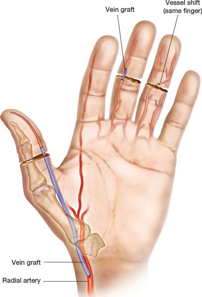
FIGURE 83.9. Techniques of bridging segmental defects in the digital arteries.
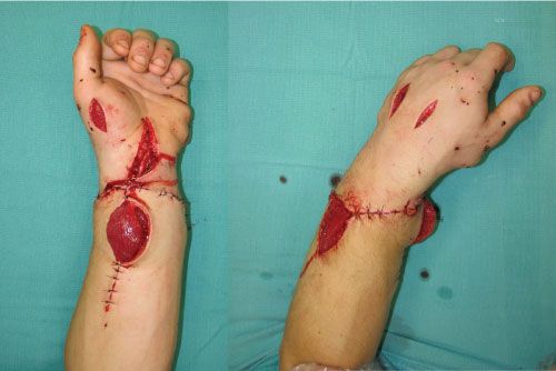
FIGURE 83.10. Appearance after carpal tunnel release and fasciotomy of muscular compartments following a distal forearm replant.
POSTOPERATIVE MANAGEMENT
I. Postoperative care: The highest risk of postoperative thrombosis is in the first 72 hours (3 days) after surgery. Arterial thrombi usually result from platelet aggregation and present on day 1, whereas venous thrombi result from fibrin clotting and usually present by day 2 or 3. Patients are restricted from eating for the first 24 hours in case they need to be taken back to the operating room.
The postoperative care is directed toward preventing external factors that may result in spasm and thrombosis. The room is kept warm and the patient well hydrated. The replanted part is elevated slightly higher than the level of the heart and kept warm using a lamp. Appropriate analgesics are used to control pain and anxiety because they can lead to an adrenergic response and vasoconstriction, especially in children. Smoking is prohibited because it leads to hypoxia, reduction of peripheral blood flow, and increases the risk of thrombosis. We also advise patients to avoid caffeinated drinks. Broad-spectrum antibiotics are given intravenously to prevent infections and maintained for 5 to 7 days. Dressing changes are minimized. Unlike digital replants where the primary dressing need not be changed for 10 days, major limb replants will need change of dressing in 24 to 48 hours depending on how soaked the dressing is. These latter dressing changes must be done under anesthesia, preferably a regional block, and if the patient requires a skin graft, it is done at the same time.
II. Anticoagulation: There are insufficient outcomes data in humans with regard to the choice of anticoagulants. Different protocols exist for perioperative anticoagulation. The commonly used anticoagulants can be divided into agents that (1) decrease platelet function (e.g., aspirin); (2) increase blood flow or decrease blood viscosity (e.g., dextran); and (3) counteract the effect of thrombin on platelets and fibrinogen (e.g., heparin). Dextran may rarely cause serious anaphylactic reaction and acute renal failure. The use of heparin is associated with an increased risk of hemorrhage and hematoma formation. Other anticoagulant agents include low molecular weight heparin; thrombolytics like streptokinase, urokinase, and tissue plasminogen activator; and hemorrheologic agents like pentoxifylline, hirudin, and iloprost. Our general protocol is to give a 100 mL bolus of dextran-40 intravenously prior to release of the vascular clamps, followed by a continuous infusion of dextran-40 at 500 mL per day for 5 days (10 mL/kg/d). We also give a one-time 5,000 unit bolus of heparin after removal of the arterial clamp. We start the patient on a once-daily dose of 100 mg of aspirin that is continued for 3 weeks. For smokers, we may give a continuous low-dose heparin infusion for 3 to 4 days that does not increase the coagulation profile but is sufficient to prevent a hypercoagulable state.
III. Monitoring: An experienced nurse should monitor the perfusion by examining color, pulp turgor, capillary refill, and temperature. This is done hourly for the first 72 hours (3 days) and once every 4 hours for the next 48 hours (2 days). A soft (flaccid), pale fingertip with a delayed capillary refill (>2 seconds) indicates arterial vasospasm or thrombosis. A swollen (turgid), blue fingertip with rapid capillary refill (≤1 second) indicates venous thrombosis. A difference in temperature between a control digit and the replanted digit greater than 2°C (3.6°F) or an absolute temperature of less than 30°C (86°F) suggests a failing replant. A pulse-oximeter probe secured to the pulp can also be used to continuously monitor the pulse and oxygen saturation, especially in more proximal replants. The loss of the pulse rate indicates arterial occlusion, whereas a fall in oxygen saturation below 90% indicates venous occlusion.
If there is a suspicion of compromised perfusion, immediate action is taken. The dressing and any sutures causing constriction are removed. If perfusion does not improve, the patient is immediately taken back to the operating room for exploration of the vascular anastomosis, unless the initial operation was deemed not improvable (rare). There is usually a thrombosis of an anastomosis that invariably requires the use of an interposition vein graft. Rarely, the thrombus is a result of a kink in the vessel that can be corrected by revising the anastomosis and positioning the vessels correctly. If adequate veins are not available one can consider the use of leeches or encourage continuous venous bleeding from the nail bed by removing a portion of the nail bed and repeatedly applying heparin-soaked pledgets. These methods are more useful in distal replants and will result in blood transfusions. The use of leeches is also associated with the risk of Aeromonas hydrophila infections that will require treatment with a third-generation cephalosporin.
IV. Therapy: Hand therapy can be started about a week after replantation, once anticoagulation is stopped. Therapy protocols depend on the level of the replant and the stability of skeletal fixation. A dorsal splint is provided and the patient started on gentle active range of motion exercises. The therapist initially sees the patient daily in the outpatient clinic and thereafter on a weekly basis. Frequent interaction between the patient, the surgeon, and the therapist is necessary to optimize outcomes. Therapy is continued until a plateau is reached, usually at 2 to 3 months postoperatively. At this stage, a decision is made regarding the need for secondary procedures.
V. Secondary surgery: Secondary surgery like tenolysis, nerve grafting, tendon transfers, bone, grafting, intrinsic muscle release, and arthrodesis are frequently required in replants to enhance function. We prefer to do these at procedures approximately 3 months after replantation. This allows adequate time for the tissues to become supple and it is safe to apply the tourniquet. Placing nonabsorbable marking sutures/metal clips at the ends of divided nerves and placing the nerve ends subcutaneously during replantation makes it easier to find these ends at a later date for nerve grafting. Extensive exposures should be avoided for tendon and nerve grafts and tendon transfers and the grafts should be tunneled. Tenolysis can be a difficult undertaking, given that tissues are scarred and vital structures such as nerves and vessels may not be in their usual locations. Furthermore, tendon rupture after tenolysis is a disastrous problem that will render the finger nonfunctional. For this reason, we prefer to avoid secondary procedures after replantation if at all possible.
TECHNICAL CONSIDERATIONS
I. Vein grafting: Suitable caliber vein grafts for digital and hand amputation can be harvested from the volar aspect of the distal forearm. These veins are marked prior to inflating the tourniquet, but must be harvested under tourniquet control. It is important to orientate the vein graft, so that the direction is reversed when the graft is sutured in position. One can place a single clip at the proximal end of the isolated vein graft, fill the vein graft with heparinized saline using a suitable cannula, and place two clips at the distal end (mnemonic: double distal) to differentiate the distal end from the proximal end of the vein graft. Alternatively, marking one end of the vein graft with a marking pen orients the flow direction. Filling the vein graft with heparinized saline serves four purposes: (1) corrects spasm; (2) identifies any leaks; (3) ensures that the graft is not twisted; and (4) lengthens the graft allowing an accurate estimate of final graft length. The length of the vein graft required must be accurately matched to the arterial gap with the digit in the resting posture. Once arterial flow is established the vein graft not only increases in diameter, but also in length and can get kinked. A Y-shaped graft can help with anastomosis of a common digital artery to the digital arteries of two adjacent digits. We prefer to do the distal anastomosis followed by the proximal anastomosis to avoid stasis in the vein graft.
II. Fingertip replantation: Many classifications of fingertip amputations have been described (Figure 83.11). We have modified the classification so that it reflects the flexor tendon zones of injury (Figure 83.12). Although the replantation of flexor zone 1 distal amputations (fingertip amputations) provides an excellent functional as well as an aesthetic outcome, it is the most technically demanding of all upper limb replantations. Not only are the arterial and venous anastomoses difficult, it is often impossible to find a suitable vein. The artery runs close to the midline and is just palmar to the distal phalanx. A small vein can be found either on the dorsum proximal to the lateral nail fold or on the palmar aspect. It may be easier to identify these veins after completion of the arterial anastomosis and release of the tourniquet. If a suitable vein cannot be found, the options to prevent venous congestion include delayed venous repair (veins get engorged after 24 hours and may be easier to find), removal of a wedge of the nail bed, and application of heparin-soaked pledgets, application of leeches, or the creation of an arteriovenous fistula between a distal artery and a proximal vein.
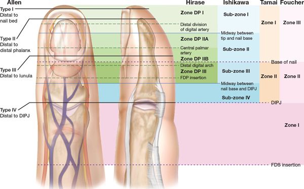
FIGURE 83.11. Classifications of distal digital amputations. DIPJ, distal interphalangeal joint; FDS, flexor digitorum superficialis; FDP, flexor digitorum profundus.
The first author prefers to perform a pocket-plasty procedure for fingertip replantations, when a suitable vein cannot be found. This involves de-epithelializing the pulp and a suitable region on the palm and suturing them together (Figure 83.13). The adhesions are separated 2 to 3 weeks later under local anesthesia. Another technique we find useful in fingertip amputations is to avoid rigid fixation initially. The Kirschner wire is passed retrograde through the amputated part, but not into the proximal stump. Preliminary stabilization of the amputated part is obtained purely by nail bed repair. This provides a certain degree of flexibility and makes placement of vascular clamps easier. The artery is quite deep and there is usually very little space available for introducing the clamps. If greater stability is desired, one or two additional skin sutures along the lateral margin can be passed. Once the vascular and nerve repairs are complete, the K-wire is driven proximally to rigidly fix the distal phalanx. Care is taken when the K-wire is driven proximally to prevent spinning as the wire turns.
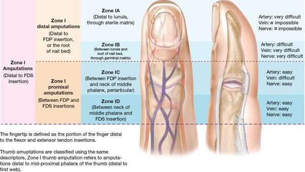
FIGURE 83.12. Modified classification of flexor zone 1 digital amputations. FDS, flexor digitorum superficialis; FDP, flexor digitorum profundus.
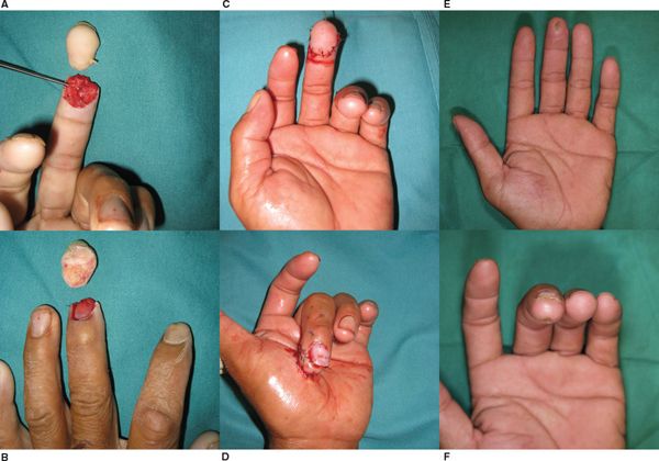
FIGURE 83.13. Pocket-plasty procedure for fingertip amputations when a vein repair cannot be performed (A–F). A, B. Preoperative appearance. C. Appearance after repair of central digital artery. D. De-epithelialization and suture of pulp to suitable region on the palm. E, F.Appearance at 2 months after separation of adhesions.
III. Thumb replantation: There is considerable variation in the anatomy of the digital arteries of the thumb proximal to the metacarpophalangeal joint as the vessels dive deeper to pass below the thenar muscle insertions. Positioning the thumb to repair the dominant ulnar digital artery is also challenging. One way is to pronate the forearm and repair the ulnar digital artery hand by viewing it through the first web space. We find it easier to use a vein graft to connect the distal ulnar digital artery to the radial artery in the anatomical snuffbox. The distal anastomosis of the vein graft may be done on the back table. In avulsion amputations of the thumb through the metacarpophalangeal joint with avulsion of the extensor and/or flexor pollicis longus tendons at the musculotendinous junctions, one can consider primary tendon transfers using the extensor indicis proprius for the extensor pollicis longus and the ring finger flexor digitorum superficialis (FDS) for flexor pollicis longus.
IV. Finger avulsion amputation: Several classification systems have been used to categorize digital avulsion amputations (Table 83.5). Complete avulsion amputation has been classically considered as a relative contraindication to replantation. A recent review found that functional outcomes of sensibility and range of motion are better than what was historically cited in literature. The authors currently recommend replantation of finger avulsion amputations with a preserved PIPJ and FDS insertion. An arthrodesis of the DIPJ is recommended because it allows shortening and solves the problem of the avulsed flexor digitorum profundus tendon. The proximal denuded stump is examined to identify the digital arteries and the head of the middle phalanx prepared for arthrodesis. The artery that has been avulsed more distally and has a longer segment on the denuded finger is not dissected, but maintained in situ to provide vascularization to the PIPJ and the flexor mechanism. The distal end of the digital artery that has been avulsed more proximally is selected for repair using an interposition vein graft.
Unlike standard digital replants where the neurovascular structures are exposed by mid-lateral incisions, one should avoid making multiple incisions in avulsion amputations to avoid necrosis of the skin between the incisions. A palmar or lateral incision is made over the selected digital artery. The distal digital artery is dissected until a healthy vessel is identified and the base of the distal phalanx is prepared for arthrodesis. To avoid the risk of soft tissue getting caught when the K-wire is passed for arthrodesis, we use the protective sleeve of a hypodermic needle as a drill guide. The K-wire is first passed retrograde, the amputated part is draped over the denuded stump, and the wire driven proximally. A reversed vein graft is used to bridge the defect between the selected digital artery and a pulsatile vessel in the palm. This vessel will perfuse the skin envelope and the distal phalanx, whereas the contralateral unrepaired vessel will perfuse the PIPJ and the flexor mechanism. It may be possible to do vein repairs primarily or a pedicled or free venous flap may be required. Nerve repairs are usually not possible and the avulsed nerve ends are draped along the phalanx. Most patients regain protective sensation.
V. Replantation of multiple digits: The decision making in multiple digital amputations involves determining the number of digits that are suitable for replantation, the position of the replanted fingers (if one or more fingers are not suitable for replantation), and the order of replantation. The replantation of the thumb always takes priority. If the thumb is not suitable for replantation, the best available finger is replanted in the thumb position. Once the thumb has been addressed, the next priority is to reconstruct the long and ring fingers. Although the index finger restores pinch, it comes at the cost of a narrow span and loss of grip strength. Restoring the long and ring fingers gives the patient a wider span that is better for overall function. The replantation may be performed in a digit-by-digit sequence or a part-by-part sequence. The part-by-part sequence is faster, but associated with a greater warm ischemia time for all the digits. It may be preferable to address the thumb first and then do the remaining fingers in a part-by-part sequence. The arterial anastomoses are done last in a part-by-part sequence to decrease bleeding that may obscure the repair of other structures and minimize swelling. Non-replantable digits are a good source of spare parts (skin, nerve, vein grafts, etc.) to reconstruct replantable digits.
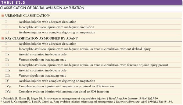
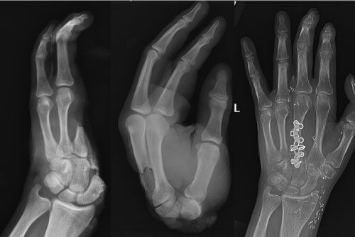
FIGURE 83.14. Bony fixation using plates and screws in an oblique transmetacarpal replantation.
VI. Transmetacarpal replantation: The intrinsic muscles (interossei, lumbrical, thenar, and hypothenar) in the amputated part are debrided. These muscles are not revascularized following replantation as their blood supply is intramuscular and derived from proximal vessels. If not debrided, these muscle fragments form a fibrotic mass of scar. In addition, the ischemic muscle can get infected, compromising the overlying vascular repairs. In transverse amputations, the bone is shortened by at least 1 cm to allow primary repair of the neurovascular structures. It is important to ligate all the branches of the deep palmar arch and the deep palmar metacarpal arteries in the amputated part as well as the stump to prevent postoperative hematoma formation. The carpal tunnel and Guyon canal are decompressed prophylactically prior to revascularization.
Bony fixation is easily achieved by multiple longitudinal K-wires. In oblique transmetacarpal amputations or crush amputations, when the fractures are not at the same level, it is difficult to shorten all bones equally. In such cases, it may be easier to fix the metacarpal with the best fracture jigsaw using plates and screws and use K-wires to maintain the alignment of the remaining metacarpals (Figure 83.14). Secondary bone grafting can be considered. Loda has classified transmetacarpal amputations into zone A and zone B based on the level of amputation in relation to the superficial palmar arch (SPA) (Figure 83.15A). Zone A amputations are proximal to the SPA, whereas zone B amputations are distal to the SPA. In zone A amputations, it is often possible to revascularize all the digits by repair of the radial or ulnar artery; however, in zone B amputations, one will need to repair multiple common digital vessels. Depending on the nature of injury, a primary repair may be possible. If not, the SPA can be divided in the middle and advanced distally to allow a primary repair (Figure 83.15B). In crush amputations, closure of dorsal skin may not be possible and a primary flap may be required. The posterior interosseous artery flap, a free lateral arm flap, or a groin flap can be considered.
VII. Major limb replantation: Replantation of amputations through the wrist or the distal forearm yields excellent results because the volume of ischemic muscle mass is small, the replantation itself is technically easy, and the rehabilitation is predictable (Figure 83.16). Bone shortening can be achieved by proximal row carpectomy, wrist arthrodesis, or shortening osteotomy of the radius and Darrach resection of the ulna depending on the level of the amputation and condition of the distal radial articular surface. The carpal tunnel and the thenar, hypothenar, and interosseous muscle compartments are decompressed.
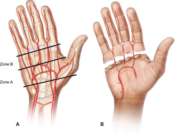
FIGURE 83.15. Loda classification of transmetacarpal amputations. A. Classification. B. Techniques to allow primary repair in zone B replantation.
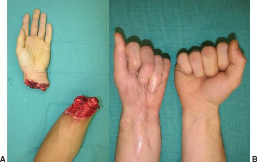
FIGURE 83.16. Late result of a distal forearm replantation (A–B). A. Prior to replant. B. Final result.
Patient selection is critical in the proximal forearm and upper arm amputations. Reconstructive options like toe transfers are available for amputations distal to the wrist; however, no such options are available for proximal amputations. Therefore, every attempt must be made to replant major limb amputations, but it must not endanger the patient’s life. Even in the best centers, it will take 2 to 3 hours from arrival of the patient until arterial inflow is reestablished. This time must be added to the time from injury to arrival at the hospital to calculate warm ischemia time. In proximal forearm amputations, arterial inflow must be established within 4 to 6 hours, whereas in upper arm amputations, one has only 3 to 4 hours to do so. The amputated part must be examined to ensure that the digits are supple. Stiff digits with firm muscles suggest ongoing rigor mortis and one must abandon replantation. In order to cut short the warm ischemia time, especially in upper arm amputations, a temporary vascular shunt can be used to connect the proximal brachial artery to the distal brachial artery or one of the forearm arteries (Figure 83.17). The shunt can be obtained from the vascular tray and is inserted into the ends of the artery and tied securely. The surgical team must be conscientious of the continued blood loss as the open veins will continue to bleed. This will result in significant blood loss and arrangements must be made for adequate replacement.
The debridement must be radical and in crush and avulsion amputations, all muscle attached to the avulsed tendons must be excised. Sufficient bone shortening to allow primary repair of the major nerves is done. The arterial anastomosis is done first and the veins are allowed to bleed (if a vascular shunt was not used initially). This will prevent return of the acidotic venous blood that has high concentrations of potassium and lactic acid. The patient can be given intravenous sodium bicarbonate prior to release of the arterial clamp to counter the effect of acidosis on the myocardium. One must try to repair the venae comitantes in addition to the superficial veins as they drain a significant amount of blood. Major limb replants must be closely monitored in the postoperative period for systemic problems, including myoglobinuria and ischemia reperfusion syndrome.
OUTCOMES
The scoring system introduced by Tamai is useful in evaluating function after hand and digital replantation (Table 83.6). It gives a numerical score (0-20) for motion, sensation, activities of daily living, subjective symptoms, patient satisfaction, and return to work (0-10). Chen’s functional criteria are useful in outcome measurement in major limb replantation (Table 83.7). It is based on four variables, namely, ability to work, range of motion, recovery of sensation, and the recovery of motor power. Paavilainen et al. compared the Tamai and Chen scoring systems in a series of transmetacarpal replantations and felt that although the Tamai scoring system was more complex, it was a more realistic description of the functional outcome.
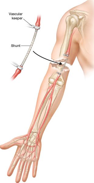
FIGURE 83.17. Preliminary shunting to decrease ischemia time in major limb replantation.
A comparison of digital replantation with completion of amputation found that replantation provided a better appearance as well as functional outcome. A recent systematic review on distal digital replantation showed an overall survival rate of 85% and the mean two-point discrimination (2-PD) averaged 7 mm. The common complications in distal replantation were nail deformity (24%) and pulp atrophy (14%). About 97% of patients returned to work. Clean-cut amputations had a better survival rate compared with crush or avulsion amputations and the repair of a vein improved survival. The outcomes are similar for more proximal digital amputations, with reported overall survival rates between 80% and 90% and an average 2-PD of 8 to 12 mm. The factors that influenced sensory recovery were age of the patient, level of amputation, and mechanism of injury. The range of motion depended on the level of amputation. Replantations proximal to the FDS insertion had on an average, 35° of motion at the PIPJ in comparison to 82° for replantation distal to the FDS insertion. Another recent systematic review of replantation of complete digital avulsion amputations showed an overall survival rate of the part of 66%, 2-PD of 10 mm, and 174° of total active motion (TAM).
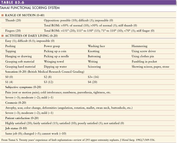
Paavilainen et al. reported a series of 43 transmetacarpal replantations with an 86% survival rate with a mean 2-PD of 14.7 mm in 78% of patients. The average TAM was 154° and the mean grip and pinch strength measured 56% and 58% of the uninjured side, respectively. Approximately half of the patients were able to return to their previous occupation. The functional outcomes of major limb replantation, especially proximal forearm, elbow, and arm, are significantly poorer compared with the more distal injuries. However, they are better than revision amputations and prostheses. In a series of 22 major limb crush avulsion amputations, Sabapathy et al. were able to obtain Chen grade I function in 3 patients and Chen grade II in 9 patients. Hierner and Berger reported that a functional upper extremity could be reconstructed in 22% to 34% of upper arm replantations, 30% to 41% of proximal forearm replantations, and 56% to 80% in distal forearm replantations.
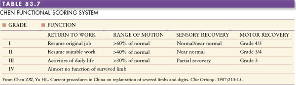
In conclusion, it is possible to achieve at least an 80% survival rate for replantation, with a range of motion equivalent to 50% of the normal side (depending on the level of injury) and nerve recovery similar to that of an isolated severed peripheral nerve. The best results are obtained in replantation of the thumb, fingers amputated distal to the insertion of the FDS, and the hand through the wrist or the distal forearm. Replantation in centers with high volumes and teams with good microsurgical skills is associated with higher success rates, better patient satisfaction, and fewer complications.
Suggested Readings
Askari M, Fisher C, Weniger FG, Bidic S, Lee A. Anticoagulation therapy in microsurgery: a review. J Hand Surg. 2006;31A:836-846.
Buncke GM. Replantation and revascularization. In: Mathes SJ, Hentz VR, eds. Plastic Surgery. Vol VII. Philadelphia, PA: Saunders-Elsevier; 2006: 565-585.
Chen ZW, Yu HL. Current procedures in China on replantation of severed limbs and digits. Clin Orthop. 1987;215:15-23.
Goldner RD, Urbaniak JR. Replantation. In: Wolfe WS, Hotchkiss RN, Pederson WC, Kozin SH, eds. Green’s Operative Hand Surgery. 6th ed., Vol I. Philadelphia, PA: Elsevier-Churchill Livingstone; 2010:1585-1602.
Graham B, Adkins P, Tsai TM, Firrell J, Breidenbach WC. Major replantation versus revision amputation and prosthetic fitting in the upper extremity: a late functional outcomes study. J Hand Surg. 1998;23A:783-791.
Hattori Y, Doi K, Ikeda K, Estrella EP. A retrospective study of functional outcomes after successful replantation versus amputation closure for single fingertip amputations. J Hand Surg. 2006;31A:811-818.
Hierner R, Berger AC. Long-term results after total and sub total macroamputations of the upper limb. Eur J Plast Surg. 2005;28:119-130.
Kleinert HE, Kasdan ML, Romero JL. Small blood-vessel anastomosis for salvage of severely injured upper extremity. J Bone Joint Surg. 1963;45A:788-796.
Kocher MS. History of replantation: from miracle to microsurgery. World J Surg. 1995;19:462-467.
Paavilainen P, Nietosvaara Y, Tikkinen KA, Salmi T, Paakkala T, Vilkki S. Long-term results of transmetacarpal replantation. J Plast Reconstr Aesthet Surg. 2007;60:704-709.
Pederson WC. Replantation. Plast Reconstr Surg. 2001;107:823-841.
Puhaindran ME, Paavilainen P, Tan DM, Peng YP, Lim AY. Dermal pocketing following distal finger replantation. J Plast Reconstr Aesthet Surg. 2010;63:1318-1322.
Sabapathy SR, Venkatramani H, Bharathi RR, Dheenadhayalan J, Bhat VR, Rajasekaran S. Technical considerations and functional outcome of 22 major replantations. J Hand Surg. 2007;32B:488-501.
Sabapathy SR, Venkatramani H, Bharathi RR, Sebastin SJ. Replantation of ring avulsion amputations. Ind J Plast Surg. 2003;36:76-83.
Sabapathy SR. Amputations and replantations. In: Guyuron B, Eriksson E, Persing JA, et al., eds. Plastic Surgery: Indications and Practice. Vol 2. Philadelphia, Saunders-Elsevier; 2008:1211-1224.
Schlenker JD, Koulis CP. Amputations and replantations. Emerg Med Clin North Am. 1993;11:739-753.
Sears ED, Chung KC. Replantation of finger avulsion injuries: a systematic review of survival and functional outcomes. J Hand Surg Am. 2011;36:686-694.
Sebastin SJ, Chung KC. A systematic review of the outcomes of replantation of distal digital amputation. Plast Reconstr Surg. 2011;128:723-737.
Sebastin SJ, Chung KC. Challenges in measuring outcomes following digital replantation. Semin Plast Surg. 2011;25(4).
Sood R, Bentz ML, Shestak KC, Browne EZ Jr. Extremity replantation. Surg Clin North Am. 1991;71:317-329.
Soucacos PN. Indications and selection for digital amputation and replantation. J Hand Surg. 2001;26B:572-581.
Tamai S. Twenty years’ experience of limb replantation—review of 293 upper extremity replants. J Hand Surg. 1982;7:549-556.
Urbaniak JR, Roth JH, Nunley JA, Goldner RD, Koman LA. The results of replantation after amputation of a single finger. J Bone Joint Surg. 1985;67A:611-619.
Yamano Y. Replantation of fingertips. J Hand Surg. 1993;18B:157-162.