PART VIII
HAND
CHAPTER 87 MANAGEMENT OF VASOCONSTRICTION
PAUL K. LIM AND ALLEN L. VAN BEEK
INTRODUCTION
Vas, from the Latin word for “vessel” or “container,” and spasmos, a Greek word meaning “convulsion,” combine to give the word vasospasm. Perhaps the more accurate word would be vasoconstriction because constrictus means “bound” or “drawn together”.1 Regardless of which term is used, the condition causes surgeons to perspire when the microsurgical tissue transfer fails to perfuse and creates painful suffering for patients with disease processes that have vasoconstriction as one of their hallmarks. This chapter discusses the management of vasoconstriction that often concerns plastic surgeons rather than the entire spectrum of diseases that have vasoconstriction as part of their presentation.
DEFINITIONS AND CLASSIFICATION
Raynaud’s Phenomenon
Maurice Raynaud, a French physician, first described the phenomenon of episodic acral pallor, cyanosis, and rubor in response to cold or emotional stress.2 The diagnosis of primary Raynaud’s phenomenon (RP), also known as Raynaud’s disease, includes multiple attacks of RP occurring over at least 2 years without gangrene or tissue loss, and no known associated vaso-occlusive or systemic diseases. The episodes are typically symmetric and bilateral involving one or more digits.
Secondary RP is the diagnosis of these events but where there is a known causal or associated disease etiology. Tissue loss and asymmetric involvement are commonly seen in secondary RP.3
Iatrogenic Vascular Stenosis
This category of vascular spasm is not usually addressed under discussions of vasoconstriction diseases but is pertinent to surgeons performing microvascular surgery. Acute partial vessel occlusions confronting a surgeon are typically not from disease processes but have to do with small vessel or microvascular constriction associated with vessel manipulation. Vasoconstriction that occurs intraoperatively, postoperatively, or postvascular intervention and results in poor tissue perfusion falls into this category. When this occurs, there are many etiologies and suggested managements but the surgeon must be certain that the “constriction” is not actually a correctable surgical error.
PHYSIOLOGY
Acute Vessel Constriction
Large- and medium-sized vessels have pliable walls that increase and decrease their lumens in response to physiological and biochemical processes, but the largest impact on vascular resistance results from activity of small vessels and the capillary bed. Arteriovenous shunts are prominent in digits, hands, and feet. The shunts are regulated by vasoconstrictors, including norepinephrine, endothelin-1, vasopressin, angiotensin II, and transforming growth factor-β, and by vasodilators including prostacyclin, nitric oxide, and estrogen. These substances or mechanisms that influence the vessel walls are the basis for pharmacologic interventions.2 When the small vessels such as the digital arteries and their capillary outflow fields have their flow shunted, as occurs in RP, the resulting ischemia may produce some or all of the “five P’s” associated with arterial thrombosis: pallor, pain, paresthesia, paralysis, and pulselessness, with the most common ones being pallor, pain, and loss of sensibility. Some vessel sclerotic processes, however, do not have an antecedent history of repeated episodes of vasospasm and present with an acute sequence of pallor, cyanosis, and rubor. Often the onset is abrupt, painful, and associated with an acute process that produces micro-emboli and even small vessel occlusion as seen with hypothenar hammer syndrome (Figures 87.1 and 87.2). Atrial fibrillation with associated mural thrombi, proximal large vessel stenosis with ulcerated plaques, and arterial aneurysms are the most common sources of emboli to distal vessels4 and are often responsible for acute onset vasospasm.
Chronic Vessel Occlusion
Vascular occlusion in proximal, larger vessels decreases the ability to respond to physiologic demands for more flow and increase the propensity for spasm to occur (Figure 87.3). Large vessel occlusions, if not recognized, may be the cause of unexpected acral vasoconstriction events. More often, distal occlusions are the reason for secondary RP to occur. Systemic sclerosis is associated with occlusion of small- and medium-sized vessels with severe fibrotic proliferation of the intima making episodic spasm events more severe as the disease progresses. Other rheumatologic diseases associated with secondary RP include systemic lupus erythematosus, mixed connective tissue disease, dermatomyositis, rheumatoid arthritis, Sjogren’s syndrome, and vasculitis.2

FIGURE 87.1. Aneurysm. (Hypothenar hammer syndrome.) Ulnar artery aneurysm producing acute onset of pain and vascular spasm to the ring and small fingers.

FIGURE 87.2. Recent thrombosis. Clot appearance confirms recent thrombosis of the ulnar artery accounting for a sudden onset of symptoms.
DIAGNOSIS
Primary Raynaud’s Phenomenon Versus Secondary Raynaud’s Phenomenon
Distinguishing between primary and secondary RP involves not only the clinical differences described above but also specific diagnostic criteria. Having any of the following rules out primary RP: 1. digital ulcerations; 2. elevated erythrocyte sedimentation rate; 3. high titer of antinuclear antibody; and 4. abnormal capillaroscopic pattern. Capillaroscopy is typically performed by rheumatologists and involves the visualization of nailfold capillaries via dynamic fluorescence videomicroscopy and obtaining 50× to 1,000× magnification images with a digital video camera.5,6

FIGURE 87.3. Chronic arterial occlusion. Chronic occlusion increases the severity and frequency of vascular spasm episodes in patients with secondary Raynaud’s. Necrosis of index finger tip from secondary Raynaud’s and vasodilation with adenosine during direct arteriography also reveal an arterial deficit.
Physiologic Assessment
Digital artery systolic blood pressure should be similar to the systolic blood pressure more proximally (segmental systolic pressures). A pressure drop at the digital level of more than 20 mm Hg compared with the brachial measurement is indicative of vascular disease.7 Blood flow in the finger can have wide variation, but flows less than 1 mL/min/100 mL tissue volume, pulp temperature lower than 30°C, and loss of ultrasound Doppler signals in the pulp of a digit are indicative of significant perfusion loss. Laser Doppler, ultrasound Doppler, finger cuffs, and temperature monitoring devices make clinical application of these parameters useful to surgeons.
Vascular Conduit Assessment
The gold standard for vascular assessment remains direct contrast angiography. Most importantly, it diagnoses or excludes proximal vascular disease or occlusion. However, in the presence of severe vasoconstriction, either present before or during angiography, the distal palm and digit vessels may not be well visualized. Improvement in magnetic resonance angiography (MRA) resolution has made distal vessel assessment possible even when vessel spasm is present and more importantly does not seem to contribute to or institute vessel spasm in those prone to attacks (Figure 87.4). Additionally, patients with profound spasm and tissue necrosis are often on anti-platelet therapy and, thus, at elevated risk for complications with direct angiography. One can start with the less invasive MRA and then proceed to direct angiogram if necessary.
TREATMENT
Management of Primary Raynaud’s Phenomenon
The phenomenon, when it is a primary process, does not manifest the severity of sequelae noted with secondary RP. Medical intervention may be instituted in some patients, but surgery is rarely required. Noninvasive treatment, when needed, is the same as for secondary RP.
MEDICAL MANAGEMENT OF RAYNAUD’S PHENOMENON
Secondary RP, by definition, has an etiology established. The most common etiologies are thromboangiitis obliterans (Buerger’s disease), connective tissue disorders, vasculitis, vascular sclerosis, thoracic outlet syndrome (TOS), vibratory mechanisms, and hematologic syndromes. The prevalence of Buerger’s disease has decreased with decreased use of tobacco products. A high percentage of scleroderma patients have secondary RP. Hypothenar hammer syndrome may be more common than reported, and proximal etiologies such as TOS and ulcerated endothelial plaques provide some difficulty in establishing diagnoses. Treatment is directed at not only the ischemic manifestations but also the underlying disease process where possible.
Typically, plastic surgeons are consulted when there is impending tissue infarction, small acute ulcerations, gangrene, or chronic ulcers. The surgeon should be familiar with the medical treatment options to confirm that maximal medical management had been attempted before proceeding with surgical interventions. Additionally, the status of the vascular conduits proximally and distally is assessed for diagnostic purposes as well as for potential operative planning.

FIGURE 87.4. Vascular imaging. Example of the vascular detail in an MRA of the hand with digital occlusive disease from hypothenar hammer syndrome, with patient’s digital vessels on the radial side.
Physical and Prevention Management
Eliminating cold exposure by using insulated containers, wearing gloves, and anticipating weather conditions may decrease the frequency of attacks. The entire body, not just the hands, should be covered, especially the forehead which is a known trigger site for attacks. Patients should avoid stimulants of vascular constriction such as caffeine, nicotine, ergots, ephedrine, amphetamines, decongestants, and β-blockers. Over-the-counter dietary, herbal, or medical products may contain these substances, so all products the patient takes are reviewed.
Patients with vasospastic tendency should be aware of the “windmill maneuver” which involves swinging the arms in a circle to use centrifugal force to push blood in the capillary bed.2
Topical Agents
In acute progressive ischemic attacks, topical 2% nitroglycerin cream is advocated with some reported success. Topical l-arginine, by theoretically supplementing nitric oxide effects, has also been utilized but with very limited success.2,8
Oral Pharmaceutical Management
Calcium Channel Blockade. Calcium channel blockers inhibit calcium ions from crossing the cell membrane which in turn reduces the ability of norepinephrine to stimulate vascular smooth muscles. The dihydropyridine calcium channel blockers are the least cardiac selective in their effects and are the most effective against RP. Agents prescribed include nifedipine, dosed from 30 to 180 mg daily, and amlodipine, dosed from 5 to 20 mg daily. Calcium channel blockers have significant side effects such as headache, lower extremity edema, and hypotension that may necessitate discontinuation of the therapy.2,8
α-Adrenergic Blockade. This class of drugs blocks vasoconstriction by competing with norepinephrine at the muscle receptor sites preventing smooth muscle constriction. Accumulation of norepinephrine at the receptor site stimulates α2 presynaptic receptors that shut down norepinephrine supply. Prazosin is often used for peripheral constriction at doses of 1 to 5 mg every 8 to 12 hours. α-Blockers have cardiac and other potential side effects. Newer, selective α2c adrenergic receptor antagonists are undergoing clinic trials showing not only improvement in patients with RP associated with systemic sclerosis but also fewer side effects.2
Angiotensin Inhibition
Angiotensin converting enzyme inhibitors have not shown clear benefit against RP. However, in a randomized, controlled trial, the angiotensin II receptor antagonist, losartan, dosed at 50 mg/day was found to be more effective and better tolerated than nifedipine at 40 mg/day for RP.9 This was a small, short-term study but did show enough promise warranting further investigation.
Phosphodiesterase Inhibition
Nitric oxide causes a relaxation of vascular smooth muscle and inhibits platelet activation by generating cyclic guanosine 5′-monophosphate (cGMP). Phosphodiesterase (PDE) inhibitors increase the amount of cGMP at the smooth muscle receptor site by blocking the enzymes that hydrolyze cGMP. A novel treatment, tadalafil, is used at a 10 to 20 mg daily dose. Sildenafil and vardenafil are other medications in this class. PDE inhibitors have significant side effects such as headache, muscle pain, flushing, rhinorrhea, and visual abnormalities and are primarily used to treat erectile dysfunction and pulmonary hypertension.8
Parenteral Pharmaceutical Management
Prostaglandin Therapy. Prostacyclin relaxes smooth muscles and dilates vessels. The use of intravenous prostacyclin analogues to decrease pulmonary hypertension associated with connective tissue disorders has also provided relief from peripheral vascular constriction. The treatment may provide relief from profound limb-threatening constriction. Epoprostenol given via a continuous intravenous route for 12 weeks at a dosage of 2 to 11 ng/kg/min has been reported to be beneficial to patients with pulmonary hypertension and RP associated with scleroderma. Side effects included jaw pain, headache, nausea, anorexia, and diarrhea, and complications related to the drug delivery system (central venous catheter): cellulitis, sepsis, hemorrhage, and pneumothorax.10 Iloprost, another prostacyclin analogue, is administered over a 6-hour infusion on a daily basis for 3 to 5 days (inpatient hospitalization) at a dose of 0.5 to 2 ng/kg/min. It is infused through a peripheral venous catheter but the patient requires frequent vital sign monitoring to ensure cardiovascular stability.11
Reserpine Venous Block
Various classes of intravenous drugs have been used to provide temporarily block of peripheral vascular smooth muscles using the equivalent of a Bier-type regional block in an attempt to provide relief of vessel constriction in profound limb-threatening ischemia. The most common pharmaceutical used is reserpine. The block is performed as an outpatient but requires cardiovascular monitoring during administration and after release of the block.12
Botulinum Toxin-A
Plastic surgeons are often consulted to administer botulinum toxin (BT)-A because the injections are directed at specific locations related to the anatomy of the digital vessels. This is further discussed in the next section on surgical management.
SURGICAL MANAGEMENT OF VASOSPASM
Aggressive medical management may prevent loss of digits, decrease painful episodes of spasm, and decrease the need for operative treatment. Typically, plastic surgeons are consulted when patients have severe symptoms, such as tissue loss and intractable pain, despite this aggressive pharmacologic therapy. The proximal vasculature is assessed to confirm patency. Any vascular occlusive pathology, such as subclavian artery stenosis and thoracic outlet obstruction, is treated. Intervention is directed at interrupting the sympathetic innervation contributing to increased vascular tone. Sympathetic blockade can occur at one of two locations: at the level of the sympathetic trunks or at the level of the perivascular tissue of the peripheral arteries.
Cervical sympathectomy has been advocated for years and has favorable short-term outcomes, but long-term results are variable. More recent reports favor thoracoscopic sympathectomy over traditional, open cervical sympathectomy.13 Lumbar sympathectomy is a potential treatment for lower extremity vessel constriction when medical management has failed and the proximal vessels have arterio-occlusive disease that cannot be reversed.
Botulinum Toxin-A Injections
The effects of BT at the skeletal neuromuscular junction (acetylcholine receptor blockade) and its clinical applications have been previously described extensively (Chapter 43). It has been used in a variety of conditions pertaining to muscular spasm, including torticollis, ocular strabismus, achalasia, and migraines.14 Regarding a potential explanation for effects on the digital arteries, Morris et al. demonstrated that BT prevents sympathetic vasoconstriction of the vascular smooth muscle of guinea pig uterine arteries by blocking exocytosis of norepinephrine vesicles at the neuromuscular junction.15 Van Beek et al reported a case series of 11 patients with severe secondary RP who were treated with perivascular injections of BT. All patients had significant pain reduction. The nine patients who had nonhealing ulcers all went on to heal their ulcers after the BT treatment.16
Injection Technique
All the fingers of an affected hand are treated (eight injection points); the thumb is treated only if symptomatic (two additional points). About 100 units of BT is reconstituted with 8 mL (10 mL if the thumb is to be treated) of 0.9% sodium chloride solution with preservative. Sodium bicarbonate may be added to balance the Ph of saline and reduce injection pain.
Targeted structures include the superficial palmar arch, common digital arteries, and proper digital arteries (Figures 87.5 and 87.6). A 30G needle is used to infiltrate the soft tissues around the arteries with the BT solution. Larger lumen needles are avoided because of potential injury to adjacent nerves and vessels. Through multiple injection sites, 1 mL of BT solution is injected to each of the 8 or 10 areas shown in Figure 87.5. Injections adjacent to the common digital arteries and the superficial palmar arch are made with the needle perpendicular to the palm and deep to the palmar fascia. The goal is to inject adjacent to the vessels without injuring them. After injection, the palm and base of the digit is massaged gently for 30 seconds to help distribute the BT throughout the region of injections. Symptomatic relief occurs from days to weeks later, whereas ulcer healing may take weeks to months. Repeat injections may be required for recurrent symptoms, usually months later, if relief is obtained from the injections.
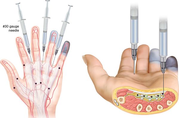
FIGURE 87.5. Technique for botulinum toxin injection. Botulinum toxin-A, 10 to 12 U, is injected into each site marked as a blue dot and in the second through fourth web spaces. Blue tinting indicates the most concentrated areas of BT diffusion. (From Van Beek AL, Lim PK, Gear AJL, Pritzker MR. Management of vasospastic disorders with botulinum toxin A. Plast Reconstr Surg. 2007;119:217-226.)

FIGURE 87.6. Clinical example to be treated with botulinum toxin. This patient has lost digits secondary to ischemia, and presently has an ulcer on the index finger. The vascular anatomy is visualized and the injections are planned along the proper and common digital arteries of the digits. (From Van Beek AL, Lim PK, Gear AJL, Pritzker MR. Management of vasospastic disorders with botulinum toxin A. Plast Reconstr Surg. 2007;119:217-226.)
PERIARTERIAL PERIPHERAL SYMPATHECTOMY
Flatt reported a limited perivascular adventitial stripping and found improvement in vascular constriction.17 Subsequent to that report, many case reports and other small series showed favorable outcomes following perivascular, direct peripheral sympathectomy. Kotsis et al. attempted to perform a meta-analysis of the digital sympathectomy data but found a lack of control groups in the majority of publications. The results appeared encouraging, but the lack of data and discrepancies in the data prevented definitive conclusions or recommendations. Of note, specific techniques (for instance, the proximal and distal extent of adventitial stripping) were not mentioned or compared.18 Zachary has reported lower extremity perivascular adventitial stripping to improve circulation to the toes and foot in selected patients.19 In our experience, the technique should be performed over a longer segment of the peripheral and distal vessels (Figure 87.7). Additionally, with collagen vascular diseases, the perivascular fibrosis should be removed along with the adventitia.
Technique of Peripheral Sympathectomy
Following failed medical management and after establishing proximal inflow, this technique may salvage digits and can produce prolonged benefits to individuals with necrosis and unhealed ulcers (Figure 87.6).
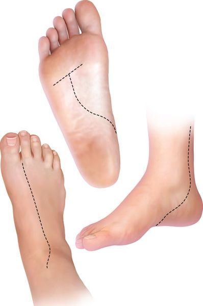
FIGURE 87.7. Perivascular sympathectomy in the foot. This illustrates an approach to manage secondary Raynaud’s involving the feet.
Because of the length of the procedure and the use of the operating microscope, general anesthesia is necessary. Guyon’s canal and the palmar fascia are opened to allow access to the vessels (Figure 87.8A). The distal ulnar artery, the superficial arch, and the common digital arteries are dissected out stripped of adventitia (Figure 87.8B). It is also important to release perivascular fibrosis if present. Spasmodic constriction may be symptomatic because of fibrotic constriction in combination with vascular spasm and obstruction. If a vessel has a wider lumen after removing encapsulating fibrosis, the sympathetic-induced constriction will have less impact. By palpation and more accurately by operating microscopic observation, the degree of fibrosis can be assessed. When the vessels are firm, thickened, without plaques, and without lateral pulsatile expansion, releasing the fibrosis on the anterior side of the vessel will allow expansion. When stripping adventitia and releasing fibrosis, the surgeon should avoid injuring the smaller vessel branches coming off the sides of the main vessels. If the vessels expand after release of the fibrosis, the incision can be extended to the proper digital arteries at the base of each finger. If vessels are damaged, they are repaired with microvascular techniques. The use of anticoagulants agents such as aspirin, heparin, dextran, and low molecular weight heparin after surgery is optional. Our patients have not been on long-term anticoagulation for their extremities but may be for other reasons. Bupivacaine 0.5% irrigated intermittently into the surgical site with an indwelling 21G catheter placed in the area of surgery will reduce pain and vessel spasm.
The catheter is left for at least 5 days after surgery and the local anesthetic instilled on an outpatient basis by patients. BT-A at 50 to 100 units reconstituted to a 5 to 10 mL volume of saline and injected around the vessels and adjacent soft tissue during surgery will provide a long-term sympathetic block, but its impact does not occur immediately. Postoperative complications include delayed healing and infection of the wounds. If improvement in perfusion does not occur, patients need to be informed that digital amputation may be necessary.

FIGURE 87.8. Perivascular sympathectomy. A. An incision is made to expose the ulnar artery for reconstruction. B. In order to obtain a successful outcome, adventitial stripping is preformed from the wrist to the end of the common digital arteries.
Vascular Spasm After Microvascular Surgery
Surgeons working with small vessels need to be aware of circumstances that mimic vessel spasm but are actually from technical issues that may be correctable (Chapter 8):
1. The incorrect rotational alignment of vessels that are being repaired is often hard to detect and may cause the vessels to appear to be in spasm. The twisted vessel allows some flow. However, because the twist reduces lumen caliber, expected perfusion may not be present, and thrombosis is likely. Twisting of vessel orientation is most likely to occur when vein grafts are being used or when pedicles are crossing under skin bridges or tunnels. Marking the front surface of vessels while they are oriented correctly with a small spot of marking ink will help prevent this error.
2. Small vessel branches that are not ligated usually stop oozing because the vessel caliber is small. However, the “self-sealing” biological technique creates small intraluminal clots in the main vessel lumen. The clots encroach on the lumen and decrease lumen size. This mechanical impact is augmented when the clot is pushed down the vascular tree by vessel stripping manipulation or by coagulation cascade propagation. Small vessels that are coagulated too close to the parent vessel also create intraluminal clots that narrow vessel diameter because of thermal injury. Coagulation is avoided in proximity to the parent vessel wall. Also, ligaclips may snag on adjacent tissue and when close to a vessel repair may cause kinking or adverse vessel rotation. Suture ligation of branch vessels when they are close to the vessel repair will avoid this problem. Removal of clips after vessel preparation and ligating them when close to the repair is recommended.
3. Excessive vessel tension across repairs leads to constriction. The linear tension directed across the repair prevents vessel expansion and can lead to a slow “cheese cutter” mechanical cutting of the vessel wall with enlargement of needle holes by sutures and delayed occlusion. When a vessel slips through the jaws of the microvascular clamp while approximating the vessel stumps, a vein graft may be the best solution.
4. Kinks in vascular conduits constrict the lumen. This is most prone to occur when vein grafts are providing additional vessel length to accommodate repair. When the vein graft is removed from its donor site, it shrinks in length and lumen diameter. When the graft is exposed to arterial pressures, it expands to its full lumen caliber and also to its original length. If the surgeon does not compensate for the expansion, the graft may kink, resulting in vasoconstriction. This malady is avoided by careful planning or by exposing the graft to the arterial pressure by performing the inflow anastomosis that will expand the graft prior to doing the outflow repair. Filling the graft prior to suturing the distal repair also helps prevent malrotation of the vessel graft.
CONCLUSION
Vasoconstriction of small vessels is a challenging problem that confronts plastic surgeons when other treatment modalities have failed. It also occurs with the most complex operations plastic surgeons perform, i.e., microvascular surgery. An understanding of the mechanisms involved and the employing of meticulous surgical technique may allow salvage of the target tissue by relieving the vasoconstriction.
ACKNOWLEDGMENT
The authors thank Dr. Mark Pritzker, MD, and Dr. Walter Dorman, MD, for their ongoing assistance and suggestions.
References
1. Venes D, ed. Taber’s Cyclopedic Medica! Dictionary Edition 20. Philadelphia, PA: F.A. Davis; 2005:2298-2302.
2. Bakst R, Merola JF, Franks AG Jr, Sanchez M. Raynaud’s phenomenon: pathogenesis and management. J Am Acad Dermatol. 2008;59:633-653.
3. Koman LA, Smith BP, Smith TL, Ruch DS, Li Z. Vascular disorders. In: Wolfe SW, Hotchkiss RN, Pederson WC, Kozin SH, eds. Green’s Operative Hand Surgery. 6th ed. Philadelphia, PA: Elsevier Churchill Livingstone; 2011:2230.
4. Henke PK. Approach to the patient with acute limb ischemia: diagnosis and therapeutic modalities. Cardiol Clin. 2002;20:513-520.
5. Lambova SN, Muller-Ladner U. The role of capillaroscopy in differentiation of primary and secondary Raynaud’s phenomenon in rheumatic diseases: a review of the literature and two case reports. Rheumatol Int. 2009;29: 1263-1271.
6. Shore A. Capillaroscopy and the measurement of capillary pressure. Br J Clin Pharmacol. 2000;50:501-513.
7. Buehner JW, Koontz CL. The examination in the vascular laboratory. Hand Clin. 1993;9:5-11.
8. Levien T. Advances in the treatment of Raynaud’s phenomenon. Vasc Health Risk Manage. 2010;6:167-177.
9. Dziadzio M, Denton C, Smith R, et al. Losartan therapy for Raynaud’s phenomenon and scleroderma. Arthritis Rheum. 1999;42:2646-2655.
10. Badesch DB, Tapson VF, McGoon MD, et al. Continuous intravenous epoprostenol for pulmonary hypertension due to the scleroderma spectrum of disease. A randomized, controlled trial. Ann Intern Med. 2000;132(6): 425-434.
11. Wigley FM, Wise RA, Seibold JR, et al. Intravenous iloprost infusion in patients with Raynaud phenomenon secondary to systemic sclerosis. A multicenter, placebo-controlled, double-blind study. Ann Intern Med. 1994;120(3):199-206.
12. Taylor LM Jr, Rivers SP, Keller FS, Baur GM, Porter JM. Treatment of finger ischemia with Bier block reserpine. Surg Gynecol Obstet. 1982;154(1): 39-43.
13. Lee AD, Agarwal S, Sadhu D. A 7-year experience with thoracoscopic sympathectomy for critical upper limb Ischemia. World J Surg. 2006;30: 1644-1647.
14. Rohrich RJ, Janis JE, Fagien S, et al. The cosmetic use of botulinum toxin. Plast Reconstr Surg. 2003;112:177S.
15. Morris JL, Jobling P, Gibbons IL. Differential inhibition of botulinumneurotoxin A of cotransmitters released from autonomic vasodilator neurons. Am J Physiol. 2001;281:H2124.
16. Van Beek AL, Lim PK, Gear AJL, Pritzker MR. Management of vasospastic disorders with botulinum toxin A. Plast Reconstr Surg. 2007;119:217-226.
17. Flatt AE. Digital artery sympathectomy. J Hand Surg (Am). 1980;5(6): 550-556.
18. Kotsis SV, Chung KC. A systematic review of the outcomes of digital sympathectomy for treatment of chronic digital ischemia. J Rheumatol. 2003;30(8):1788-1792.
19. Agarwal J, Zachary L. Digital sympathectomy of the lower extremity: a novel approach to toe salvage. Plast Reconstr Surg. 2005;116(4): 1098-1102.