
Dirk Van Raemdonck and Ivan Bravio
 INDICATIONS/CONTRAINDICATIONS
Indications
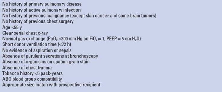
Every potential organ donor should be considered as a potential lung donor. The ideal lung donor criteria as defined in the early years of lung transplantation are listed in Table 1.1. The perfect lung donor matching all these criteria has become very rare in Western Europe because of the constant change in donor profile with a rising proportion of older donors becoming brain death from cerebrovascular disease. The scientific evidence to rely on these strict donor criteria was extensively reviewed by a panel of the Pulmonary Council of the International Society for Heart and Lung Transplantation and found to be very low. Recommendations were made to relax the acceptance criteria. The lung yield from all available donors, therefore, varies between countries and between lung transplant centers within the same country from 10% to nearly 50%. This percentage depends on (1) the expertise of the local donor team with active donor management; (2) the confidence by the recipient team to relax the preset lung donor criteria accepting pulmonary grafts from extended criteria donors and from donors after cardiocirculatory death; and (3) the willingness of the lung retrieval team to travel to the donor hospital to verify whether the initial information regarding lung oxygenation capacity, appearance on chest x-ray, and bronchoscopy findings provided at the time of organ offer, truly reflects lung quality witnessed at final evaluation in the donor after endotracheal suctioning and maximal alveolar recruitment.
Contraindications
As stated above, many potential lung donors will present with one of the more parameters that do not match the ideal criteria listed in Table 1.1. Many of these should be considered as relative contraindications as good immediate outcome is often possible. Nevertheless donors with older age, significant smoking history, inferior oxygenation, and radiographic infiltrates are considered to carry potential risk factors for the onset of chronic allograft dysfunction resulting in impaired long-term survival although strong evidence is missing in published literature. Some donor factors, however, should be considered as absolute contraindications for lung use such as pneumonia or sepsis, significant underlying parenchymal or vascular lung disease, history of recent malignancy (except skin cancer and some brain tumors), ABO incompatibility, and seropositivity for human immunodeficiency virus or hepatitis B or C virus in case of seronegative recipients.
TABLE 1.1 Ideal Lung Donor Selection Criteria
 PREOPERATIVE PLANNING
 We prefer to send an experienced lung surgeon to the donor hospital as the decision process whether or not to accept the lung is as important as proper lung preservation and excision.
Before leaving, the surgeon in charge should check with the scrub nurse and donor coordinator whether all surgical instruments and preservation solutions are available.
Upon arrival in the donor hospital, the recipient team is quickly informed on the estimated timing of cross clamp in the donor as unexpected delays in transport or start of donor operation may have occurred.
An advice that is greatly appreciated by younger colleagues when joining for their first lung retrieval is to take off their socks before entering the operating room. Otherwise they risk returning home with a soaked pair after organ flush and additional topical cooling!
It is important to introduce the lung team to all members of the local donor team including scrub nurse and anesthesiologist and to retrieval surgeons from other transplant centers.
In case of simultaneous heart procurement, a plan is discussed and agreed in advance with the heart surgeon on the surgical steps to be taken. Some may not be familiar with combined heart and lung retrieval!
It is the surgeon’s responsibility to check all relevant donor medical history, to verify the blood group, and to check whether the death certificate was completed according to state legislation.
In case the donor is not yet fully installed on the operating table, we prefer to have both arms next to the body to get more working space for both abdominal and thoracic teams. The shoulders are lifted up with towels and the head is extended backwards to have the neck free for better exposure to the trachea.
A broad-spectrum antibiotic and 1 g methylprednisolone is administered IV to the donor if not done already.
We routinely ask the anesthesiologist in charge of the donor to switch the gas mixture on the ventilator to 100% oxygen and to increase positive end-expiratory pressure to 5 cm H2O if needed. Ventilatory parameters are checked paying attention to the tidal volume and peak airway pressure so to have an idea on pulmonary compliance. A new arterial blood gas sample can be taken after 5 minutes although we prefer to wait for intrathoracic pulmonary vein blood sampling.
First a tracheal aspirate for culture is taken through the endotracheal tube followed by a quick flexible bronchoscopy whenever possible to verify the correct position of the endotracheal tube and to appreciate the amount and color of airway secretions, presence of blood or stomach content, and degree of mucosal inflammation. After cleaning the airways, we routinely perform a bronchoalveolar lavage with 2 × 50 mL of saline solution. The returned fluid is aspirated for culture and cellular and biomolecular analysis. Purulent secretions persistently bubbling up into the central airways are a bad sign indicative for the presence of pneumonia.
 SURGERY
A standard median sternotomy is performed usually in combination with median laparotomy in case abdominal organ retrieval is scheduled. In case sternotomy is left to be done by our own team (often the chest cavity is already opened by the abdominal team), we prefer to split the sternum in apnea in order not to damage lung parenchyma protruding into the retrosternal plane in a fully ventilated donor.
Lung Inspection and Evaluation
If not done yet by others, we open the pleural cavity on both sides by incising the mediastinal pleura. Care must be taken not to damage lung parenchyma on its medial aspect when using cautery. At this moment, we do not yet open the pericardium in order not to destabilize the heart too much when inspecting the lungs.
Transection of the diaphragmatic muscles is a tremendous help to gain access with easier exposure of the lower lobes.
The first step is to take a blood sample with a heparinized syringe by puncturing the extrapericardial part of the lower or middle lobe vein on both sides to assess oxygenation capacity (partial oxygen pressure in pulmonary vein = PpvO2) of left and right lungs individually.
The next step is to inspect the lungs for abnormalities that may preclude safe transplantation. We, therefore, ask the anesthesiologist to disconnect the endotracheal tube from the ventilator after initial preoxygenation as described above. First, it offers the possibility to observe the elasticity of both lungs (“collapse” test). If the lungs do not collapse instantly or symmetrically, this may be indicative of retained secretions or presence of interstitial lung pathology (edema, hemorrhage, pneumonia, emphysema). Deflated lungs are easier to be eviscerated from the pleural cavity without too much cardiac compression. We then quickly palpate both lungs looking for abnormal findings (nodules, blebs, adhesions) that may need further attention during and after retrieval. Significant structural abnormalities may finally exclude the lungs for transplantation.
The anesthesiologist is then asked to manually reinflate both lungs with 50% oxygen to a sustained 30 cm H2O pressure for recruitment of all atelectactic lung segments helped with gentle massage by the surgeon (Fig. 1.1). This maneuvre is also very indicative of lung compliance.
Once the blood gas results have returned and PpvO2 values fit with macroscopic findings, the recipient team is called to inform that lungs fulfill quality requirements and what the estimated cross-clamp time will be. Depending on the transport time and estimated operative time needed to explant the native lungs, it is now about the moment to call the recipient to the operating room and to prepare him for induction of anesthesia.
In case of discrepancy between healthy looking lungs and low oxygenation capacity (PpvO2/FiO2 <300 mm Hg), blood gas analysis should be repeated to verify.
If any doubt about lung performance and quality, lungs can always be explanted for further testing during ex vivo perfusion in the donor hospital or in the recipient hospital after being transported back on ice in case the expertise and equipment is available.
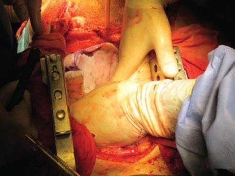
Figure 1.1 All atelectatic parenchymal zones are recruited by pressure ventilation and gentle massage.
Lung Dissection
If not previously done by others, the next step is to open the pericardium and to suspend both edges to the skin with heavy stitches.
In case the heart is retrieved for separate cardiac transplantation, much of the dissection is usually carried out by the heart surgeon. The ascending aorta is freed from the main pulmonary artery and encircled with a tape. The superior vena cava is mobilized from its pericardial attachments and encircled with a heavy ligature distal to both innominate veins. We do not favor to dissect or ligate the azygos vein as this maneuvre may cause bleeding or result in inadvertent ligation of the upper lobe branch of the right pulmonary artery. We have witnessed lobar infarction when arterial transection was not recognized or ignored at implantation. The intrapericardial inferior vena cava is mobilized from its pericardial attachments to facilitate adequate clamping. Passing a ligature is not needed as many abdominal teams like to vent the liver into the pericardium.
A horizontal mattress suture is then placed in the anterior midportion of the ascending aorta to secure the cardioplegia cannula once it is inserted.
For lung procurement without the heart, we advise to restrict the intrapericardial dissection to a minimum to speed up the retrieval and to avoid too much cardiac manipulation risking arrhythmias and premature cardiac arrest in an unstable donor. If no heart surgeon is around, the help of a thoracic assistant is greatly appreciated for suctioning during lung flush.
The placement of a purse-string suture on the main pulmonary artery is the only important act that is needed in a beating heart procedure. We prefer a 4-0 polypropylene suture widely placed for the introduction of a large-sized (18 to 24 Fr) pulmoplegia cannula allowing a high-volume flush at low pressure. If the heart is being used, care should be taken to stay distal to the pulmonary valve (>1.5 cm) but away from the pulmonary artery bifurcation to prevent inadvertent selective cannulation of the left main pulmonary artery. In case the heart is not used and not retrieved for the homograft bank, cannulation close to the pulmonary valve is recommended (Fig. 1.2A).
Lung Preservation
Once the dissection of all organs is completed, the donor is heparinized with 25,000 units through central venous access or direct intracaval injection. We prefer the abdominal surgeons to insert their cannulas first, one in the abdominal aorta for flushing and a sump in the abdominal vena cava for venting. Next the cardioplegia needle or thin cannula is introduced. We ask the heart surgeon to secure his cannula with a ligature, so there is less risk for dislocation during manipulation of heart and lungs while verifying adequate flush especially in atelectatic lower lobes.
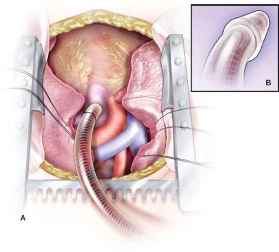
Figure 1.2 A: A large pulmoplegia cannula is inserted in the main pulmonary artery through a purse string proximal to the pulmonary valve for antegrade flush. B: The pulmoplegia cannula has a wider tip at the end preventing from inadvertent decannulation during flush.
A pulmonary artery incision is then made in the middle of the purse string with a sharp blade no. 15 and the arteriotomy is sufficiently dilated to allow easy entrance of the flush cannula. We prefer a cannula with a broader tip at the end, so that it stays intraluminal when pulled back to verify the correct position in the main pulmonary artery (Fig. 1.2B). This maneuvre may sometimes lead to significant blood loss and should, therefore, be done at the very end when all teams are ready to start flushing the organs.
The purse string is snugged and a ligature is tied around the cannula for fixation. With the cannula in the upright position, the height of the blood column is an indicator of the arterial pressure in the pulmonary circulation. A three-way stopcock is connected to the side hole in the cannula. This greatly helps to de-air once connected to the purged pulmoplegia line.
Before initiating the flush, it is wise to reconfirm full expansion of the lung and if needed, to recruit the alveoli in atelectatic zones.
A bolus of a strong vasodilator prostaglandin E1 alprostadil (Prostin VR 0.5 mg/mL, Pfizer), prostaglandin I2 epoprostenol (Flolan 0.5 mg, GlaxoSmithKline), or nitroglycerine (Nitro “Pohl”, 1 mg/mL, Pohl-Boskamp BV) is injected with a 50-mL syringe directly into the pulmonary artery via the three-way stopcock on the cannula. It is wise to inform other members that this bolus injection may result in a significant drop in systemic arterial pressure.
After circulation of the vasodilator through the lungs, inflow to the right heart is stopped by ligating or clamping both vena cava. The inferior vena cava is hemitransected proximal to the clamp for venting of the right heart and the tip of the left atrial appendage is transected to decompress the left atrium. Alternatively, the left atrium can be incised in the middle of the atrioventricular groove and the left pulmonary vein after the apex of the heart is lifted in the cephalad position. The latter procedure is, however, less effective in draining the large volume of lung preservation solution.
The aortic clamp is then placed and both cardioplegia and pulmoplegia lines are opened verifying that both left and right ventricles are adequately decompressed once the solutions start filling both atria. Larger incisions may be needed for adequate venting. If the heart is not used and the abdominal team has put a clamp on the infradiaphragmatic part of the descending aorta, no clamp on the ascending aorta is needed. In this way, the pulmoplegia solution is injected by the left ventricle into the aorta flushing the bronchial arteries, so the airways may be better preserved. The heart will stop beating by cold perfusion running into the coronary arteries and by the additional topical cooling. The vascular clamp on the inferior vena cava is then released for better venting of the liver.
Attention should be paid to the position of the tip of the pulmoplegia cannula. Inadvertent cannulation of the left pulmonary artery is possible leading to inadequate flush of the right lung. Gentle traction on the cannula may help to keep the cannula in the midline position with the tip directed toward the pulmonary valve.
We flush both lungs by gravity (60 cm H2O) with one large bag (2.8 L) and one small bag (1 L) of cold Perfadex preservation fluid (XVIVO AB, Gothenburg, Sweden) running simultaneously. This will result in a total flush volume of ±40 to 70 mL/kg body weight (45 to 95 kg donor). The free outflowing perfusate from the left atrium should gradually clear and must be monitored during the flush assuring equal distribution of Perfadex to both lungs.
The pericardial suspension stitches are released and cold saline solution is poured on the heart and lungs for additional topical cooling.
During the flush, ventilation is continued with the same tidal volume, PEEP 5 cm H2O and FiO2 0.5. If needed, recruitment of atelectatic zones will result in more equal blanching of the lungs.
When retrieving lungs from donors after cardiocirculatory death (Maastricht category III or IV), the steps for lung procurement are somewhat reversed. After quick sternotomy, both pleural cavities are widely opened and cold saline solution is poured over deflated lungs. The pericardium is opened; a large sump drain is inserted in the inferior vena cava through an opening in the right atrial appendage for decompression of the right heart. The pulmoplegia cannula is inserted in the main pulmonary artery through the pulmonary valve after incising the right ventricular outflow tract. The cannula is then secured by tying a heavy ligature passed around the main pulmonary artery distal to the valve. The cannula is de-aired and connected to the flushed pulmoplegia line. The left atrial appendage is transected and antegrade flush with Perfadex is started while ventilation of both lungs is regained. In experienced hands the whole procedure will not take longer than 3 to 5 minutes after skin incision.
In addition to the antegrade flush, lung preservation should be completed by an additional retrograde flush with 1 L Perfadex (250 mL via each of the four pulmonary veins) once the heart is extracted. This can be done with the same cannula gently wedged in the orifice of the four veins (Fig. 1.3). Small clots and debris may flow out from the pulmonary artery. It is believed that adding a retrograde flush improves airway preservation via bronchopulmonary collaterals. We prefer to do this while the lungs are still in the body attached to the trachea, so that ventilation can continue during the flush allowing better distribution of the preservation solution. If performed on the back table the Perfadex used for retrograde perfusion can be collected and utilized for lung storage.
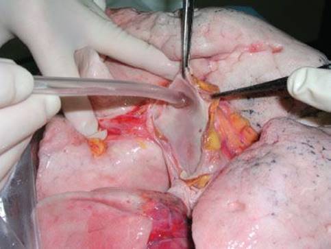
Figure 1.3 The same pulmoplegia cannula is wedged in the orifice of the right superior pulmonary vein for retrograde flush.
Lung Extraction
Once both lungs and heart are sufficiently flushed and cooled, the organs can be extracted.
Decannulation is performed, the ventilator is disconnected leaving the endotracheal tube in place, and pleural and pericardial cavities are emptied with heavy suction devices.
In case of planned combined heart–lung transplantation, the organs are not split but dissected and extracted en bloc. If the heart is used for a separate heart recipient or for the homograft bank, this organ will be extracted first.
The first step is to completely transect the inferior vena cava and to dissect its attachment up to the level of the right inferior pulmonary vein. Thereafter, the heart can be lifted and turned completely with the apex pointing in the cephalad direction. In this way the left atrium is maximally distended. A horizontal cut with a sharp blade is made in the midportion of the left atrium in between the atrioventricular groove and the left pulmonary vein. The left atrial incision is further extended parallel to the atrioventricular groove toward the base of the left atrial appendage on the left side and toward the inferior edge of the inferior vena cava on the right. The surgeon standing on the left side of the donor has the best view from inside the left atrium on the orifices of both right-sided pulmonary veins. The left atrial incision on the right side is then continued along the interatrial groove (Waterston). We do not dissect the groove from the outside. A sufficient rim of left atrium (minimum 1 cm) should be left as cuff for both the heart and the lungs. It is important not to overstretch the heart as this may result in insufficient atrial cuff on the lung side once it retracts. The scissors should always be kept in a horizontal position when completing circular cuff excision. Attention should be paid not to incise the pulmonary arteries at this stage.
The heart is put back in its normal position and the superior vena cava and ascending aorta are transected at sufficient length needed for the heart recipient. Both vessels are then freed from its attachments with the right pulmonary artery. Finally, the main pulmonary artery is usually transected at the cannulation site unless a longer cuff is needed for a heart recipient with a congenital abnormality. In case the lung will be hooked up to a device for ex vivo lung perfusion, a longer segment of main pulmonary artery will facilitate arterial cannulation.
After heart extraction, dissection of the double-lung bloc is to be continued by the lung surgeon. The inferior pericardium is transected in a U-shape taking care not to injure the lung at its attachment with the pulmonary ligament. This structure is transected carefully by cephalad traction on the double-lung bloc. We leave the esophagus attached to the vertebral body, so sharp dissection is carried out between the esophagus and posterior pericardium. With the right lung moved over into the left pleural cavity, the azygos vein is now transected. The same maneuvre is done on the left side now dividing the descending aorta distal to the arch. The dissection in the posterior mediastinum is continued separating the esophagus from the trachea as high as possible. Thereafter the innominate veins are transected as well as all supra-aortic vessels and pleural attachments. The trachea is then freed and vagal and recurrent nerves are transected.
Once all attachments are loosened, the double-lung bloc remains connected to the trachea only. The endotracheal cannula is pulled back by the anesthesiologist (who has often left the room by that time) guided by the surgeon, so that a sufficient length of trachea can be harvested, especially when the airway will be hooked up to a device for ex vivo perfusion and ventilation. Before dividing the trachea between two parallel linear stapler lines (TA 45-4.8 disposable stapler, Covidien, Mansfield, MA), the endotracheal tube is suctioned and both lungs are manually and gently reinflated with 50% oxygen until all atelectatic zones have been recruited. The staple lines are whipped with betadine solution. The inflated double-lung bloc is then extracted out of the body after maximal spreading of the sternal incision and pulling down the diaphragm.
Lung Packing
The double-lung bloc is immersed in cold Perfadex solution and covered with soaked towels. Arterial and venous cuffs are inspected for sufficient length and iatrogenic tears that may need repair on the bench prior to implantation (Fig. 1.4). Parenchymal abnormalities like apical blebs, scarring, or tears may be stapled (ILA 100 reusable stapler, Covidien, Mansfield, MA).
This is about the moment to give a third call to the recipient surgeon informing him on the exact cross-clamp time and on any surgical issues discovered in the donor lungs.
We prefer to split the double-lung bloc already in the donor hospital, so that both lungs can be packed and stored on ice individually as long as needed for sequential implantation.
The posterior pericardium is transected first followed by the left atrial cuff on the midline (Fig. 1.5A). The pulmonary artery is transected at its bifurcation (Fig. 1.5B). The subcarinal nodes and peritracheal fatty tissue are dissected freeing the orifice of both left and right main bronchi. (The peribronchial tissue of the left and right main bronchi should be preserved to avoid bronchial damage and for future buttressing of the anastomosis.) Lung splitting is finished by stapling the left main bronchus close to the trachea using two additional cartridges of the same stapler (Fig. 1.5C). If desired, the trachea can be resected for culture by adding another stapler line on the orifice of the right main bronchus.
The individual left and right lungs are then triple bagged (Fig. 1.6). The first bag is filled with cold Perfadex and the second bag with cold saline and crushed ice. We take care to completely de-air the first bag, so that the lung is entirely surrounded by preservation solution. Finally, the double-bagged lung is then secured inside a third sterile plastic bag. Both lungs are then submerged in the cool box filled with ice. A label is attached clearly indicating the position of the left and right lungs in the box that is now securely closed ready for transport.
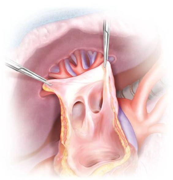
Figure 1.4 The left atrial cuff is inspected. Sufficient muscular tissue surrounding the pulmonary veins is present to safely perform the left atrial anastomosis.
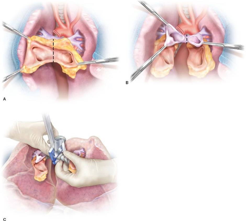
Figure 1.5 A: The posterior pericardium is transected first followed by the left atrial cuff on the midline. B: The pulmonary arteries are divided at their bifurcation. C: Lung splitting is finished by double-stapling of the left main bronchus with a linear stapler close to the trachea.
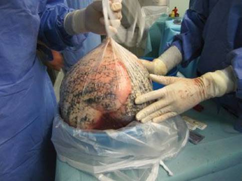
Figure 1.6 The lungs are each packed in three sterile bags. The first bag is filled with Perfadex solution and maximally de-aired, so that the lung is completely surrounded by cold preservation solution. The second bag contains cold saline solution and crushed ice. The third bag is dry and will protect the organs when covered by ice during storage in the cool box.
 POSTOPERATIVE MANAGEMENT
Before leaving the donor operating room, it is important to check for all surgical instruments used during lung procurement and to gather all personal belongings.
Donor blood samples, lymphoid tissue, and chest x-ray whenever possible are collected and stored to accompany the lungs to the donor hospital for further analysis and tissue typing.
It is very important to thank the local team for their help and hospitality. This is a token of appreciation and motivates the local team members to offer potential donors in future.
When leaving the donor hospital, a fourth call is given to the recipient team with the exact time of departure and the estimated time of arrival in the recipient operating room.
 COMPLICATIONS
From experimental and clinical experience, it is well known that the lungs tolerate warm ischemia up to about 1 hour, much longer compared to other solid organs. This results from the alveolar oxygen reserve in ventilated or inflated lungs enabling cellular aerobic metabolism to continue. Therefore, there is no reason whatsoever to panic during lung retrieval in case an unexpected event happens to the donor (premature cardiac arrest, major bleeding, or malposition of flush cannula). Even if the cardiac or abdominal team decides to abort the procurement, lungs can still be harvested safely. We refer to the procedure previously described for retrieving lungs from donors after cardiocirculatory death. Lungs can always be cooled topically with cold saline and a flush cannula can be quickly inserted via the right ventricular outflow tract through the pulmonary valve into the pulmonary artery for antegrade flush. Lungs can also be flushed on the back table after extraction. Deflated lungs can also be ventilated or inflated on the back table using a sterile endotracheal tube connected to the ventilator or to a portable Ambu bag.
Structures at risk to be damaged during lung procurement are vascular cuffs and parenchyma. The most frequent complication encountered is a short venous cuff especially on the right side around the inferior pulmonary vein. Surgical techniques have been described how to enlarge the venous cuff with surrounding donor pericardium and how to reconstruct individual pulmonary veins inadvertently transected. A small tear in the pulmonary artery may happen occasionally during dissection as the wall of the vessel is often thin and fragile in a young donor. This can easily be repaired with a 6-0 polypropylene stitch. Tears in lung parenchyma resulting from pleural adhesions or inadvertent cuts can usually be sealed with a linear stapler.
 RESULTS
The success rate of lung donor procurement largely depends on the willingness of the surgical team to travel to the donor hospital and on the acceptance rate of the retrieving surgeon when donor lungs do not fit the ideal criteria. Once the decision is taken to accept the organs, the technical success rate of lung procurement is very high in case all sequential steps and principles of lung preservation are well respected. It is unusual that a donor lung is rejected after retrieval. This may be the case if a tear is technically unrepairable or if significant structural damage is observed once the lungs are inspected on the back table (edema, contusion, pneumonia, or emphysema). If a suspicious nodule is found that can be wedged out, a frozen section may help to make a final decision whether or not to accept the organs. Occasionally, lungs have to be turned down during transport when an unexpected solid tumor is found in liver or kidney after explantation. If proven to be malignant, the presence of undetectable micrometastases in the lung may be a too high risk for the immunosuppressed patient to receive the organs.
 CONCLUSIONS
Every potential organ donor should be considered a potential lung donor. All efforts should be made to check lung donor quality during multiorgan retrieval in the donor hospital. The lungs are unique organs as they tolerate long warm ischemic periods. Therefore, even if the retrieval of other organs is stopped prematurely because of unexpected cardiac arrest or massive bleeding, the lungs can still be harvested with the help of simple topical cooling and subsequent antegrade and retrograde flush in the body or on the back table.
Recommended References and Readings
Brodman RE, Goldsmith J, Veith FJ, et al. A technique for donor lung procurement and preservation after completion of cardiac donation. Surg Gynecol Obstet. 1988;166:363–366.
Casula RP, Stoica SC, Wallwork J, et al. Pulmonary vein augmentation for single lung transplantation. Ann Thorac Surg. 2001;71:1373–1374.
Gamez P, Alvarez R, Hernández H, et al. Lung transplantation: How to do the venous anastomosis when the pulmonary graft has no auricular cuff. J Heart Lung Transplant. 2005;24:1123–1125.
Orens JB, Boehler A, de Perrot M, et al. A review of lung transplant donor acceptability criteria. J Heart Lung Transplant. 2003; 22:1183–1200.
Oto T, Rabinov M, Negri J, et al. Techniques of reconstruction for inadequate donor left atrial cuff in lung transplantation. Ann Thorac Surg. 2006;81:1199–1204.
Parekh K, Patterson GA. Technical considerations in adult lung transplantation. Semin Thorac Cardiovasc Surg. 2004;16:322–332.
Pasque MK. Standardizing thoracic organ procurement for transplantation. J Thorac Cardiovasc Surg. 2010;139:13–17.
Shigemura N, Bhama J, Nguyen D, et al. Pitfalls in donor lung procurements: How should the procedure be taught to transplant trainees? J Thorac Cardiovasc Surg. 2009;138:486–490.
Sundaresan S, Trachiotis GD, Aoe M, et al. Donor lung procurement: Assessment and operative technique. Ann Thorac Surg. 1993;56:1409–1413.
Todd TR, Goldberg M, Koshal A, et al. Separate extraction of cardiac and pulmonary grafts from a single organ donor. Ann Thorac Surg. 1988;46:356–359.
Van Raemdonck D. Thoracic organs: Current preservation technology and future prospects; part 1: Lung. Curr Opin Organ Transplant. 2010;15:150–155.
Van Raemdonck D, Neyrinck A, Verleden GM, et al. Donor selection and management. Proc Am Thorac Soc. 2009;6:28–38.
Van Raemdonck DE, Rega FR, Neyrinck AP, et al. Non-heart-beating donors. Semin Thorac Cardiovasc Surg. 2004;16:309–321.
Yarbrough WM, Bates MJ, Deuse T, et al. Alternative technique for salvage of donor lungs with insufficient atrial cuffs. Ann Thorac Surg. 2009;88:1374–1376.