Niall D. Ferguson
Arthur S. Slutsky
Mechanical ventilation practices continue to evolve. In particular, the ventilatory care of patients with acute lung injury (ALI) and acute respiratory distress syndrome (ARDS) has been refocused on avoiding or minimizing further damage to the fragile, injured lung. Conventional ventilators have been used successfully to limit this ventilator-induced lung injury (VILI) (1,2,3), but questions remain regarding the optimal method for lung protection during mechanical ventilation. The recognition of the critical role that VILI can play in the outcome of adults with ALI/ARDS has, in part, spawned a renewed interest in alternative modes of ventilation that may be more lung protective.
High-frequency ventilation (HFV) is just such a modality. In this chapter, we briefly review the different subtypes of HFV and then concentrate on high-frequency oscillation as the mode with the most supporting data and promise. As will be seen, a great deal of experience with HFV has been accrued in the neonatal and pediatric settings. We will draw on this knowledge where relevant, but focus this chapter from the viewpoint of HFV as an emerging therapy in the adult intensive care unit (ICU).
Definitions, Terminology, and Subtypes of High-frequency Ventilation
High-frequency ventilation is a collection of ventilatory modes, grouped together by their common property of employing high respiratory rates, all greater than 60 breaths per minute (4,5). In addition to high-frequency oscillation (HFO), other modes in this group include high-frequency jet ventilation (HFJV) and high-frequency percussive ventilation (HFPV).
High-frequency Jet Ventilation
HFJV is a mode of ventilation in which gas is delivered through a small-bore catheter into the lungs at rates of 100 to 150 breaths per minute (5,6). Delivered tidal volume is still small, but higher than just the volume exiting the jet, as the jets entrain an additional flow of gas by the Venturi effect. Exhalation is passive during HFJV, and gas trapping or dynamic hyperinflation can be an issue. In practice, a conventional ventilator is set up as a “slave” to the jet to provide positive end-expiratory pressure, along with basic monitoring and alarms.
HFJV is a very efficient mode for removing CO2. Additionally, because of HFJV's very high flow rates and the differing pulmonary time constants in the clinical setting of bronchopleural fistulae, this mode of ventilation is purported to have beneficial effects in the presence of this disorder; however, these benefits have not been verified during objective testing (7,8). Concerns with HFJV relate to the delivery of high pressures (10–50 pounds per square inch), unpredictable tidal volumes, and the development of dynamic hyperinflation, all of which may worsen VILI rather than minimize it. In addition, problems with adequate humidification of inspired gas and the subsequent risks of tracheobronchitis have been noted and documented.
High-frequency Percussive Ventilation
HFPV is the newest and least well studied of the HFV modes. It combines a high-frequency rate of 200 to 900 breaths per minute superimposed on a conventional pressure mode of ventilation (9). HFPV is reported to enhance the clearance of respiratory secretions and has been successfully used in this regard in patients with burns and inhalational injury (9).
High-frequency Oscillation
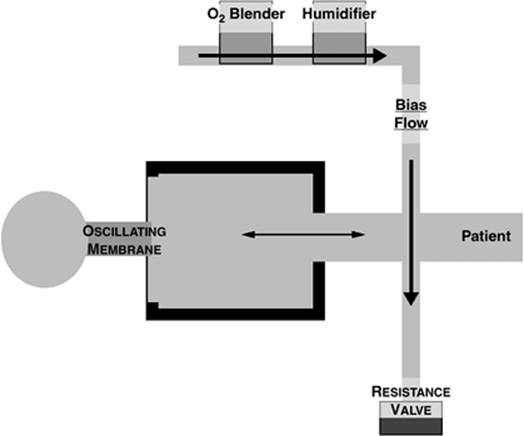
HFO is a mode of mechanical ventilation that delivers very small tidal volumes around a set mean airway pressure at high respiratory rates of 3 to 15 Hz (equivalent to 180–900 breaths per minute, as 1 Hz = 60 breaths per minute) (10). HFO has been widely and effectively used in neonates and children for close to 20 years (11,12,13,14,15), but only recently has become available in the adult ICU; previous versions of this ventilator were only capable of generating sufficient flow so as to provide adequate CO2 elimination in patients under 35 kg. In contrast to other HFV modes, humidification is less of an issue during HFO, as a continuous bias flow of humidified gas is passed in front of an oscillating membrane (Fig. 133.1). This oscillating membrane pushes the humidified gas into the patient and also provides active expiration, a factor that likely accounts for the lack of important gas trapping that is observed when HFO is employed at adequate airway pressures (16). The elegance of HFO is that it allows for “decoupling” of oxygenation and ventilation. Alveolar ventilation, and thus carbon dioxide elimination, are dependent on the frequency and tidal volume but are relatively independent of lung volume (17). In contrast, oxygenation is proportional to mean airway pressure and lung volume (18,19).
|
|
|
Figure 133.1. Schematic overview of the high-frequency oscillation (HFO) circuit. (Reused with permission from Ferguson ND, Stewart TE. New therapies for adults with acute lung injury: high-frequency oscillatory ventilation. Crit Care Clin. 2002;18:91–106.) |
Rationale for the Use of High-frequency Ventilation in Acute Lung Injury
Ventilator-induced Lung Injury
Mechanisms of Ventilator-induced Lung Injury
VILI is histologically indistinguishable from ARDS; three decades of experimental research has shown it to occur through a number of mechanisms, including (a) overdistention injury (volutrauma) (20,21,22,23,24,25,26,27,28), (b) collapse–reopening injury (atelectrauma) (1,2,20,29,30,31,32,33,34), and (c) oxygen toxicity (35,36,37). Each of these can lead, in turn, to further injury—termed biotrauma, the release of inflammatory mediators that may worsen pulmonary injury and propagate systemically to harm distant organs (1,2,31,32,38,39,40,41,42,43,44,45).
Numerous studies, performed using both small and large animals, consistently show that ventilatory high end-inspiratory stretch can cause a clinical and histologic picture similar to ARDS even in the absence of any other noxious stimulus (20,21,22,23,24,25,26,27,28). Patients with ARDS are at increased risk of regional lung overdistention because of the patchy nature of ARDS (46,47); the small areas of relatively normal lung (the so-called “baby lung”) receive the bulk of the tidal volume and are at particular risk of volutrauma (48,49). Repeated opening and closing of alveolar units can also cause VILI. In the injured lung, alveolar damage and absolute, or qualitative, deficiencies in alveolar surfactant lead to alveolar instability and localized lung unit collapse. Through each respiratory cycle, these unstable alveoli undergo collapse and reopening, a process that generates injurious mechanical forces and causes further lung injury. There is a substantive body of animal evidence showing that efforts to limit lung unit closing on expiration by maintaining an adequate positive end-expiratory pressure (PEEP) are relatively protective against atelectrauma (1,2,20,30,31,32,33,34). Here, the paradigm is one of “opening the lung and keeping it open,” thereby avoiding cyclic collapse and recruitment/derecruitment (2,50).
The major cause of death among ARDS patients is not refractory hypoxemia, as one might expect, but rather multiorgan dysfunction syndrome (MODS) (51). One hypothesis to explain the link between VILI and multiorgan failure is biotrauma (38,39)—the release of inflammatory mediators in response to injurious ventilation; these mediators may enter the systemic circulation, and hence lead to the development of MODS. Inflammatory mediators, such as interleukins, tumour necrosis factor-β, and platelet-activating factor, are released in response to VILI, oxygen-free radicals, and alveolar shearing. These mediators perpetuate the cascade of lung inflammation and worsen lung injury (31,32). Additionally, injurious ventilation has been shown to increase these inflammatory mediators in the peripheral blood of patients with ARDS (44,52). Through a variety of mechanisms, this inflammatory injury can lead to nonpulmonary organ dysfunction (41,42,43).
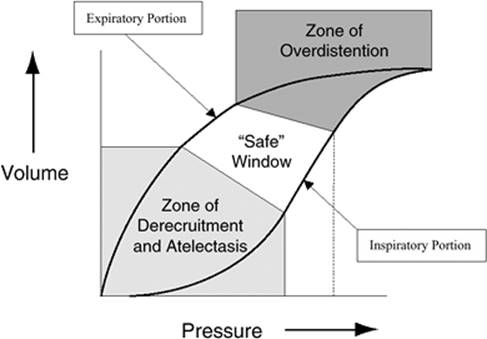
With an increased appreciation of the mechanisms of VILI, the next logical step is to employ ventilatory strategies that attempt to decrease overdistention, minimize shear injury, and limit oxygen toxicity (53,54,55). However, these aims may be competing; increasing PEEP may increase the risks of regional alveolar overdistention, while lowering tidal volumes can result in progressive alveolar collapse, a reduction in total lung volume, and higher oxygen requirements. The goals of mechanical ventilation in a patient at risk of VILI therefore should be to ventilate and oxygenate the patient while staying within a “safe window,” avoiding both overdistention and derecruitment—collapse—as illustrated in the volume–pressure curve of the lung shown in Figure 133.2 (56).
Impact of Limiting Ventilator-induced Lung Injury with Conventional Ventilation
In the mid-1990s, given the expanding animal data on VILI outlined above and in light of initial promising, but uncontrolled, human studies of lung-protective ventilation (57,58), a call was made for randomized trials (53). Initial trials focused on avoiding volutrauma, comparing strategies that limited tidal volumes and inspiratory pressures with traditional strategies, both using conventional ventilation. Three smaller studies did not find mortality differences between these approaches (59,60,61). However, a large, methodologically rigorous, and adequately powered trial conducted by the National Institutes of Health (NIH) ARDS Network did show important differences in mortality (3). In this study, 861 patients were randomly assigned to receive a low-stretch strategy with a targeted tidal volume of 6 mL/kg predicted body weight (PBW) and a plateau pressure limit of 30 cm H2O, or to a higher-stretch strategy using a targeted tidal volume of 12 mL/kg PBW and a plateau pressure limit of up to 50 cm H2O. The low-stretch strategy was associated with a mortality reduction from 40% in the control group to 31% in the experimental group (relative risk [RR] 0.78; 95% confidence interval [CI] 0.65–0.93). This trial clearly indicates that avoiding volutrauma saves lives in patients with acute lung injury. The trial has subsequently generated significant discussion regarding its mechanisms of benefit (62) and its choice of control strategy (63,64,65,66), but nevertheless, 6 mL/kg PBW has emerged as a standard for tidal volume limitation against which other strategies are compared (67,68).
|
|
|
Figure 133.2. Volume–pressure curve of the injured lung. (Reused with permission from Froese AB. High-frequency oscillatory ventilation for adult respiratory distress syndrome: let's get it right this time. Crit Care Med. 1997;25[6]:906–908.) |
Another early randomized controlled trial (RCT) published in the late 1990s demonstrated dramatic reductions in mortality using a lung-protective strategy whose goal was to limit both volutrauma and atelectrauma using low tidal volumes and higher PEEP compared with traditional ventilation (69). Amato et al. found a statistically significant reduction in 28-day mortality (11/29 [38%] vs. 17/24 [71%] deaths; RR 0.53; 95% CI 0.31–0.91) favoring patients exposed to the lung-protective strategy. In light of the subsequent positive ARDS Network trial noted above, interpretation of this trial is confounded by the use of both lower PEEP levels and higher tidal volumes and inspiratory pressures in the control group; the relative contribution of efforts to avoid cyclic collapse and reopening is unclear.
Drawing on the promise of the Amato trial, three RCTs have now been completed analyzing the effects of higher versus lower levels of PEEP, while limiting tidal volumes in all study patients. Two of these—the ExPress trial by Mercat et al. and the Lung Open Ventilation Study conducted by Meade et al.—are very recently completed and not yet published; both were presented at the 2006 European Society of Intensive Care Meeting. The one fully published trial, the ALVEOLI study, was conducted by the NIH ARDS Network investigators (70) and was stopped early, which likely contributed to the large baseline imbalance in age favoring the lower PEEP group. When we consider the relative risk after adjusting for baseline imbalances in prognostic factors (including age) from this trial, along with the preliminary findings from the two recent trials, a consistent trend favoring higher PEEP begins to emerge. Therefore, while no single trial has definitively shown an incremental mortality benefit with higher PEEP and attempts to limit atelectrauma while already avoiding overdistention injury, when viewed together, these trials suggest that this benefit may well exist. They also suggest that a higher level of PEEP with or without other maneuvers to open the lung, along with limited tidal volumes and inspiratory pressures, may be considered a very reasonable comparison strategy in future ventilation trials; this approach may be superior, and there is no suggestion of harm compared with lower PEEP levels. These studies clearly demonstrated that ventilatory strategy is important in patients with ARDS, and that lung-protective strategies can minimize VILI and decrease mortality in humans with ARDS. As such, and given the proposed mechanisms of lung protection (Fig. 133.2), a strategy that minimizes overdistention and allows the use of high PEEP should be the ideal mode in patients with ARDS. This is where HFO may have great clinical benefit.
Animal Studies Comparing Conventional Ventilation to High-frequency Oscillation in Acute Lung Injury
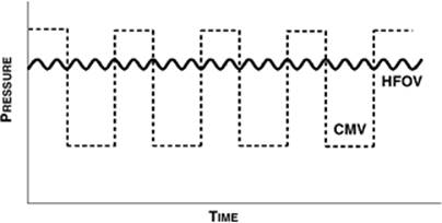
The very small delivered tidal volumes are the key to the lung-protective potential of HFO. Because cyclic alveolar stretch is minimal, clinicians are able to set the mean airway pressure (mPAW) on HFO significantly higher than they are able to set PEEP on conventional ventilation, thereby avoiding cyclic collapse and atelectrauma—and yet they are still able to avoid very high peak inspiratory pressures and subsequent volutrauma (Fig. 133.3). This should allow a larger margin of error and make it easier to stay within the “safe window” of lung protection (Fig. 133.2) (56). Since the first descriptions of HFO (71,72), numerous studies have examined the lung-protective potential of HFO using animal models (Table 133.1) (73,74,75,76,77,78,79,80,81,82,83,84,85,86,87,88,89). Perhaps not surprisingly, earlier studies that compared HFO with potentially injurious conventional ventilation uniformly found improvements in physiology, decreased inflammatory markers, and/or improved histology in animals treated with HFO (73,74,75,76,77,78,79,80,81,82,83). More relevant today, however, are recent studies that have compared HFO with lung-protective conventional ventilation, the majority of which continue to favor HFO in terms of physiology, inflammation, and pathology (Table 133.1) (86,87,88,89).
|
|
|
Figure 133.3. Pressure–time curve contrasts tidal variations in airway pressure associated with conventional ventilation (dashed line) and high-frequency oscillation (solid line). HFOV, high-frequency oscillation ventilation; CMV, conventional mechanical ventilation. (Reused with permission from Ferguson ND, Stewart TE. New therapies for adults with acute lung injury: high-frequency oscillatory ventilation. Crit Care Clin. 2002;18:91–106.) |
Mechanisms of Gas Transport during High-frequency Oscillation
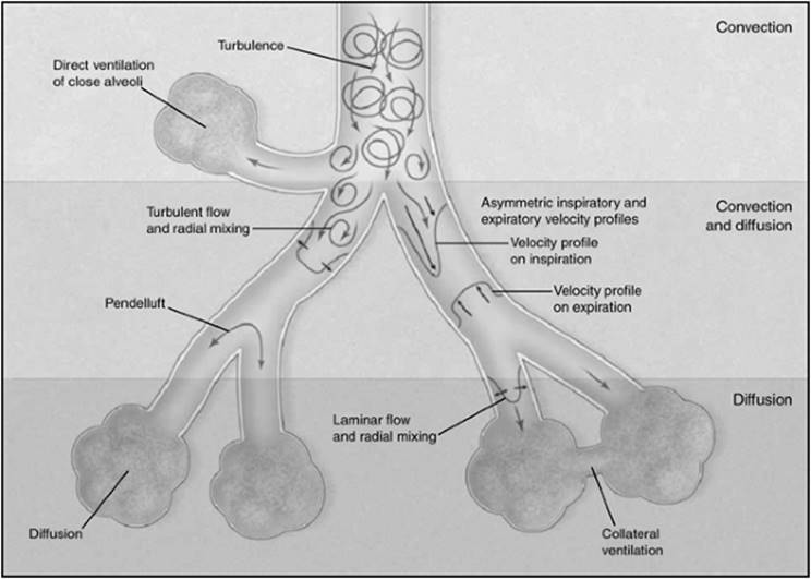
Tidal volumes are extremely small with HFO, often smaller than the anatomic dead space, in the range of 1 to 2 mL/kg (90). In addition to relying on bulk flow, adequate CO2 removal during HFO is achieved through a number of alternative mechanisms including convective streaming due to asymmetric velocity profiles, Pendelluft, cardiogenic mixing, and diffusion (Fig. 133.4). A full explanation of these physiologic principles is outside the scope of this chapter; the reader is referred to a number of papers reviewing this physiology in detail (5,91,92,93,94,95,96,97,98,99). The major message from these theoretical and experimental studies is that CO2 elimination—which is inversely proportional to PaCO2—is described by the relationship faVTb, where a = ~1 and b = ~2. This is important clinically in that it indicates that increases in frequency may lead to increased PaCO2 if VT falls (see below).
Implementing and Optimizing High-frequency Oscillation in Adults
Because of the lack of an appropriate surrogate end point that correlates with mortality (3,100), a large multicenter trial will be needed to definitively determine the relative effects of HFO compared with conventional mechanical ventilation (CMV) in terms of lung protection and mortality (101,102). Similarly, when making decisions regarding the best methods for applying HFO, we must draw inferences from multiple alternate sources (including animal studies, neonatal RCTs, and adult case series), since large RCTs comparing different HFO strategies are unavailable and would be, frankly, difficult to justify ethically in the absence of demonstrable efficacy over conventional ventilation.
Animal Studies
Too many animal studies have been conducted on high-frequency ventilation to list them in this review (4). Rather, in Table 133.2, we outline a number of these studies that are informative when considering how best to employ HFO in adults (72,103,104,105,106,107,108,109,110,111,112,113,114,115,116,117,118). Several key messages for HFO can be gleaned from these studies, including the following:
1. At adequate mPAW, gas trapping is not an issue despite very high respiratory rates.
2. Increasing mPAW leads to improved oxygenation through lung recruitment.
3. Tidal pressure swings measured in the trachea can be increased with either underrecruitment or overdistention of the lung.
4. Recruitment maneuvers (sustained inflations) may be required to adequately recruit severely injured lungs.
Taken together, and in keeping with the current understanding of VILI mechanisms, these studies suggest that HFO in adults should be employed using an open-lung strategy, facilitated by higher mean airway pressures and lung recruitment maneuvers (56,119,120).
Neonatal Studies
The longest clinical experience and the most rigorous evaluation of HFO have both been in the neonatal population (121,122,123,124,125,126,127,128,129,130). The initial large RCT evaluating HFO in this setting raised safety concerns, but it later became evident that here, too, the safe and effective application of HFO requires lung volume recruitment (121,131,132). Subsequent RCTs of HFO with an open-lung approach have demonstrated HFO to be safe and effective in improving oxygenation and, as indicated by the current Cochrane systematic review, may reduce the risk of death or chronic lung disease (15). Interpreting the neonatal HFO literature is challenging due to differences in study populations (preterm vs. term), interventions (degree of lung recruitment targeted), the timing of HFO (immediately after birth vs. later), and by the introduction of exogenous surfactant as a standard therapy in the 1990s. In addition, it is important to realize that the baseline mortality rate in infant respiratory distress syndrome is an order of magnitude less than that seen in adults with severe ARDS. All of these factors mean that results from neonatal studies cannot be directly extrapolated to adult populations. Important lessons can be learned from the neonatal HFO literature, however, including the importance of thoroughly understanding the underlying physiologic mechanisms of the therapy, and the recognition that a learning curve may exist in the initiation of an HFO program (131,132).
|
Table 133.1 Animal studies comparing VILI with HFO vs. CMV |
||||||||||||||||||||||||||||||||||||||||||||||||||||||||||||||||||||||||||||||||||||||||||||||||||||||||||||||||||||||||||||||||||||||||||||||||||||||||||||||||||||||||||||||||||||||||||||||||||||||||||||||||||
|
||||||||||||||||||||||||||||||||||||||||||||||||||||||||||||||||||||||||||||||||||||||||||||||||||||||||||||||||||||||||||||||||||||||||||||||||||||||||||||||||||||||||||||||||||||||||||||||||||||||||||||||||||
P.2034
|
|
|
Figure 133.4. Alternative mechanisms of gas exchange with high-frequency oscillation (HFO). (Reused with permission from Slutsky AS, Drazen JM. Ventilation with small tidal volumes. N Engl J Med. 2002;347[9]:630–631.) |
Uncontrolled Adult Studies
The studies reporting the clinical experience with HFO in adults, which are summarized in Table 133.3 (133,134,135,136,137,138,139,140,141,142,143), are also informative for optimizing an HFO protocol. Almost all of them studied HFO as rescue therapy for patients with extremely severe disease who were “failing” conventional ventilation. Despite being limited by their uncontrolled nature and by selection bias, consistent messages that arise from these rescue series include:
1. HFO is usually effective in improving oxygenation.
2. HFO appears safe, with no obvious increased rates of complications.
3. Both baseline oxygenation index and duration of CMV prior to HFO are associated with increased mortality.
One prospective, multicentered study from our group is unique in that it tested an explicit protocol for applying HFO early in the course of ARDS, incorporating lung recruitment maneuvers (RMs) and a descending titration of mean airway pressure to optimize lung volumes for gas exchange and lung protection (140). RMs—sustained inflation maneuvers with 30 to 40 cm H2O pressures for 30 to 40 seconds—have been found to be safe in adults on CMV, but studies have shown mixed results in terms of efficacy and duration of their oxygenation effects (144,145,146,147,148,149,150,151,152). Because of the small tidal volumes generated with HFO, there is very little tidal recruitment of the lung, creating a more compelling rationale for RMs during HFO compared with CMV (19,50,102,153,154). The main finding from a physiologic standpoint in our study was that the combination of HFO and RMs was well tolerated and resulted in rapid and sustained lung recruitment (140). Furthermore, the explicit HFO protocol appeared feasible; adherence was excellent, and the method for weaning HFO and transitioning to CMV appeared practical and safe.
Tidal volume during HFO has always been assumed to be very low, but it is not routinely measured with currently available oscillators. Tidal volume is known to be inversely related to frequency due to a decreasing inspiratory time with increasing frequency (17), and in adults, typical frequencies have been significantly lower than those used in neonates (3–6 vs. 12–15 Hz) (132,139). Sedeek et al. explored this issue in a lung-injured sheep model, measuring tidal volumes of up to 4 mL/kg at high-pressure amplitudes and low frequencies (Table 133.2) (118). These tidal volume measurements may be overestimated because of technical reasons (155), but they do highlight the importance of efforts to minimize delivered tidal volumes with adult HFO, and they have spurred further investigation. Tidal volumes on HFO measured in adults using a very accurate device (156) were small—1.1 to 2.5 mL/kg—and strongly influenced by frequency (90). Cumulatively, these data are reassuring in that HFO in adults can be set to deliver very small tidal volumes, but at the same time they highlight the potential importance of targeting the lowest tidal volume possible (155). This could be achieved by increasing frequency as high as tolerated while avoiding severe respiratory acidosis, a strategy that has been proposed as physiologically sensible (157) and recently shown to be feasible in most patients (158).
|
Table 133.2 Animal studies informing optimal HFO implementation |
||||||||||||||||||||||||||||||||||||||||||||||||||||||||||||||||||||||||||||||||||||||||||||||||||||||||||||||||||||||||||||||||||||||||||||||||||||||||||||||||||||||||||||||||||||||||||||||||
|
||||||||||||||||||||||||||||||||||||||||||||||||||||||||||||||||||||||||||||||||||||||||||||||||||||||||||||||||||||||||||||||||||||||||||||||||||||||||||||||||||||||||||||||||||||||||||||||||
|
Table 133.3 Uncontrolled studies HFO for adult ARDS |
||||||||||||||||||||||||||||||||||||||||||||||||||||||||||||||||||||||||||||||||||||||||||||||||||||||||||||||||||||||||||||||||||||||||||||||||||||||||||||||||||||||||||||||||||||||||||||||||
|
||||||||||||||||||||||||||||||||||||||||||||||||||||||||||||||||||||||||||||||||||||||||||||||||||||||||||||||||||||||||||||||||||||||||||||||||||||||||||||||||||||||||||||||||||||||||||||||||
Randomized Controlled Trials of High-frequency Oscillation in Adults
To date, only two RCTs have compared HFO with CMV in adults (101,159). Both of these trials were planned and started prior to the completion of the first ARDS Network study that showed benefit from strict control of tidal volumes (3). Neither RCT demonstrated safety concerns (the primary outcome); rates of barotrauma and other complications were similar between groups in both studies. The larger of the two RCTs shows an impressive trend toward a mortality benefit with HFO (RR 0.72; 95% CI 0.50–1.04) despite more than 10% of the control group crossing over to HFO (101). The second trial, which began accrual at the same time, was stopped because of slow enrollment. In fact, the enrollment rates per center per month were identical in both studies; the latter study simply highlights the need for extensive multicentered collaboration for a successful HFO RCT. Mortality results from the smaller study are less encouraging, but these are significantly confounded by large baseline differences favoring the control group, and by an almost 20% crossover rate. Nevertheless, pooled results from these trials still suggest a potentially important survival benefit with HFO (RR 0.899, 95% CI 0.51–1.58; random effects model). Inferences from these RCTs are limited, however, by their methodology, small sample sizes, and, importantly, by use of now dated, potentially injurious conventional ventilation strategies.
Future Directions
A research focus on the ventilatory care of ARDS patients has, to date, paid significantly greater dividends than the disappointing results of pharmacotherapy testing (160). Following the completion of the three conventional ventilation RCTs comparing a “lung open” approach with a lower PEEP approach, we believe that the study of HFO represents the next logical and important question to be addressed in ARDS research. We ground this belief in:
1. A strong physiologic rationale and database of animal studies
2. Experience from the neonatal and pediatric arenas
3. An expanding clinical experience with adult HFO
4. Promising results from small nondefinitive RCTs
Despite the very strong physiologic rationale and the encouraging clinical data to date, there are potential detrimental consequences of HFO. First, the physiologic benefit of HFO is likely derived from recruitment of the lung with higher mean airway pressures, while still not overdistending alveoli because of the very small tidal volumes. Hence, patients with mild disease and minimal collapsed or consolidated lung may not be good candidates for HFO (151). Second, the high mean airway pressures may negatively impact hemodynamics. Third, because the bias flow rate is insufficient to meet the inspiratory flow demands of adults in respiratory distress, all adults on HFO must have their respiratory efforts suppressed with intravenous sedation. This means that the majority of adults will be heavily sedated, and many may need transient neuromuscular blockade. Due to these concerns, and to target a population in need of recruitment, we believe that future studies should enroll patients with severe ARDS who are likely to have significant lung collapse and who frequently require significant amounts of sedation, with or without paralysis, on conventional ventilators.
In summary, we believe that HFO—and other forms of high-frequency ventilation—should currently be reserved for “rescue therapy” in patients who are failing conventional ventilation. We do not believe that HFO can be recommended for routine use in adults with ARDS at the current time because:
· There are potential detrimental physiologic and clinical consequences as described above
· This would represent premature dissemination of a complex technology without rigorous evaluation of its risks, benefits, and indications
A definitive RCT to establish the impact of HFO versus best current conventional ventilation on mortality is needed.
References
1. Pinhu L, Whitehead T, Evans T, et al. Ventilator-associated lung injury. Lancet. 2003;361:332–340.
2. Tremblay LN, Slutsky AS. Ventilator-induced lung injury: from the bench to the bedside. Intensive Care Med. 2006;32(1):24–33.
3. The Acute Respiratory Distress Syndrome Network. Ventilation with lower tidal volumes as compared with traditional tidal volumes for acute lung injury and the acute respiratory distress syndrome. N Engl J Med. 2000;342(18):1301–1308.
4. Froese AB, Bryan AC. High frequency ventilation. Am Rev Resp Dis. 1987;135(6):1363–1374.
5. Drazen JM, Kamm RD, Slutsky AS. High-frequency ventilation. Physiol Rev. 1984;64(2):505–543.
6. Klain M, Smith RB. High frequency percutaneous transtracheal jet ventilation. Crit Care Med. 1977;5(6):280–287.
7. Roth MD, Wright JW, Bellamy PE. Gas flow through a bronchopleural fistula. Measuring the effects of high-frequency jet ventilation and chest-tube suction. Chest. 1988;93(1):210–213.
8. Bishop MJ, Benson MS, Sato P, et al. Comparison of high-frequency jet ventilation with conventional mechanical ventilation for bronchopleural fistula. Anesth Analg. 1987;66(9):833–838.
9. Salim A, Martin M. High-frequency percussive ventilation. Crit Care Med. 2005;33(3 Suppl):S241–S245.
10. Chan KP, Stewart TE. Clinical use of high-frequency oscillatory ventilation in adult patients with acute respiratory distress syndrome. Crit Care Med. 2005;33(3 Suppl):S170–S174.
11. Froese AB, Butler PO, Fletcher WA, et al. High frequency oscillatory ventilation in premature infants with respiratory failure: a preliminary report. Anesth Analg. 1987;66:814–824.
12. Bryan AC. The Oscillations of HFO. Am J Resp Crit Care Med. 2001;163:816–817.
13. Bollen CW, Uiterwaal CS, van Vught AJ. Cumulative metaanalysis of high-frequency versus conventional ventilation in premature neonates. Am J Respir Crit Care Med. 2003;168(10):1150–1155.
14. Arnold JH, Truog WE, Thompson JE, et al. High-frequency oscillatory ventilation in pediatric respiratory failure. Crit Care Med. 1993;21:272–278.
15. Henderson-Smart DJ. Elective high frequency oscillatory ventilation versus conventional ventilation for acute pulmonary dysfunction in preterm infants. Cochrane Database Syst Rev. 2006;2.
16. Bryan AC, Slutsky AS. Lung volume during high frequency oscillation. Am Rev Resp Dis. 1986;133(5):928–930.
17. Slutsky AS, Kamm RD, Rossing TH, et al. Effects of frequency, tidal volume, and lung volume on CO2 elimination in dogs by high frequency (2–30 Hz), low tidal volume ventilation. J Clin Invest. 1981;68(6):1475–1484.
18. Suzuki H, Papazoglou K, Bryan AC. Relationship between PaO2 and lung volume during high frequency oscillatory ventilation. Acta Paediatr Jpn. 1992;34(5):494–500.
19. Kolton M, Cattran CB, Kent G, et al. Oxygenation during high-frequency ventilation compared with conventional mechanical ventilation in two models of lung injury. Anesth Analg. 1982;61(4):323–332.
20. Webb HH, Tierney DF. Experimental pulmonary edema due to intermittent positive pressure ventilation with high inflation pressures. Protection by positive end-expiratory pressure. Am Rev Resp Dis. 1974;110(5):556–565.
21. Dreyfuss D, Soler P, Basset G, et al. High inflation pressure pulmonary edema. Respective effects of high airway pressure, high tidal volume, and positive end-expiratory pressure. Am Rev Resp Dis. 1988;137(5):1159–1164.
22. Parker JC, Hernandez LA, Longenecker GL, et al. Lung edema caused by high peak inspiratory pressures in dogs. Role of increased microvascular filtration pressure and permeability. Am Rev Resp Dis. 1990;142(2):321–328.
23. Dreyfuss D, Basset G, Soler P, et al. Intermittent positive-pressure hyperventilation with high inflation pressures produces pulmonary microvascular injury in rats. Am Rev Resp Dis. 1985;132(4):880–884.
24. Kolobow T, Moretti MP, Fumagalli R, et al. Severe impairment in lung function induced by high peak airway pressure during mechanical ventilation. An experimental study. Am Rev Resp Dis. 1987;135(2):312–315.
25. Tsuno K, Miura K, Takeya M, et al. Histopathologic pulmonary changes from mechanical ventilation at high peak airway pressures. Am Rev Resp Dis. 1991;143(5 Pt 1):1115–1120.
26. Dreyfuss D, Saumon G. Ventilator-induced lung injury: lessons from experimental studies. Am J Resp Crit Care Med. 1998;157:294–323.
27. Hernandez LA, Peevy KJ, Moise AA, et al. Chest wall restriction limits high airway pressure-induced lung injury in young rabbits. J Appl Physiol. 1989;66(5):2364–2368.
28. Steinberg J, Schiller HJ, Halter JM, et al. Tidal volume increases do not affect alveolar mechanics in normal lung but cause alveolar overdistension and exacerbate alveolar instability after surfactant deactivation. Crit Care Med. 2002;30(12):2675–2683.
29. Slutsky AS. Lung injury caused by mechanical ventilation. Chest. 1999;116:9S–15S.
30. Muscedere JG, Mullen JB, Gan K, et al. Tidal ventilation at low airway pressures can augment lung injury. Am J Resp Crit Care Med. 1994;149(5):1327–1334.
31. Tremblay L, Valenza F, Ribeiro SP, et al. Injurious ventilatory strategies increase cytokines and c-fos m-RNA expression in an isolated rat lung model. J Clin Invest. 1997;99(5):944–952.
32. Tremblay L, Govindarajan A, Veldhuizen R, et al. TNFa levels are both time and ventilation strategy dependent in ex vivo rat lungs. Am J Resp Crit Care Med. 1998;157(3):A213.
33. Steinberg JM, Schiller HJ, Halter JM, et al. Alveolar instability causes early ventilator-induced lung injury independent of neutrophils. Am J Resp Crit Care Med. 2004;169(1):57–63.
34. Dos Santos CC, Slutsky AS. Cellular responses to mechanical stress: invited review: mechanisms of ventilator-induced lung injury: a perspective. J Appl Physiol. 2000;89(4):1645–1655.
35. Bryan CL, Jenkinson SG. Oxygen toxicity. Clin Chest Med. 1988;9(1):141–152.
36. Nash G, Blennerhassett JB, Pontoppidan H. Pulmonary lesions associated with oxygen therapy and artificial ventilation. N Engl J Med. 1967;276(7):368–374.
37. Davis WB, Rennard SI, Bitterman PB, et al. Pulmonary oxygen toxicity. Early reversible changes in human alveolar structures induced by hyperoxia. N Engl J Med. 1983;309(15):878–883.
38. Tremblay LN, Slutsky AS. Ventilation-induced lung injury: from barotrauma to biotrauma. Proc Assoc Am Phys. 1998;110:482–488.
39. Slutsky AS, Tremblay LN. Multiple system organ failure. Is mechanical ventilation a contributing factor? Am J Resp Crit Care Med. 1998;157:1721–1725.
40. Chiumello D, Pristine G, Slutsky AS. Mechanical ventilation affects local and systemic cytokines in an animal model of acute respiratory distress syndrome. Am J Respir Crit Care Med. 1999;160(1):109–116.
41. Guery BP, Welsh DA, Viget NB, et al. Ventilation-induced lung injury is associated with an increase in gut permeability. Shock. 2003;19(6):559–563.
42. Choi WI, Quinn DA, Park KM, et al. Systemic microvascular leak in an in vivo rat model of ventilator-induced lung injury. Am J Respir Crit Care Med. 2003;167(12):1627–1632.
43. Imai Y, Parodo J, Kajikawa O, et al. Injurious mechanical ventilation and end-organ epithelial cell apoptosis and organ dysfunction in an experimental model of acute respiratory distress syndrome. JAMA. 2003;289:2104–2112.
44. Ranieri VM, Suter PM, Tortorella C, et al. Effect of mechanical ventilation on inflammatory mediators in patients with acute respiratory distress syndrome: a randomized controlled trial. JAMA. 1999;282(1):54–61.
45. Dos Santos CC, Slutsky AS. The contribution of biophysical lung injury to the development of biotrauma. Annu Rev Physiol. 2006;68:585–618.
46. Gattinoni L, Pelosi P, Vitale G, et al. Body position changes redistribute lung computed-tomographic density in patients with acute respiratory failure. Anesthesiology. 1991;74(1):15–23.
47. Gattinoni L, Caironi P, Pelosi P, et al. What has computed tomography taught us about the acute respiratory distress syndrome? Am J Respir Crit Care Med. 2001;164(9):1701–1711.
48. Roupie E, Dambrosio M, Servillo G, et al. Titration of tidal volume and induced hypercapnia in acute respiratory distress syndrome. Am J Resp Crit Care Med. 1995;152:121–128.
49. Gattinoni L, Pesenti A. The concept of “baby lung”. Intensive Care Med. 2005;31(6):776–784.
50. Lachmann B. Open up the lung and keep it open. Intensive Care Med. 1992;18:319–321.
51. Montgomery AB, Stager MA, Carrico CJ, et al. Causes of mortality in patients with the adult respiratory distress syndrome. Am Rev Resp Dis. 1985;132(3):485–489.
52. Parsons PE, Eisner MD, Thompson BT, et al. Lower tidal volume ventilation and plasma cytokine markers of inflammation in patients with acute lung injury. Crit Care Med. 2005;33(1):1–6.
53. Slutsky AS. Mechanical ventilation. American College of Chest Physicians' Consensus Conference. Chest. 1993;104(6):1833–1859.
54. Stewart TE, Slutsky AS. Mechanical ventilation: a shifting philosophy. Curr Sci. 1995;1:49–56.
55. Stewart TE. Lung protection during mechanical ventilation. Ontario Thorac Rev. 1997;9:1–4.
56. Froese AB. High-frequency oscillatory ventilation for adult respiratory distress syndrome: let's get it right this time. Crit Care Med. 1997;25(6):906–908.
57. Hickling KG, Henderson SJ, Jackson R. Low mortality associated with low volume pressure limited ventilation with permissive hypercapnia in severe adult respiratory distress syndrome. Intensive Care Med. 1990;16(6):372–377.
58. Hickling KG, Walsh J, Henderson S, et al. Low mortality rate in adult respiratory distress syndrome using low-volume, pressure-limited ventilation with permissive hypercapnia: a prospective study. Crit Care Med. 1994;22(10):1568–1578.
59. Stewart TE, Meade MO, Cook DJ, et al. Evaluation of a ventilation strategy to prevent barotrauma in patients at high risk for acute respiratory distress syndrome. Pressure- and Volume-Limited Ventilation Strategy Group. N Engl J Med. 1998;338(6):355–361.
60. Brochard L, Roudot-Thoraval F, Roupie E, et al. Tidal volume reduction for prevention of ventilator-induced lung injury in acute respiratory distress syndrome. The Multicenter Trial Group on Tidal Volume reduction in ARDS. Am J Resp Crit Care Med. 1998;158(6):1831–1838.
61. Brower RG, Shanholtz CB, Fessler HE, et al. Prospective, randomized, controlled clinical trial comparing traditional versus reduced tidal volume ventilation in acute respiratory distress syndrome patients. Crit Care Med. 1999;27(8):1492–1498.
62. de Durante G, del Turco M, Rustichini L, et al. ARDSNet lower tidal volume ventilatory strategy may generate intrinsic positive end-expiratory pressure in patients with acute respiratory distress syndrome. Am J Resp Crit Care Med. 2002;165:1271–1274.
63. Eichacker PQ, Gerstenberger EP, Banks SM, et al. Meta-analysis of acute lung injury and acute respiratory distress syndrome trials testing low tidal volumes. Am J Resp Crit Care Med. 2002;166:1510–1514.
64. Brower RG, Matthay M, Schoenfeld D. Meta-analysis of acute lung injury and acute respiratory distress syndrome trials. Am J Resp Crit Care Med. 2002;166:1515–1517.
65. Steinbrook R. How best to ventilate? Trial design and patient safety in studies of the acute respiratory distress syndrome. N Engl J Med. 2003;348:1393–1401.
66. Drazen JM. Controlling research trials. N Engl J Med. 2003;348:1377–1380.
67. Tobin MJ. Culmination of an era in research on the acute respiratory distress syndrome. N Engl J Med. 2000;342(18):1360–1361.
68. Stewart TE. Controversies around lung protective ventilation. Am J Resp Crit Care Med. 2002;166:1421–1422.
69. Amato MB, Barbas CS, Medeiros DM, et al. Effect of a protective-ventilation strategy on mortality in the acute respiratory distress syndrome. N Engl J Med. 1998;338(6):347–354.
70. Brower RG, Lanken PN, MacIntyre N, et al. Higher versus lower positive end-expiratory pressures in patients with the acute respiratory distress syndrome [see comment]. N Engl J Med. 2004;351(4):327–336.
71. Lunkenheimer PP, Rafflenbeul W, Keller H, et al. Application of transtracheal pressure oscillations as a modification of “diffusion respiration”. Br J Anaesth. 1972;44:627.
72. Bohn DJ, Miyasaka K, Marchak BE, et al. Ventilation by high-frequency oscillation. J Appl Physiol. 1980;48(4):710–716.
73. Hamilton PP, Onayemi A, Smyth JA, et al. Comparison of conventional and high-frequency oscillatory ventilation: oxygenation and lung pathology. J Appl Physiol. 1983;55:131–138.
74. Tamura M, Kawano T, Fitz-James I, et al. High-frequency oscillatory ventilation and pulmonary extravascular water. Anesth Analg. 1985;64 (11):1041–1046.
75. McCulloch PR, Forkert PG, Froese AB. Lung volume maintenance prevents lung injury during high frequency oscillatory ventilation in surfactant-deficient rabbits. Am Rev Resp Dis. 1988;137(5):1185–1192.
76. Bond DM, Froese AB. Volume recruitment maneuvers are less deleterious than persistent low lung volumes in the atelectasis-prone rabbit lung during high-frequency oscillation. Crit Care Med. 1993;21(3):402–412.
77. Imai Y, Kawano T, Miyasaka K, et al. Inflammatory chemical mediators during conventional ventilation and during high frequency oscillatory ventilation. Am J Resp Crit Care Med. 1994;150(6 Pt 1):1550–1554.
78. Matsuoka T, Kawano T, Miyasaka K. Role of high-frequency ventilation in surfactant-depleted lung injury as measured by granulocytes. J Appl Physiol. 1994;76(2):539–544.
79. Sugiura M, McCulloch PR, Wren S, et al. Ventilator pattern influences neutrophil influx and activation in atelectasis-prone rabbit lung. J Appl Physiol. 1994;77(3):1355–1365.
80. Takata M, Abe J, Tanaka H, et al. Intraalveolar expression of tumor necrosis factor-alpha gene during conventional and high-frequency ventilation. Am J Resp Crit Care Med. 1997;156(1):272–279.
81. Gommers D, Hartog A, Schnabel R, et al. High-frequency oscillatory ventilation is not superior to conventional mechanical ventilation in surfactant-treated rabbits with lung injury. Eur Resp J. 1999;14(4):738–744.
82. Kerr CL, Veldhuizen RA, Lewis JF. Effects of high-frequency oscillation on endogenous surfactant in an acute lung injury model. Am J Resp Crit Care Med. 2001;164(2):237–242.
83. Noda E, Hoshina H, Watanabe H, et al. Production of TNF-alpha by polymorphonuclear leukocytes during mechanical ventilation in the surfactant-depleted rabbit lung. Pediatr Pulmonol. 2003;36(6):475–481.
84. Vazquez de Anda GF, Hartog A, Verbrugge SJ, et al. The open lung concept: pressure-controlled ventilation is as effective as high-frequency oscillatory ventilation in improving gas exchange and lung mechanics in surfactant-deficient animals. Intensive Care Med. 1999;25(9):990–996.
85. Rimensberger PC, Pache JC, McKerlie C, et al. Lung recruitment and lung volume maintenance: a strategy for improving oxygenation and preventing lung injury during both conventional mechanical ventilation and high-frequency oscillation. Intensive Care Med. 2000;26(6):745–755.
86. Rotta AT, Gunnarsson B, Fuhrman BP, et al. Comparison of lung protective ventilation strategies in a rabbit model of acute lung injury. Crit Care Med. 2001;29(11):2176–2184.
87. Imai Y, Nakagawa S, Ito Y, et al. Comparison of lung protection strategies using conventional and high-frequency oscillatory ventilation. J Appl Physiol. 2001;91(4):1836–1844.
88. Sedeek KA, Takeuchi M, Suchodolski K, et al. Open-lung protective ventilation with pressure control ventilation, high-frequency oscillation, and intratracheal pulmonary ventilation results in similar gas exchange, hemodynamics, and lung mechanics. Anesthesiology. 2003;99(5):1102–1111.
89. von der Hardt K, Kandler MA, Fink L, et al. High frequency oscillatory ventilation suppresses inflammatory response in lung tissue and microdissected alveolar macrophages in surfactant depleted piglets. Pediatr Res. 2004;55(2):339–346.
90. Hager DN, Fessler HE, Fuld MK, et al. Effects of frequency and pressure amplitude on tidal volumes in adults with ARDS during high-frequency oscillatory ventilation. Proc Am Thorac Soc. 2006;3:A376.
91. Villar J, Slutsky AS. Alternative modalities for ventilatory support. In: Vincent JL, ed. Update in Intensive Care and Emergency Medicine. Brussels: Springer-Verlag; 1991:345–354.
92. Chang HK. Mechanisms of gas transport during ventilation by high-frequency oscillation. J Appl Physiol. 1984;56(3):553–563.
93. Ribeiro SP, Tremblay LN, Slutsky AS. High-frequency ventilation. In: Marini JJ, Slutsky AS, eds. Physiological Basis of Ventilatory Support. New York: Marcel Dekker; 1998:889–920.
94. Schmid ER, Knopp TJ, Rehder K. Intrapulmonary gas transport and perfusion during high-frequency oscillation. J Appl Physiol Respir Environ Exerc Physiol. 1981;51(6):1507–1514.
95. Slutsky AS. Gas mixing by cardiogenic oscillations: a theoretical quantitative analysis. J Appl Physiol Respir Environ Exerc Physiol. 1981;51(5):1287–1293.
96. Slutsky AS, Brown R. Cardiogenic oscillations: a potential mechanism enhancing oxygenation during apneic respiration. Med Hypotheses. 1982;8(4):393–400.
97. Cybulsky IJ, Abel JG, Menon AS, et al. Contribution of cardiogenic oscillations to gas exchange in constant-flow ventilation. J Appl Physiol. 1987;63(2):564–570.
98. Slutsky AS, Drazen JM. Ventilation with small tidal volumes. N Engl J Med. 2002;347(9):630–631.
99. Slutsky AS, Drazen FM, Ingram RH Jr, et al. Effective pulmonary ventilation with small-volume oscillations at high frequency. Science. 1980;209(4456):609–671.
100. Schoenfeld DA, Bernard GR, and the ARDS Network. Statistical evaluation of ventilator-free days as an efficacy measure in clinical trials of treatments for acute respiratory distress syndrome. Crit Care Med. 2002;30:1772–1777.
101. Derdak S, Mehta S, Stewart TE, et al. High frequency oscillatory ventilation for acute respiratory distress syndrome: a randomized controlled trial. Am J Resp Crit Care Med. 2002;166:801–808.
102. Froese AB. The incremental application of lung-protective high-frequency oscillatory ventilation. Am J Resp Crit Care Med. 2002;166:786–787.
103. Bryan AC, Slutsky AS. Long volume during high frequency oscillation. Am Rev Resp Dis. 1986;133(5):928–930.
104. Bancalari A, Gerhardt T, Bancalari E, et al. Gas trapping with high-frequency ventilation: jet versus oscillatory ventilation. J Pediatr. 1987;110(4):617–622.
105. Courtney SE, Weber KR, Spohn WA, et al. Cardiorespiratory effects of changing inspiratory to expiratory ratio during high-frequency oscillation in an animal model of respiratory failure. Pediatr Pulmonol. 1992;13(2):113–116.
106. Pillow JJ, Neil H, Wilkinson MH, et al. Effect of I/E ratio on mean alveolar pressure during high-frequency oscillatory ventilation. J Appl Physiol. 1999;87(1):407–414.
107. Boynton BR, Villanueva D, Hammond MD, et al. Effect of mean airway pressure on gas exchange during high-frequency oscillatory ventilation. J Appl Physiol. 1991;70(2):701–707.
108. Goddon S, Fujino Y, Hromi JM, et al. Optimal mean airway pressure during high-frequency oscillation: predicted by the pressure-volume curve. Anesthesiology. 2001;94(5):862–869.
109. van Genderingen HR, van Vught JA, Jansen JR, et al. Oxygenation index, an indicator of optimal distending pressure during high-frequency oscillatory ventilation? Intensive Care Med. 2002;28(8):1151–1156.
110. Dembinski R, Max M, Bensberg R, et al. High-frequency oscillatory ventilation in experimental lung injury: effects on gas exchange. Intensive Care Med. 2002;28(6):768–774.
111. Luecke T, Meinhardt JP, Herrmann P, et al. Setting mean airway pressure during high-frequency oscillatory ventilation according to the static pressure-volume curve in surfactant-deficient lung injury: a computed tomography study. Anesthesiology. 2003;99(6):1313–1322.
112. Sakai T, Kakizawa H, Aiba S, et al. Effects of mean and swing pressures on piston-type high-frequency oscillatory ventilation in rabbits with and without acute lung injury. Pediatr Pulmonol. 1999;27(5):328–335.
113. van Genderingen HR, van Vught AJ, Duval EL, et al. Attenuation of pressure swings along the endotracheal tube is indicative of optimal distending pressure during high-frequency oscillatory ventilation in a model of acute lung injury. Pediatr Pulmonol. 2002;33(6):429–436.
114. Byford LJ, Finkler JH, Froese AB. Lung volume recruitment during high-frequency oscillation in atelectasis-prone rabbits. J Appl Physiol. 1988;64(4):1607–1614.
115. Sznajder JI, Nahum A, Hansen DE, et al. Volume recruitment and oxygenation in pulmonary edema: a comparison between HFOV and CMV. J Crit Care. 1998;13(3):126–135.
116. Walsh MC, Carlo WA. Sustained inflation during HFOV improves pulmonary mechanics and oxygenation. J Appl Physiol. 1988;65(1):368–372.
117. Suzuki H, Papazoglou K, Bryan AC. Relationship between PaO2 and lung volume during high frequency oscillatory ventilation. Acta Paediatr Jpn. 1992;34(5):494–500.
118. Sedeek KA, Takeuchi M, Suchodolski K, et al. Determinants of tidal volume during high-frequency oscillation. Crit Care Med. 2003;31(1):227–231.
119. Derdak S. High-frequency oscillatory ventilation for acute respiratory distress syndrome in adult patients. Crit Care Med. 2003;31:S317–S323.
120. Fessler HE, Brower RG. Protocols for lung protective ventilation. Crit Care Med. 2005;33(3 Suppl):S223–S227.
121. HIFI Study Group. High-frequency oscillatory ventilation compared with conventional mechanical ventilation in the treatment of respiratory failure in preterm infants. N Engl J Med. 1989;320(2):88–93.
122. Ogawa Y, Miyasaka K, Kawano T, et al. A multicenter randomized trial of high frequency oscillatory ventilation as compared with conventional mechanical ventilation in preterm infants with respiratory failure. Early Hum Dev. 1993;32(1):1–10.
123. HiFO Study Group. Randomized study of high-frequency oscillatory ventilation in infants with severe respiratory distress syndrome. J Pediatr. 1993;122(4):609–619.
124. Gerstmann DR, Minton SD, Stoddard RA, et al. The Provo multicenter early high-frequency oscillatory ventilation trial: improved pulmonary and clinical outcome in respiratory distress syndrome. Pediatrics. 1996;98 (6 Pt 1):1044–1057.
125. Moriette G, Paris-Llado J, Walti H, et al. Prospective randomized multicenter comparison of high-frequency oscillatory ventilation and conventional ventilation in preterm infants of less than 30 weeks with respiratory distress syndrome. Pediatrics. 2001;107:363–372.
126. Thome U, Kossel H, Lipowsky G, et al. Randomized comparison of high-frequency ventilation with high-rate intermittent positive pressure ventilation in preterm infants with respiratory failure. J Pediatr. 1999;135(1):39–46.
127. Rettwitz-Volk W, Veldman A, Roth B, et al. A prospective, randomized, multicenter trial of high-frequency oscillatory ventilation compared with conventional ventilation in preterm infants with respiratory distress syndrome receiving surfactant. J Pediatr. 1998;132(2):249–254.
128. Johnson AH, Peacock JL, Greenough A, et al. High-frequency oscillatory ventilation for the prevention of chronic lung disease of prematurity. N Engl J Med. 2002;347(9):633–642.
129. Courtney SE, Durand DJ, Asselin JM, et al. High-frequency oscillatory ventilation versus conventional mechanical ventilation for very-low-birth-weight infants. N Engl J Med. 2002;347(9):643–652.
130. Plavka R, Kopecky P, Sebron V, et al. A prospective randomized comparison of conventional mechanical ventilation and very early high frequency oscillatory ventilation in extremely premature newborns with respiratory distress syndrome. Intensive Care Med. 1999;25(1):68–75.
131. Bryan AC, Froese AB. Reflections on the HIFI trial. Pediatrics. 1991;87:565–567.
132. Froese AB, Kinsella JP. High-frequency oscillatory ventilation: lessons from the neonatal/pediatric experience. Crit Care Med. 2005;33(3 Suppl):S115–S121.
133. Fort P, Farmer C, Westerman J, et al. High-frequency oscillatory ventilation for adult respiratory distress syndrome-a pilot study. Crit Care Med. 1997;25(6):937–947.
134. Claridge JA, Hostetter RG, Lowson SM, et al. High-frequency oscillatory ventilation can be effective as rescue therapy for refractory acute lung dysfunction. Am Surg. 1999;65(11):1092–1096.
135. Mehta S, Lapinsky SE, Hallett DC, et al. A prospective trial of high frequency oscillatory ventilation in adults with acute respiratory distress syndrome. Crit Care Med. 2001;29:1360–1369.
136. Andersen FA, Guttormsen AB, Flaatten HK. High frequency oscillatory ventilation in adult patients with acute respiratory distress syndrome—a retrospective study. Acta Anaesthesiol Scand. 2002;46(9):1082–1088.
137. David M, Weiler N, Heinrichs W, et al. High-frequency oscillatory ventilation in adult acute respiratory distress syndrome. Intensive Care Med. 2003;29(10):1656–1665.
138. Cartotto R, Ellis S, Gomez M, et al. High frequency oscillatory ventilation in burn patients with the acute respiratory distress syndrome. Burns. 2004;30(5):453–463.
139. Mehta S, Granton J, MacDonald RJ, et al. High-frequency oscillatory ventilation in adults: the Toronto experience. Chest. 2004;126(2):518–527.
140. Ferguson ND, Chiche JD, Kacmarek RM, et al. Combining high-frequency oscillatory ventilation and recruitment maneuvers in adults with early acute respiratory distress syndrome: The Treatment with Oscillation and an Open Lung Strategy (TOOLS) Trial pilot study. Crit Care Med. 2005;33:479–486.
141. David M, Karmrodt J, Weiler N, et al. High-frequency oscillatory ventilation in adults with traumatic brain injury and acute respiratory distress syndrome. Acta Anaesthesiol Scand. 2005;49(2):209–214.
142. Pachl J, Roubik K, Waldauf P, et al. Normocapnic high-frequency oscillatory ventilation affects differently extrapulmonary and pulmonary forms of acute respiratory distress syndrome in adults. Physiol Res. 2006;55(1):15–24.
143. Finkielman JD, Gajic O, Farmer JC, et al. The initial Mayo Clinic experience using high-frequency oscillatory ventilation for adult patients: a retrospective study. BMC Emerg Med. 2006;6:2.
144. Villagra A, Ochagavia A, Vatua S, et al. Recruitment maneuvers during lung protective ventilation in acute respiratory distress syndrome. Am J Resp Crit Care Med. 2002;165(2):165–170.
145. Grasso S, Mascia L, Del Turco M, et al. Effects of recruiting maneuvers in patients with acute respiratory distress syndrome ventilated with protective ventilatory strategy. Anesthesiology. 2002;96:795–802.
146. Lapinsky SE, Aubin M, Mehta S, et al. Safety and efficacy of a sustained inflation for alveolar recruitment in adults with respiratory failure. Intensive Care Med. 1999;25(11):1297–1301.
147. Lim CM, Jung H, Koh Y, et al. Effect of alveolar recruitment maneuver in early acute respiratory distress syndrome according to antiderecruitment strategy, etiological category of diffuse lung injury, and body position of the patient. Crit Care Med. 2003;31(2):411–418.
148. The ARDS Clinical Trials Network. Effects of recruitment maneuvers in patients with acute lung injury and acute respiratory distress syndrome ventilated with high positive end-expiratory pressure. Crit Care Med. 2003;31:2592–2597.
149. Tugrul S, Akinci O, Ozcan PE, et al. Effects of sustained inflation and postinflation positive end-expiratory pressure in acute respiratory distress syndrome: focusing on pulmonary and extrapulmonary forms. Crit Care Med. 2003;31(3):738–744.
150. Crotti S, Mascheroni D, Caironi P, et al. Recruitment and derecruitment during acute respiratory failure: a clinical study. Am J Resp Crit Care Med. 2001;164(1):131–140.
151. Gattinoni L, Caironi P, Cressoni M, et al. Lung recruitment in patients with the acute respiratory distress syndrome. N Engl J Med. 2006;354(17):1775–1786.
152. Borges JB, Okamoto VN, Matos GFJ, et al. Reversibility of lung collapse and hypoxemia in early acute respiratory distress syndrome. Am J Respir Crit Care Med. 2006;174(3):268–278.
153. Snyder JV, Froese AB. The open lung approach: concept and application. In: Snyder JV, Pinsky MR, eds. Oxygen Transport in the Critically Ill. Chicago: Yearbook Medical Publishers; 1987:358–373.
154. Froese AB. Role of lung volume in lung injury: HFO in the atelectasis-prone lung. Acta Anaesthesiol Scand. 1989;90(Suppl):126–130.
155. Gerstmann D. Major benefit of small tidal volumes during high-frequency ventilation. Crit Care Med. 2003;31(1):328–329.
156. Hager DN, Fuld M, Kaczka DW, et al. Four methods of measuring tidal volume during high-frequency oscillatory ventilation. Crit Care Med. 2006;34(3):751–757.
157. Froese AB. The incremental application of lung-protective high-frequency oscillatory ventilation. Am J Resp Crit Care Med. 2002;166:786–787.
158. Fessler HE, Hager DN, Brower RG. Feasibility of very high frequencies during high-frequency oscillation in adults with acute respiratory distress syndrome. Proc Am Thorac Soc. 2006;3:A378.
159. Bollen CW, van Well GT, Sherry T, et al. High frequency oscillatory ventilation compared with conventional mechanical ventilation in adult respiratory distress syndrome: a randomized controlled trial. Crit Care London. 2005;9:R430–R439.
160. Ware LB, Matthay MA. The acute respiratory distress syndrome. N Engl J Med. 2000;342(18):1334–1349.