William E. Winter
Introduction
This chapter will focus on three aspects of laboratory testing in the intensive care unit (ICU) setting: (a) choice of point-of-care (POC) tests, (b) quality assurance in POC testing (POCT), and (c) regulatory issues germane to POCT.
Definition of Point-of-care Testing
Point-of-care testing is the performance of laboratory tests in the immediate physical vicinity of the patient (1). A synonym for POCT is “near-patient testing.” By definition, samples for POCT are not sent by courier or tube system to another geographically distant site.
POCT can be performed in the patient's home, business, or school; in a physician's office (e.g., a physician office laboratory [POL]); in a clinic; or near the patient's bedside in the hospital or emergency room. POC tests can essentially be performed anywhere where trained personel are present to provide patient care such as ICUs, operating rooms, ambulances, helicopters, ships, and airplanes. The most common POCT performed by patients is outpatient self-monitoring of blood glucose (SMBG) (2). In the outpatient setting, another commonly performed test—but far less common than SMBG—is self-testing for the prothrombin time-international normalized ratio (PT-INR) that is used to monitor and adjust warfarin doses in chronically anticoagulated patients (3).
Concerning inpatients, testing that is performed by medical technologists using central laboratory-type instruments near the patient's bedside can—geographically—qualify as POCT. However, such testing is outside the scope of this chapter, as such testing is really central laboratory testing in a noncentral laboratory location.
There are several strengths to POCT:
· Better sample stability between the time of sample drawing and analysis—often seconds in duration
· Shorter turnaround time (TAT); some results are available within a minute or less of sample acquisition
· Reduced sample volume requirements
· Immediate result availability to the respiratory therapist, nurse, or physician caring for the patient
· Opportunity for instantaneous notification of staff in cases of critical (e.g., “panic”) values
Choosing which Tests to Run at the Point of Care
POCT is most valuable when such test results immediately influence acute patient management (Tables 30.1 and 30.2) (1). An alternative way to provide similar rapid TATs is the placement of a satellite laboratory adjacent to the ICU or a tube system with direct sample delivery to a rapid response laboratory. Nevertheless, in resuscitations, it is difficult to argue against POCT being immediately adjacent to the patient.
If the test result will not immediately affect patient care, the higher cost of POCT compared with central laboratory testing is usually not justified. Also, POCT is usually not as accurate or precise as central laboratory testing, making central laboratory testing advantageous. In addition, if a nurse or respiratory therapist is performing POCT, this takes time away from his or her direct patient care activities. Other experts argue that the time to perform POCT is no longer than the time it takes to draw and label a sample for transit to a central or satellite laboratory.
A list of tests appropriate for POCT in the ICU include arterial blood gas (ABG) analysis, potassium, ionized calcium, glucose, and lactate. In addition to pH, pO2, pCO2, and calculated bicarbonate and hemoglobin saturation—either estimated from the pO2 or measured directly via co-oximetry—ABG analysis provides a measurement of hemoglobin (g/dL) that can be of critical importance in postoperative patients or other patients who develop acute hypotension or manifest external evidence of bleeding, such as melena. If carbon monoxide poisoning or methemoglobinemia is present, pulse oximetry will not reflect the true hemoglobin saturation. In such instances, hemoglobin saturation must be directly determined by co-oximetry.
Sodium measurements are frequently performed concurrent with potassium measurements. However, unless a patient is suffering a seizure and hyponatremia must be excluded on an emergency basis, sodium measurements from a central laboratory are usually adequate to meet the clinical urgency of (nonseizure) circumstances. However, many POCT devices that measure potassium will concurrently and automatically measure sodium using ion-selective electrodes.
It can be argued that POCT for coagulation studies is not justified in the ICU setting because no immediate anticoagulation or procoagulation decisions need be made that cannot wait until coagulation studies are available from the central laboratory. This assumes a TAT for prothrombin time (PT), activated partial thromboplastic time (aPTT), and fibrinogen of less than 60 minutes. However, if cardiovascular intervention procedures are carried out in the ICU, as opposed to the radiology suite, where heparin is administered in moderate to large doses, activated clotting times (ACTs) must be available within the unit to monitor heparin's effects. The ACT is monitored in such settings because such high doses of heparin will prolong the aPTT to infinity, as no clot forms. ACT is then monitored in place of the aPTT to determine when the arterial sheath can be removed. If an intravascular sheath is in place, the ACT is monitored to confirm that excessive anticoagulation is not present prior to sheath removal. The intensivist must be aware that while the term ACT is generic, ACT measurements performed on devices produced by different manufacturers are most often not equivalent. Thus, clotting time guidelines from one device are not necessarily transferable to another device, and such clotting guidelines must be determined for each manufacturer's ACT instrument.
|
Table 30.1 Point-of-care testing recommendations for intensive care units (ICUs) |
||||||||||||||||||||
|
||||||||||||||||||||
|
Table 30.2 Examples of laboratory tests and the decisions based on their results |
||||||||||||||||||||
|
While POC measurements of cardiac markers [myoglobin, MB isoenzyme of creatine kinase (CK-MB), troponine I, or troponin-T], markers of cardiac failure [BE type natriuretic peptide (BNP) or NT-proBNP (N-terminal-pro-B-type natriuretic peptide)], and emergency toxicology testing (ethanol, opiates, cocaine, PCP, and so forth) may be justified in the emergency room where patients require immediate triage (4), the performance of these tests at the POC in the ICU is not justified. The advantages of superior accuracy and reproducibility available through the central laboratory outweigh any TAT advantage of POCT. It is unlikely in the ICU that a cardiac marker result TAT of less than 30 to 60 minutes will improve patient care. Likewise, there are no emergency decisions concerning renal function assessment, such as creatinine, blood urea nitrogen, and urinalysis, or liver function assessment, such as total protein, albumin, total and direct bilirubin, alanine aminotransferase (ALT), aspartate aminotransferase (AST), and alkaline phosphatase, that warrant routine POCT for these analytes in the ICU.
Pearls
· Every hospital and ICU is distinctive and has unique laboratory needs.
· The nursing staff, intensivists, POCT coordinators, and clinical pathologists should work together to define the appropriate testing mix for their institution.
Quality Assurance in Point-of-care Testing
Because so many ICU decisions are based upon the results of laboratory analyses (Table 30.1), the intensivist must understand the strengths and the limitations of laboratory testing, whether performed in a central lab, in a satellite lab, or at the POC (1).
In order to provide quality results, an overview of quality assurance (QA) concepts in laboratory testing follows. The National Committee for Clinical Laboratory Standards (now the Clinical Laboratory Standards Committee [CLSC]) defined quality assurance as “the practice which encompasses all endeavors, procedures, formats and activities directed towards ensuring that a specified quality or product is achieved and maintained.” QA programs encompass assessments of analytical quality control; monitoring of turnaround times, regulatory compliance, and success of proficiency testing; and supervision of personnel training and competency.
To provide quality results:
1. Standard operating procedures (SOPs) must be developed and followed
2. Systems must be in place to recognize and solve random and systematic problems
3. Result reliability must be defined in terms of suitable precision and accuracy
A QA program assesses all aspects of testing: Preanalytical, analytical, and postanalytical events. Preanalytical issues concern proper patient identification and tube labeling, proper sample acquisition, appropriate transport to the central lab or to the POCT device (e.g., cooling of ABG samples), and timing of the test (e.g., proper timing for therapeutic drug monitoring). Analytical matters concern the instrument performance, and postanalytical issues concern proper result reporting (e.g., the correct result is reported on the correct patient).
Theoretically, the goal of laboratory testing is to produce timely and reliable (e.g., quality) measurements of analytes that assist in the diagnosis, management, and prevention of human diseases. Analyte is a generic term for any substance that is measured in any fluid. Testing is most commonly carried out on blood or urine samples, but other fluids can be examined, such as cerebrospinal fluid (CSF), stool, semen, vitreous fluid, saliva, cyst fluids, exudates, and transudates. Blood tests can be performed on whole blood, serum, or plasma. The source of blood can be arterial, capillary, or venous. Essentially all capillary tests are performed on whole blood.
When whole blood is drawn into a tube containing an anticoagulant (e.g., heparin, potassium oxalate, or sodium citrate) and the tube is centrifuged, plasma results and floats above the level of the red blood cells. Platelet-poor plasma results when the anticoagulated blood is spun hard enough to sediment the platelets. For example, platelet-poor citrated plasma is used in the PT, aPTT, thrombin time (TT), and fibrinogen assays. On the other hand, when plasma is not spun vigorously (i.e., a “soft” spin), platelet-rich plasma results (i.e., plasma in which the platelet count is greater than 10,000 platelets/µL), which is used when certain types of specialized platelet function tests are carried out, such as platelet aggregation studies in response to agents such as thrombin, epinephrine, adenosine diphosphate (ADP), thromboxane A2, or ristocetin.
When whole blood is placed into a tube containing no anticoagulant (e.g., a red top tube) and the blood is allowed to clot, serum results after centrifugation. Plasma contains clotting factors, whereas these are absent from serum. Plasma is what is present in living people; serum results when blood clots and serum is present in the bloodstream postmortem as blood clots following death.
In the ICU setting, the sample of choice is usually whole blood drawn from an artery when measuring blood gases, or arterial or venous whole blood when, for example, measuring sodium, potassium, glucose, lactate, or ionized calcium. If patients are not in shock and display satisfactory peripheral perfusion, a warmed finger or toe can be lanceted to obtain a capillary whole-blood sample for glucose measurement. In the ICU setting, besides hematocrit and glucose measurements, there are no other common reasons to obtain capillary blood.
The Value of Laboratory Test Results
The value of a test result can be conceptualized as the quality of the result divided by the TAT (Equation 1). This assumes that the laboratory data can be acted upon as soon as the data become available. Certainly, physiologic parameters that change the most rapidly attract and demand our attention, such as pH, pCO2, pO2, glucose, and potassium.
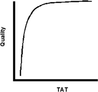
Quality results are accurate, and repeated measurements of the same sample demonstrate reproducibility (e.g., high precision; Equation 2). The central laboratory's ability to provide both quality results and a short TAT are often at odds with one another; more accurate and precise complex assays are usually more time-consuming, and such tests may not be available on POCT devices. Figure 30.1 depicts a theoretical curve for the relationship of result quality (y axis) and TAT (x axis). If assay time is reduced below a certain limit, the quality of the assay will be reduced. On the other hand, significant delays in making critical clinical decisions can adversely affect patient outcome. We must also acknowledge that POC tests rarely, if ever, will be as accurate or precise as tests accomplished in the central laboratory.
|
|
|
Figure 30.1. Relationship of quality to TAT. TAT, turnaround time. |
Equation 1:
Value of a test ≈ Quality of the result × Turnaround time-1
Note: Higher-quality results and lower turnaround times can provide higher-value tests.
Equation 2:
Quality of the result ≈ Bias-1 × Coefficient of variation-1
Note: Reduced bias (e.g., higher accuracy) and reduced coefficients of variation (e.g., higher precision) improve the quality of the test result.
Desirable intrinsic characteristics of the assay for the diagnosis, management, or prediction of disease are a high sensitivity and specificity. In epidemiologic terms, sensitivity is the number of true positive results divided by the number of observations in a diseased population.
Sensitivity = True Positives ÷ (True Positives + False Negatives)
Specificity is the number of true negative results divided by the number of observations undertaken in a nondiseased population.
Specificity = True Negatives ÷ (True Negatives + False Positives)
In its broadest sense, TAT is the “vein to brain” time: The time it takes between sample acquisition (e.g., venipuncture: the vein time) to result recognition by the treating physician (e.g., the brain time). Usually TAT is defined as the duration of time between sample acquisition and result reporting. Unfortunately, the laboratory often has little control over factors that determine when a sample is delivered to the central laboratory after acquisition. Similarly, preanalytical problems frequently develop because the sample is not properly drawn, labeled, or preserved prior to delivery to the lab. To be of value, the correct sample must be drawn from the correct patient at the correct time in the correct volume and placed in the correct tube.
If the analysis produces the most accurate result possible, but the TAT is unacceptably long, the value of the result in patient management is significantly degraded. TAT is most important in the ICU setting when the test results are used to immediately alter the patient's care. Examples of such tests include arterial blood gases for ventilator patients and glucose measurements in tight glycemic control protocols. There are many instances, however, where a TAT of several hours or more may be appropriate when the test is not used for immediate patient management (e.g., a karyotype result in a patient with suspected Down syndrome). Thus, the required TAT for any test result is relative. On the other hand, an instantaneous result that is not sufficiently accurate will not help—and may even hurt—the patient. It is wise to remember that bad data are worse than no data at all; physicians using bad data are misled. While the patient's true status is not the status reflected by the test result, recognizing that the patient lacks an important test result may stimulate the physician to repeat the test or undertake another method of patient assessment.
Assay Performance: Precision
Precision is synonymous with reproducibility; for example, if aliquots of the original sample are retested, will the same result as the original result be observed (5)? Precision can be defined in terms of the assay's standard deviation (SD) and coefficient of variation (CV). When aliquots of a single sample are measured repeatedly, the histographic distribution of results will represent a bell-shaped curve. Other descriptions for such a distribution include a Gaussian distribution or parametric distribution.
The SD for an assay is the square root of the variance. The variance is calculated as follows: The difference between each individual value and the mean is squared, these values are summed, and the sum of the squares is then divided by the number of repeats minus one. Sixty-eight percent of the repeats will fall within ±1 SD of the mean. Approximately 95% of the repeats will fall within ±2 SD of the mean, and approximately 99% of the repeats will fall within ±3 SD of the mean. This concept will be used in developing rules that will help us determine when an analysis and analyzer are or are not working properly.
CV is expressed as a percent: The SD is divided by the mean multiplied by 100. While SDs have values with units—mg/dL for glucose or mm Hg for pO2—and are difficult to remember, CVs are unitless and allow easy comparisons among various analyses without needing to recall the specific SD or units. For example, electrolyte measurements using ion-selective electrodes usually display CVs of 1% to 2%. In comparison, analyses that use chemical reactions with spectrophotometric or electrical detection typically have CVs of 4% to 5%. As a consequence of their complex nature involving antigen–antibody interactions, immunoassays can show even greater variability, with CVs of 5% to 10%.
Precision can be further described as intra-assay or interassay reproducibility. Intra-assay precision is assessed when the same sample is run 10, 20, or more times in a single run. A “run” is the series of same analyses that are accomplished in a single day, shift, or other period of time during which the analyzer is believed to be analytically stable (e.g., does not require recalibration; many modern analyses are so stable that calibration may not be required for many days or longer). Intra-assay comparisons would not exceed 1 day.
Intra-assay precision is almost always superior to interassay precision. Interassay precision is determined by measuring the same sample serially on different days (e.g., measuring the same sample once per day for 20 or more workdays in a row). For a typical chemical analysis, the intra-assay CV might be 5% and the interassay CV might be 7%. Clinicians do need to know the total imprecision—the combined intra-assay (e.g., same-day or same-shift reproducibility) and interassay (e.g., reproducibility over several days) imprecision—because some patients may be, for example, on ventilatory support for days or weeks with various degrees of pulmonary failure. While CVs and SDs cannot be added together to determine total imprecision, the intra-assay and interassay variances can be added together. The square root of the total variance then provides the SD, and the SD divided by the sample mean (multiplied by 100) provides the percent CV.
Assay Performance: Accuracy
Accuracy is a measure of bias. Bias is the difference between the “real” (or “true”) result and the measured result; bias can be positive or negative. A positive bias is present when the measured result exceeds the true result. A negative bias is found when the measured result is less than the true result. Bias must not be excessive; the bias that does exist must not lead to incorrect diagnosis, management, or disease prediction.
The true result of an assay may be difficult to define or determine. This is especially true when there is only one basic method available for the measurement of an analyte. For many measurements, the only method of analysis is the field method (i.e., the analytical procedure that is used in the central laboratories or at POC). For example, PO2 can only be measured using an oxygen-sensitive electrode. Reference methods, by definition, are more specific for the measurement of the analyte in question than the field method. Definitive methods are the best available methods of measurement with the highest specificity. Ideally, reference and definitive methods also have better precision than field methods. A review of cholesterol measurements can be instructive in comparing field, reference, and definitive methods.
The field method for cholesterol measurements is a spectrophotometric enzymatic assessment using cholesterol esterase and cholesterol oxidase. The definitive cholesterol measurement involves direct acidic oxidation of cholesterol with spectroscopic assessment of the oxidation products. The definitive measurement is isotopic dilution mass spectroscopy. Certainly, isotopic dilution mass spectroscopy will never be carried out in central laboratories or at the POC. Even acid oxidation of cholesterol will not be carried out in central laboratories or at the POC because concentrated acids are highly toxic and dangerous to work with, and such methods are not automated. However, the definitive method can be used to create standards that can be used as calibrators for reference methods. The reference methods are then employed to develop calibrators for the field methods.
Because reference intervals (i.e., the “normal” ranges) are based on such calibrators, if there is a significant bias in calibration between the method used to establish the reference interval and the method in real-time use in the care of the patient, errors may be made in the interpretation of the result as to whether or not it falls within the reference interval, and to what degree the result may exceed or fall below the reference interval. On the other hand, relative change (i.e., the present result compared to a previous result) will not be affected by bias if instrument calibration is stable and the assay is precise. However, a lack of precision can have a major misleading effect on the interpretation of serial results. A lack of precision (i.e., imprecision) requires that larger absolute differences occur between serial measurements to ensure that any difference occurring is an actual difference in the patient, rather than a variation (e.g., imprecision) in the assay. With a highly precise assay, small serial differences are more likely to represent a true difference in the patient's condition. With a highly imprecise assay, larger serial differences are required to indicate a true difference in the patient's condition. To further complicate the consideration of a normal versus an abnormal result, we must consider biologic variation: The normal variation in a biologic measurement that can represent minute-to-minute or hour-to-hour fluctuations: Ultradian rhythms (e.g., luteinizing hormone [LH] or follicle-stimulating hormone [FSH] secretion); daily variations: Circadian rhythms (e.g., a.m. vs. p.m. levels of cortisol); or variations greater than a day: Infradian rhythm (e.g., the menstrual period).
Analytical Sensitivity and Specificity
In analytical terms, sensitivity is the lowest concentration of an analyte that can reliably be measured. As measurements approach zero concentration of the analyte, the uncertainty of the measurement increases. At a certain point with a progressive decline in analyte concentration, the uncertainty of the measurement is so great that to report a lower number becomes meaningless. Analyzer manufacturers should define their lower limit of detection (LLD) to inform the user of the analyzer's expected analytical sensitivity. In addition, it is routine policy for laboratories to define their own LLD or, at a minimum, to confirm the manufacturer's stated LLD. In the ICU setting, LLD is most probably important in the measurement of glucose: “How low a glucose concentration can our POCT analyzer reliably report?” This must be carefully defined.
Analytical specificity is the certainty that the assay only measures the analyte of interest and does not measure other unintended substances in solution (e.g., “What is the assay's cross-reactivity to other analytes?”). Cross-reactivity is not usually an issue for POCT in ICUs based on the types of assays run in such situations. However, in the central laboratory, cross-reactivity can be a significant issue. For example, cardiac troponin-T or troponin-I measurements should not cross-react with skeletal muscle troponin-T or troponin-I. On the other hand, assay cross-reactivity is desirable if one wishes to test for a class of drugs (e.g., drug abuse testing for benzodiazepines, opiates, sympathomimetics, or barbiturates).
Quality Control Testing
For all inpatient testing, whether waived-regulated testing, moderate complexity testing, or high-complexity testing (see below), quality control must be assessed at least daily for all analytes measured on the device. For certain types of testing, such as radioimmunoassays or enzyme-linked immunosorbent assays (ELISAs), control testing may need to be performed with each run of patient samples.
To perform quality control testing, a sample of known concentration is measured with the device in question (5). This is the “control material” or, simply, the “control.” The control material is usually available in a large volume and is prepared in many aliquots (e.g., greater than 100) in a stable (e.g., frozen) form, so that the control material can be used over the course of many months to even longer than 1 year.
If the control result for a run of samples falls within previously defined limits, the device is said to be “in control,” and patient results can be reported. If the control result is outside defined limits, the device is said to be “out of control,” meaning an analytical error has occurred and patient results cannot be reported. Another way to express an out-of-control run is to state that the run was rejected. Thus, before any patient results can be reported, the operator must ensure that the analyzer is functioning correctly. Clearly, the control material must be measured prior to the release of any patient results. Many times, the control is placed at the beginning and at the end of the run. For moderate- and high-complexity testing, at least two levels of control are usually assessed. For example, the mean value of one control can be near a clinical decision point, while the mean value of the other control value can be considerably above the clinical decision point.
If the assay is out of control, the operator must troubleshoot the problem. Possible causes of out-of-control runs include:
· Machine mechanical errors (e.g., pipetting too little or too much liquid)
· Outdated reagents
· Reagents that have lost potency due to heating or lack of refrigeration
· Degraded control materials
· Operator error (e.g., mislabeled or switched controls, as in reversing the low-level and high-level controls)
· Spectrophotometric error (e.g., bulb loss or degraded function)
· Detector error
Fortunately, most POCT devices, even if moderately complex, are self-contained, are fairly robust, and can be simply “fixed” by replacing the reagent cartridge.
James Westgard created a series of rules that can be used to determine if a run or device is in control or is out of control (5). These “Westgard rules,” or their variations, are used essentially universally throughout the laboratory community. For each control material, the performance of the material is initially established by running this sample daily over the course of 20 to 30 days when the assay is otherwise known to be in control by using previously characterized control materials. From these data, the mean and the SD for the sample's measurement—the performance—on the device in question can be calculated.
Once the performance of the control material is known (i.e., its mean value and SD are established), this material can then be used to determine if subsequent runs are in control. If a single control value is 3 or fewer SDs away from the mean, the assay is in control and the results can be released. While, strictly speaking, being in control—a control result of more than +2 to +3 SDs above or less than -2 to -3 SDs below the mean—is a warning, the operator should review previous control data and confirm that other instrument parameters are functioning normally.
If the control result exceeds the mean value ±3 SDs, this is such an unlikely event (e.g., this should occur at random no more than ~1% of the time) as to suggest that the run is out of control. A single out-of-control run represents the consequence of a random error. On the other hand, if in two sequential runs a control displays a warning result each time, the second run is out of control. This is the 22S rule1and demonstrates a probable systemic (i.e., nonrandom) error. The Westgard quality control errors are summarized in Table 30.3. Systematic errors reflect recurrent errors such as short sampling and a degraded reagent, a constant interference, or loss of calibration.
For many POCT assays, the mechanics of the measuring device (e.g., electrodes) are designed into a single-use, disposable cartridge. In such cases, individual cartridges cannot be quality controlled, as measurement of a control material in the cartridge expends the cartridge. However, when such cartridges are manufactured using highly automated and monitored systems, the reproducibility of the manufacturing process can be so highly regulated that minimal variation exists among cartridges within a single manufacturing run, batch, or lot. While individual cartridges cannot be tested for quality control, the batch of cartridges can be assessed upon receipt by the health care institution by measuring a control material in one or more cartridges chosen at random from the batch received. Devices that use disposable cartridges can have their electronics or optics checked daily or more often via electronic quality control. In electronic quality control, a cartridge simulator is placed into the instrument to test if the instrument reports the proper result as defined for the simulator.
|
Table 30.3 Westgard quality control rules for a single level of control |
||||||||||||||
|
Pearls
· The take-home message for the intensivist and patient care staff is that POCT must be quality controlled to ensure a high reliability of test results.
Regulatory Issues in Point-of-care Testing
Laboratory testing, both at the POC and in satellite or central laboratories, is highly regulated by the Clinical Laboratory Improvement Amendments (CLIA) passed by the U.S. Congress in 1988. The shorthand term for the subsequent regulations is “CLIA 88” or, more simply, “CLIA.” Laboratories that perform ex vivo tests on any human tissue or body fluid must be certified by the Secretary of Health and Human Services (HHS).
Analyses where a biologic sample is not intentionally removed from the body does not fall under CLIA regulations. These types of analyses reflect monitoring and not testing according to CLIA. Examples of such analyses include measurement of the partial pressure of exhaled carbon dioxide, alcohol breathalyzers, exhalation of 13CO2 after oral administration of 13C-urea in search of Helicobacter pyloriinfection, transcutaneous bilirubinometers, pulse oximetry, and intermittent arterial sampling via indwelling canula for blood gases when the blood is returned to the patient's body. Incidentally, workplace drug abuse testing does not fall under the CLIA regulations.
The CLIA laboratory certification program is operated by the Centers for Medicare and Medicaid Services (CMS) (formerly the Health Care Financing Administration [HCFA]), the Food and Drug Administration (FDA), and the Centers for Disease Control and Prevention (CDC). Specific information on CLIA can be found at http://www.fda.gov/cdrh/clia (the FDA CLIA website that addresses complexity test categorizations and waivers); http://www.cms.hhs.gov/clia (the CMS CLIA website concerning program information, statistics, etc.); and http://www.phppo.cdc.gov/clia/ (the CDC CLIA website regarding regulations).
Currently the FDA determines whether an in vitro diagnostic test (including the test system) is waived or not waived (e.g., nonwaived test). Nonwaived tests are further classified as moderate complexity or high complexity. Therefore, there are three major CLIA regulatory categories: Waived testing, moderate-complexity testing, and high-complexity testing. Strictly speaking, the location of testing—POC versus satellite or central laboratory—does not define the complexity of the testing. Moderate-complexity testing can be performed immediately adjacent to the POC in a satellite laboratory, while, alternatively, a waived test (e.g., BNP, BioSite Incorporated, San Diego, CA) can be performed in a central laboratory.
Waived Testing
Waived tests are defined as determinations that can be performed at any site by any operator (including the patients themselves) following the manufacturer's recommendations. Theoretically, a waived test is a test that is so simple to perform that it is believed to carry little risk of error. CLIA describes waived tests as “simple procedures with little chance of negative outcomes if performed inaccurately.” However in the real world, experience teaches us that even waived tests can be performed improperly. Furthermore, erroneous results from certain waived tests in various situations can undoubtedly lead to potentially serious or fatal adverse outcomes (e.g., underestimation or overestimation of the PT-INR in patients being treated with warfarin for anticoagulation).
Waived testing that a person performs on him- or herself is not directly regulated by CLIA. However, whenever a health care provider performs a waived test on a patient, the testing is regulated. This is a common situation in ICUs when, for example, blood glucose testing is carried out by nursing personnel. Thus, CLIA has implications for waived testing in inpatient settings.
According to CLIA, hospitals must develop procedures and policies that specify the circumstances in which waived-test results are employed in patient management, services, and treatment. To achieve this, waived-test results must be placed in the clinical record, along with the appropriate reference interval (i.e., the normal range). The need for confirmatory testing must be defined; for example, if the POCT blood glucose is less than 60 mg/dL or greater than 500 mg/dL, a blood sample is sent to the central laboratory for confirmation. Finally, the test usage must be consistent with other hospital policies and the manufacturer's recommendations.
For inpatient waived testing to be performed properly, CLIA mandates that the staff executing the test must be identified, supervised, and qualified to perform the test. This requires adequate specific training and orientation to test performance and documentation of a satisfactory level of competence. This applies to all health care providers, including physicians. Competency must be demonstrated at the time of orientation training and yearly thereafter. Determination of competency must include at least two of the following four assessments:
1. Performing a test on an unknown specimen
2. Observation by a supervisor or qualified delegate
3. Monitoring the user's quality control performance
4. Written testing relevant to the waived-test method
Other CLIA standards for inpatient waived testing include that written policies and procedures are readily available and kept up to date; quality control checks are defined and conducted on each procedure; and the quality control results are recorded and maintained for review.
Blood glucose testing at the POC is an example of a waived and regulated test. Other examples of waived tests are given in Table 30.4. Presently, there are at least 40 waived tests; the current list of waived tests and devices can be found at http://www.accessdata.fda.gov/scripts/cdrh/cfdocs/cfClia/testswaived.cfm.
|
Table 30.4 Examples of waived tests |
||
|
It is important to recognize that CLIA approval of a test as being waived is device-specific: The test and the device are together approved as being waived for a specific analysis. For example, just because one manufacturer's test for blood glucose is waived does not mean that all single-use strip measurements of glucose by all manufacturers are waived. In the outpatient setting, the most commonly performed waived tests include urine pregnancy tests, blood glucose measurements, urine dipstick/tablet chemistries, ovulation tests, and fecal occult blood tests.
A laboratory or health care unit performing only waived tests must obtain a certificate of waiver (COW). COW laboratories are required to follow manufacturers' test instructions, participate in the CLIA program, and pay applicable biennial certificate fees. This is relevant outside the formal boundaries of the hospital and ICU setting.
Moderate- and High-Complexity Testing
CLIA defines moderate-complexity tests as being more intricate than waived tests. Moderate-complexity testing is typically carried out on automated analyzers. Examples of such tests include blood counts and routine chemistries. High-complexity tests are still more complicated, usually involving nonautomated or complicated analyses requiring considerable technologist or laboratory professional judgment, such as cross-matching of blood or microbiology testing.
Seven categories are considered when classifying the complexity of a nonwaived test:
1. Required operator knowledge
2. Operator training and experience
3. Preparation of reagents and materials
4. Characteristics of the operational steps
5. Calibration, quality control, and proficiency testing
6. Test system troubleshooting and equipment maintenance
7. Test result interpretation and judgment
For each category, the complexity of the test is scored: A score of 1 indicates the lowest level of complexity, and a score of 3 indicates the highest level. A score of 2 indicates complexity intermediate between 1 and 3. If the total score for the seven criteria is 12 or less, the test/device system is categorized as moderate complexity, whereas those test/device systems receiving scores above 12 are codified as high complexity.
Moderate- and high-complexity tests require quality control, proficiency testing, a quality assurance program, and so forth. There are also specific and detailed requirements regarding personnel qualifications and, as an aside, we point out that the experienced laboratorian recognizes that their most important resource is a highly skilled staff. Excluding provider-performed microscopy (PPM), which is a subset of moderate-complexity testing, all nonwaived tests are generally the purview of the pathologist and the clinical laboratory, or supervised by the pathologist and the clinical laboratory. Similar to waived, but regulated, POC tests, all moderate-complexity tests performed at the POC are regulated and, at a minimum, require similar training, supervision, quality control, and proficiency testing as waived inpatient tests.
Many POC testing devices applicable to ICUs are of moderate complexity. Some POCT devices applicable to ICUs perform only blood gas measurements (pH, pO2, pCO2), while the option to perform co-oximetry may also be available. Other devices will measure Na+, K+, glucose, ionized Ca2+, and/or lactate, in addition to blood gases. At least one device on the market has the capacity to measure cardiac markers and PT-INR in addition to the above parameters. Some devices can perform a wide battery of non–blood gas analyses and may not even be of moderate complexity (e.g., the Abraxis Piccolo POC Chemistry Analyzer [Abraxis North America, Union City, CA] is CLIA waived and regulated).
While not attempting to provide an exhaustive list of all available blood gas analyzers, robust blood gas analyzers are available from a variety of manufacturers, including the following:
· Abbott Point of Care Inc., East Windsor, NJ: iStat
· Bayer Diagnostics, MA: Rapidpoint 400
· ITC, Edison, NJ: IRMA TRUpoint Blood Gas Analysis System
· Nova Biomedical, Waltham, MA: Stat Profile CCX, pHOx and pHOx Plus
· Radiometer America Inc., Westlake, OH: Radiometer ABL80 FLEX, ABL77, and NPT7
Some of these devices are completely mobile and hand-held (e.g., iStat), while other analyzers can be pole-mounted (Radiometer ABL80 FLEX) or only require a small amount of bench space (e.g., they have a small “footprint”) (Radiometer NPT7).
Pearls
· The “take-home” message here is that intensivists and patient care staff must ensure that all regulatory rules are followed and enforced. This provides the best environment possible for the provision of accurate and precise laboratory test results.
Further Considerations in Point-of-care Testing in the Intensive Care Unit
Intensivists should work closely with their hospital's clinical pathologists and POCT coordinators in determining what type of acute testing should be available in their ICU. With the emergence of tight glycemic control, blood glucose testing must be available at the POC in ICUs. Such testing should be robust, accurate, and precise, and suffer from few, if any, critical interferences. These characteristics must also be sought in any POCT device that is brought into the ICU.
The need for POCT for blood gases and other tests depends on the ease of sample delivery to a satellite or central hospital laboratory and result TAT. In our satellite “STAT” laboratory located immediately outside the operating room complex and adjacent to many of the ICUs, 90% of blood gas results are reported in 7 minutes or less.
If blood gas analyses are performed in the ICU, unless personnel are added to the ICU staff, the work load is transferred, to some extent, to the ICU nursing or respiratory care staffs from the laboratory staff. However, there may be time savings in the ICU if a sample does not need to be prepared for transit to the satellite or central laboratory, and it is this immediacy of the test result that may improve patient care. Nevertheless, it is very difficult to find evidence-based medicine studies that clearly demonstrate better patient outcomes that result from reduced laboratory turnaround times. Even if patient care is not markedly improved, reduced TATs may aid in transferring patients to the floor or home more quickly. Patients may be more rapidly weaned from ventilation, which may decrease the use of resources. After improving patient outcomes, the next most important outcome variable for most hospital administrators is the expense of care and the need to reduce those costs (6).
If the decision has been made to proceed with POCT in the ICU, the analyzer and the support system must be carefully chosen. Ideally, the POCT device should have the ability to easily interface with the laboratory information system (LIS) to enable laboratory data transfer, billing, quality control data management, and tracking of operator competency (7). The nursing staff or respiratory care staff that will perform the testing should have a voice in the analyzer choice. The device's reproducibility must be examined (i.e., the precision of the device) (5), and the device results should be correlated with those of the central laboratory in search of biases (i.e., the accuracy of the device) (5).
Just as important as the analyzer is the quality of the blood sample that will be used for testing. For example, blood for a glucose measurement that is drawn through a line though which glucose has been infused may give falsely elevated values unless a sufficient “blank” sample is drawn through the line—in other words, “clearing the line” beforehand. Recall that D5 has a glucose concentration of 500 mg/dL (five times greater than normal) and D10 has a glucose concentration of 1,000 mg/dL (ten times greater than normal). Another example of such a preanalytical error is the exposure of blood to room air when blood gas testing is warranted. Blood samples exposed to room air can exhibit an increased PO2, decreased PCO2, and increased pH. When POCT devices that require cartridges are used, proper filling of the cartridge is essential to obtain a valid result. Wasting cartridges is expensive; in some systems, individual cartridges may cost several dollars, as opposed to pennies per test in a central laboratory.
Analytical interferences must be considered in the choice of ICU POCT instruments. An interference is a characteristic of a patient sample that may bias the result. Examples of interferences affecting certain central laboratory tests include hyperlipidemia, hyperbilirubinemia, and hemolysis. At the POC, blood glucose testing devices that use glucose dehydrogenase and the PQQ reagent (pyrroloquinolinequinone) display positive interferences when maltose is present in the patient's bloodstream. Maltose is used as a stabilizer in drugs such as intravenous immunoglobulin, and icodextrin used in dialysis is metabolized to maltose. This positive interference can lead to an overestimate of the blood glucose and subsequent overtreatment of “hyperglycemia,” with severe or even fatal hypoglycemia as the consequence. Glucose oxidase devices that use the patient's blood as a source of oxygen exhibit negative biases in the glucose measurement in cases of hypoxia or where the elevation is over approximately 5,000 feet. On the other hand, glucose oxidase devices that use ferricine or ferricyanide display positive biases in blood glucose when the patient is hypoxic and negative biases when the patient is hyperoxic, such as a ventilated patient receiving supplemental oxygen.
Quality control for the POCT must be carried out and monitored as part of an overall quality assurance program. All device operators will require initial training and competency testing. Cost per test must also be a consideration. POCT can be ten or more times as expensive as central laboratory testing. In considering any POCT in the ICU, the pathologist and POCT coordinator must be involved in this process from the start, as this fosters a collegial relationship. In the end, the ultimate goal is to provide excellent patient care. New laboratory tests will continue to appear that will influence ICU care such as test panels for stroke and sepsis and improved cardiac risk panels. Prudent review of the medical literature and cooperation between the ICU staff and the laboratory staff will help determine where such testing is best carried out.
References
1. Nichols JH. Point-of-care testing. In: Nichols JH, ed. Point-of-Care Testing, Performance Improvement and Evidence-Based Outcomes. New York: Marcel Dekker, Inc.; 2003:1.
2. Winter WE. Point-of-care testing in the management of diabetes mellitus. In: Nichols JH, ed. Point-of-Care Testing, Performance Improvement and Evidence-Based Outcomes. New York: Marcel Dekker, Inc.; 2003:235.
3. Nuttall GA, Santrach P. Hemoglobin and coagulation. In: Nichols JH, ed. Point-of-Care Testing, Performance Improvement and Evidence-Based Outcomes. New York: Marcel Dekker, Inc.; 2003:353.
4. Christenson RH, Azzazy El-Badaway HME. Point-of-care testing for biochemical markers of acute coronary syndromes. In: Nichols JH, ed. Point-of-Care Testing, Performance Improvement and Evidence-Based Outcomes. New York: Marcel Dekker, Inc.; 2003:379.
5. Westgard JO, Klee GG. Quality management. In: Burtis CA, Ashwood ER, Bruns DE, eds. Tietz Textbook of Clinical Chemistry and Molecular Diagnostics. St. Louis: Elsevier Saunders; 2006:485.
6. Schallom L. Point of care testing in critical care. Crit Care Nurs Clin North Am. 1999;11:99.
7. Halpern NA. Point of care diagnostics and networks. Crit Care Clin. 2000;16:623.