
 UISS-UCLA INTERNATIONAL KIDNEY CANCER STAGING SYSTEM
DESCRIPTION A prognostic system for renal cell carcinima (RCC) to differentiate survival; the system integrates the 3 most commonly used prognostic factors: Cancer TNM stage, Furman grade, and patient performance status. Patients are categorized after nephrectomy into 3 risk groups: Low-, intermediate-, and high-risk for localized and metastatic disease.
REFERENCE
Escudier B, Eisen T, Porta C, et al. Renal cell carcinoma: ESMO Clinical Practice Guidelines for diagnosis, treatment and follow-up. Ann Oncol. 2012;23(Suppl 7):vii65-vii71.
UNDERVIRILIZED MALE SYNDROME (MILD ANDROGEN INSENSITIVITY)
DESCRIPTION A disorder of androgen receptor function caused by androgen receptor gene mutation. Patients with androgen receptor mutations have a 46, XY karyotype and present with a spectrum of phenotypes from complete external feminization, to ambiguous genitalia, to phenotypically infertile male, which is also known as undervirilized male syndrome. These patients present with gynecomastia at puberty, and may have scarce body hair, small penis, and complaints of impotence. Spermatogenesis may or may not be impaired. Patients may have elevated luteinizing hormone, normal to slightly elevated testosterone, and high estradiol. Treatment may not be necessary, however, breast reduction surgery at puberty is sometimes necessary. Infertile men may benefit from assisted reproductive techniques (ART’s). (See also Section II: “Androgen Insensitivity Syndrome [AIS or Androgen Resistance Syndrome], Complete and Partial.”)
REFERENCE
Gottlieb B, Lombroso R, Beitel LK, et al. Molecular pathology of the androgen receptor in male (in)fertility. Reprod Biomed Online. 2005:42–48.
UNINHIBITED DETRUSOR CONTRACTION
DESCRIPTION Uninhibited detrusor contraction leads to an overactive bladder. Bladder overactivity can result from damage to central inhibitory pathways, sensitization of peripheral afferent terminals in the bladder that unmask primitive voiding reflexes, or changes in bladder smooth muscle cells. Cystometry is essential if a definitive diagnosis is required. (See Section I: “Overactive Bladder.”)
SYNONYMS
• Overactive bladder
• Unstable bladder
TREATMENT
• Traditionally centers on the use of anticholinergic medications (oxybutynin, tolterodine, others)
• Estrogens may help in the postmenopausal woman.
REFERENCE
Anonymous. The overactive bladder: From basic science to clinical management consensus conference. Urology. 1997;50(Suppl 6A):1–114.
URACHAL ABNORMALITIES
DESCRIPTION The urachus is a tubular connection between the allantoic stalk and the dome of the bladder. Faulty embryologic resolution of this connection results in urachal abnormalities. Microscopic urachal remnants are common, appearing in 3% of autopsy specimens, and are almost always asymptomatic. Except for the asymptomatic urachal diverticulum, the treatment of all urachal abnormalities is surgical; ie, complete excision of the abnormal structure, including a cuff of bladder. Congenital urachal abnormalities can be divided into 4 types:
• Urachal sinus: The most common urachal abnormality. The urachal sinus arises from a persistent patent urachus that drains to the umbilicus; may present with wetness, purulence, or malodorous discharge.
• Urachal cyst: Persistence of part of this channel between the bladder and umbilicus, lacking communication to either structure. The 2nd most common urachal anomaly. Most commonly presents in an older child with signs of suppuration (Latin “calor, rubor, dolor”) in the lower abdominal wall. Occasionally, a urachal cyst will present as an asymptomatic midline lower abdominal mass or tenderness.
• Patent urachus: Persistence of the urachal channel between the bladder and umbilicus; an uncommon type of urachal abnormality.
• Urachal diverticulum of the bladder: May result from drainage of a urachal cyst to the bladder; presents with UTI
Urachal carcinoma is a malignant adenocarcinoma that presents later in life. (See also Section I: “Urachal Carcinoma” and “Umbilical Abnormalities, Urologic Considerations” and (Image  ).)
).)
REFERENCE
Mesrobian H, Zacharias A, Balcom AH, et al. Ten years of experience with isolated urachal anomalies in children. J Urol. 1997;158:1316–1318.
URACHAL CARCINOMA STAGING SYSTEMS
DEFINITION
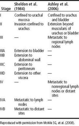
Two staging systems have been reported by Sheldon and Ashley. No formal TNM classification exists specifically for urachal carcinoma. (See also Section I: “Urachal carcinoma.”)
REFERENCE
Mohile SG, Schleicher L, Petrylak DP. Treatment of metastatic urachal carcinoma in an elderly woman. Nat Clin Pract Oncol. 2008;5:55–58.
URATE, DIETARY
DESCRIPTION Foods rich in urate should be restricted in patients with hyperuricosuric calcium oxalate stone disease and in patients with uric acid stone disease. The intake of urate should not exceed 500 mg/day. Urate rich foods include:
• Calf thymus, 900 mg urate/100 g
• Liver, 260–360 mg urate/100 g
• Kidneys, 210–255 mg urate/100 g
• Poultry skin, 300 mg urate/100 g
• Herring with skin, sardines, anchovies, sprats, 260–500 mg urate/100 g.
REFERENCE
Tiselius HG, et al. EAU Guidelines on Urolithiasis. http://www.uroweb.org/fileadmin/user_upload/Guidelines/Urolithiasis.pdf, Accessed April 6, 2014.
UREAPLASMA UREALYTICUM
DESCRIPTION Common bacterial inhabitant of the lower GU tract in adult men and women who are sexually active. As an STI/STD it can also be transmitted venereally and from mother to offspring. It is the most common cause of nongonococcal and nonchlamydial urethritis, and can cause chorioamnionitis, pyelonephritis, and septic arthritis. It is implicated in chronic prostatitis in men and urgency–frequency symptoms in women and in HIV-related acute epididymitis. It is also sometimes associated with decreased fertility. Diagnosis is by culture, but specific media and growth conditions are necessary. Treatment is doxycycline 100 mg/d for 2 wk or a single dose of azithromycin 1 g PO. (See also Section I: “Sexually Transmitted Infections [STIs] (Sexually Transmitted Diseases [STDs]), General.”)
REFERENCE
Frenkl TL. Sexually transmitted infections. Urol Clin N Am. 2008;35(1):33–46.
URETER, AGENESIS/ATRESIA
DESCRIPTION Bilateral ureteral agenesis is incompatible with life. Unilateral ureteral agenesis indicates failure of ureteral bud development and is often accompanied by ipsilateral renal agenesis or multicystic kidney. Ureteral atresia is caused by varying degrees of failure in ureteral bud development. When either atresia or agenesis is unilateral, it is usually asymptomatic and of no clinical significance. However, it can be associated with infection (UTI) on occasion.
REFERENCE
Morozumi M, Ogawa Y, Fujime M, et al. Distal ureteral atresia associated with crossed renal ectopia with fusion: Recovery of renal function after release of a 10 yr ureteral obstruction. Int J Urol. 1997;4(5):512–515.
URETER, DEVIATION
DEFINITION
The ureter is a retroperitoneal structure in adults and is typically 25–30 cm in length. The ureters are in close proximity to abdominal and pelvic viscera, lymph nodes, and vessels. The course of the ureter can be altered by extrinsic pathologic processes as well as congenital and normal anatomic variation. Normal course of ureters: Proximally lateral to the lumbar vertebral pedicles and distally medial to pelvic brim in the true pelvis. Lateral ureteral deviation is more common than medial deviation.
• Medial deviation:
– Normal anatomic variant in up to 15%
– Retroperitoneal fibrosis; usually involving the lower lumbar and sacral regions
– Retrocaval ureter: “shepherd’s crook” configuration; the right ureter passes behind the IVC at approximately the level of L4 (most common location). The distal ureter lies medial to the dilated proximal ureteral segment
– Retroiliac ureter (very rare)
– Pelvic lipomatosis; associated with elevation and elongation of the bladder, rectum, and sigmoid and increased pelvic wall lucency
– After abdominal perineal resection
– Iliac lymphadenopathy
– Aneurysmal dilation of the iliac vessels
– Perianeurysmal fibrosis due to chronic leakage associated with aortoiliac aneurysms
– Lower pole renal mass; usually medial deviation of proximal ureter
• Lateral deviation
– Psoas muscle hypertrophy; more common in young men; lateral and/or anterior deviation of the middle 3rd of 1 or both ureters
– Retroperitoneal adenopathy (paracaval/paraaortic lymphadenopathy): Metastatic disease, lymphoma, testicular cancer
– Pelvic mass (fibroids, ovarian tumor and cysts)
– Aortic aneurysm
– Malrotated and horseshoe kidney
– Patent umbilical artery (children)
– Retroperitoneal tumors
– Neurogenic tumors
– Fluid collection (abscess, urinoma, lymphocele, hematoma)
REFERENCES
Bamberger MH. Ureteral Deviation: A sign of retroperitoneal anatomy and pathology. Available at http://www.medscape.com/viewarticle/410210_4, Accessed March 29, 2014.
Chapter 8.39 Deviated Ureters. In: Davies, SG ed. Chapman & Nakielny’s Aids to Radiological Differential Diagnosis. 6th ed. Philadelphia, PA: Saunders Elsevier; 2013.
URETER, DIVERTICULUM
DESCRIPTION Diverticula can be congenital or acquired, although most have been discovered in adults. Most diverticula are solitary outpouchings involving the distal ureters and upper portions of the pelvis. They are true diverticula composed of a muscular wall, which is lined by transitional cell epithelium. Renal pelvic diverticula tend to be larger than those found in the ureter. Diverticula may be associated with other pathology, such as Ask-Upmark kidney. The most common complications are infection and/or stone formation. Unlike diverticula found in the bladder and urethra, major complications and development of urothelial carcinoma (TCC) are rare (Image ).
REFERENCE
McLoughlin LC, Davis NF, Dowling C, et al. Ureteral diverticulum: A review of the current literature. Can J Urol. 2013;20(5):6893–6896.
URETER, DUPLICATED AND BIFID
DESCRIPTION Duplication of ureters is a common anomaly. Duplication may be either complete or incomplete. Complete duplication is most often associated with vesicoureteral reflux, ectopic ureteral insertion, and ectopic ureterocele, all of which are more commonly found in females than in males. Incomplete duplication is most often associated with UPJ obstruction of the lower pole of the kidney. Common clinical presentations include UTIs and urinary incontinence. Diagnosis is made usually in childhood by US, excretory urography, and voiding cystourethrography.
Also known as bifid ureter (if partial duplication) or double ureters (if complete duplication). Treatment is ureteroneocystostomy in the presence of persistent reflux or ureteropyelostomy if obstructed without reflux (Image ).
REFERENCE
Fernbach SK, Feinstein KA, Spencer K, et al. Ureteral duplication and its complications. Radiographics. 1997;17(1):109–127.
URETER, ECTOPIC (URETERAL ECTOPIA)
DESCRIPTION Ectopic ureters open in a position other than on the trigone and can be associated with both reflux and obstruction and with ureteroceles. They predispose to UTI and may cause hematuria or abdominal/flank pain. Up to 80% are associated with a duplicated collecting system in females and ∼20% are nonduplicated ureters usually in males with an absent hemi-trigone. In females, the ureter typically inserts in the urethra or vagina (distal to the sphincter) and cause incontinence or constant dribbling. In males the sites include the posterior urethra or seminal vesicles (no incontinence is seen) and often present later in life. Treatment is usually partial nephroure-terectomy of the nonfunctioning upper pole moiety with nephrectomy often necessary for single ureter systems.
REFERENCE
Hanson GR, Gatti JM, Gittes KG. Diagnosis of ectopic ureter as a cause of urinary incontinence. J Ped Urol. 2007;3(1):53–57.
URETER, FIBROEPITHELIAL POLYPS
DESCRIPTION Fibroepithelial polyps are rare benign neoplasms. The majority of these polyps are found at the UPJ. Signs and symptoms usually associated with ureteral obstruction include flank pain and hematuria. In addition, varying degrees of hydroureteronephrosis and ureteral intussusception have been described. Grossly, ureteral polyps are intraluminal lesions, most commonly covered with transitional epithelium. The bulk of the polyp is composed of vascularized collagenous fibrous tissue, with or without areas of chronic inflammation and edema. Ureteroscopy is often necessary to confirm the diagnosis.
SYNONYMS
• Fibromyxoma
• Myxoma
• Fibroma
• Vascular fibrous polyps
TREATMENT
• Ureteroscopic resection
• Open ureterotomy with polypectomy or partial ureterectomy is a viable conservative treatment option if the diagnosis can be confirmed preoperatively
• Many patients undergo nephroureterectomy for suspected malignancy
REFERENCE
Wang ZJ, et al. Ureteral Fibroepithelial Polyp. J Ultrasound Med. 2008;27(11):1647–1649.
URETER, FISH HOOK (REVERSE J)
DESCRIPTION A radiographic appearance of the type I (low-loop) circumcaval ureter, in which the dilated proximal part of the ureter takes a characteristic fish hook or reverse J course. Ureteral dilation usually ends 1–2 cm lateral to the inferior vena cava, where the ureter turns upward at the border of the psoas muscle.
REFERENCE
Kenawi MM, Williams DI. Circumcaval ureter: A report of four cases in children with a review of the literature and a new classification. Br J Urol. 1976;48:183.
URETER, HEMANGIOMA
DESCRIPTION Hemangiomas are benign ureteral tumors. They may be the most common cause of chronic unilateral hematuria. Symptomatology may include hematuria, pain, hydronephrosis, bladder irritations, and palpable tumor. Varicoceles have also been found, but less frequently. Like other ureteral tumors, hemangiomas usually cause incomplete obstruction and may eventually cause complete obstruction with dilation of the urinary tract. They present as red, slightly elevated structures, fairly diffusely, and demarcated from their surroundings. Urothelial malignancies must be excluded, especially in the elderly. Flexible ureteropyeloscopy is considered a good diagnostic and therapeutic option in selected patients with unilateral hematuria of uncertain etiology.
REFERENCE
Biyani CS, Mackay AM, Sissions G, et al. An unusual filling defect in the ureter. Urol Int. 1998;61(2):124–125.
URETER, J HOOKING
DESCRIPTION With progressive benign prostatic hypertrophy, elevation of the trigone occurs, resulting in a characteristic J hooking of the distal ureters. This is a reliable sign on IVP of significant prostatic hypertrophy.
REFERENCE
Amis ES, Newhouse JH, eds. Essentials of Uroradiology. 1st ed. Boston, MA: Little Brown, 1991:320.
URETER, LEIOMYOMA
DESCRIPTION Leiomyomas of the urinary tract are rare (neoplasms of mesenchymal origin comprise <3% of all primary ureteral tumors). These benign tumors are seen predominantly in the 4th–5th decades of life. The left ureter is more frequently affected. Immunohistochemical studies confirm the diagnosis. Conservative management (urethroscopic or partial ureterectomy) is the treatment of choice.
REFERENCE
Naruse K, Yamada Y, Aoki S, et al. A case of primary leiomyoma of the ureter. Int J Urol. 2007;14(3):248–250.
URETER, LEIOMYOSARCOMA
DESCRIPTION Leiomyosarcoma originating from the ureters is exceedingly rare, with only 13 reported cases of primary leiomyosarcoma of the ureter. It is a disease that is very difficult to diagnose, and furthermore, it has a poor 5-yr disease-specific survival. Patients present with flank pain, hematuria, and/or UTI. Radiographic exam includes IVP, retrograde pyelogram, and CT. Light microscopy immunohistochemical staining and electron microscopy should be used to confirm the diagnosis of leiomyosarcoma.
TREATMENT
• Tumor resection
• Possible nephroureterectomy, depending on tumor grade and stage
• Adjuvant radiation therapy may be helpful.
REFERENCE
Griffin JH, Waters WB. Primary leiomyosarcoma of the ureter. J Surg Oncol. 1996;62(2):148–152.
URETER, METASTASIS TO
DESCRIPTION Metastatic tumors of the ureter are uncommon, and 1 of the rarest causes of ureteric obstruction. Since their 1st mention in the literature in 1909 only ∼400 cases are reported. Among 1/2 of the cases, the breast or gastrointestinal tract (colorectum) is the site for primary cancer. Prostate cancer and uterine cervical cancer are responsible for 30–40% of cases, with stomach and lung cancer being reported in the remaining case. A predilection exists for the lower 1/3 of the ureter. Many are asymptomatic with the majority autopsy reports.
REFERENCE
Arvind NK, Singh O, Gupta S, et al. Ureteral Metastasis as the presenting manifestation of pancreatic carcinoma. Rev Urol. 2013;15(3):124–130.
URETER, NEPHROGENIC ADENOMA (NA)
DESCRIPTION Nephrogenic adenoma (NA) is a rare metaplastic lesion of the ureter and assumed to be secondary to chronic irritation of the urothelium. It is a benign papillary and tubular proliferation in response to trauma, infection, or ionizing radiation. Although it can be seen anywhere in the urinary tract, it is most commonly observed in the bladder (55%). The incidence in ureter is ∼4%. Biopsy and fulgeration are appropriate, as it is treated as a low-grade urothelial malignancy. Malignant transformation has not been described.
REFERENCE
Doluoglu OG, Acarer EY, Yavuz A, et al. Nephrogenic adenoma of the ureter. Rare Tumors. 2012;4(2):e28.
URETER, NEUROFIBROMA
DESCRIPTION Grossly, neurofibromas may be single or multiple and comprise different sized nodules. Histologically, they are composed of fascicles of elongated, spindle-shaped cells with thin, wavy nuclei in a collagenized background. Neurofibromas in the ureter are very rare but have an increased incidence in von Recklinghausen disease. Neurofibromas frequently recur and can cause death by urinary obstruction and renal failure. Endoscopic or open excision is the treatment of choice.
REFERENCE
Bostwick DG, ed. Urologic Surgical Pathology. 1st ed. St. Louis, MO: Mosby; 1997.
URETER, PIPE-STEM
DESCRIPTION A radiographic appearance seen in late stages of tuberculous involvement of the ureter. On IVP, the ureter appears straight with a narrow lumen, due to diffuse fibrotic changes of the wall.
REFERENCE
Murphy DM, Fallon B, Lane V, et al. Tuberculous stricture of ureter. Urology. 1982;20:382–384.
URETER, RADIATION INJURY TO
DESCRIPTION Clinically, radiation injury to the ureter will present as obstruction. Upper urinary tract obstruction secondary to the effects of radiation is generally reported to occur in ∼5% of patients with ureteral encroachment, and in <1% of all treated patients. The ureters are relatively resistant to the effects of radiation, although some factors are postulated to increase the chance of injury after radiation exposure, such as infection of the ureter, necrosis of the tumor invading the ureteral wall, and direct radiation injury to the ureteral wall. Radiation therapy of ≥8,000 Gy results in a 40% urologic complication rate. A dose of ≤6,000 Gy results in a <2% complication rate. (See also Section I: “Ureter, Obstruction.”)
REFERENCE
Resnick MI, Kursh ED. Extrinsic obstruction of the ureter. In: Walsh PC, Retik AB, Vaughan ED, et al., eds. Campbell’s Urology. 7th ed. Philadelphia, PA: Saunders; 1998:409–410.
URETER, RETROCAVAL (CIRCUMCAVAL, POSTCAVAL)
DESCRIPTION A retrocaval ureter is a congenital anomaly in which the problem arises from the inferior vena cava rather than the ureter. Normally, the vena cava derives from the supracardinal vein, which lies posterior to the ureter. If it derives from either the persistent right subcardinal or postcardinal vein, both of which lie anterior to the ureter, a portion of the lumbar ureter becomes trapped behind the vena cava. Clinically, a retrocaval ureter may present as ureteral obstruction with a Shepards crook deformity. Males are affected more than females and the presentation usually relates to the ureteral obstruction (pain infection, stones).
Two types have been described. Type 1 is the more common where the ureter crosses behind IVC at the level of 3rd lumbar vertebra assuming the appearance of a shephards crook or sickle shaped at the point of obstruction. Marked hydronephrosis usually present. Type 2 retrocaval ureter is less common and the ureter tends to cross at a much higher level relative to the renal pelvis and the degree of hydronephrosis is usually mild. Treatment is surgery with transection of the ureter and reanastomosis in front of the inferior vena cava. (See “Ureter, Shepherd’s Crook.”)
REFERENCE
Chung BI, Gill IS. Laparoscopic dismembered pyeloplasty of a retrocaval ureter: Case report and review of the literature. Eur Urol. 2008;54(6):1433–1436.
URETER, SHEPHERD’S CROOK
DESCRIPTION Characteristic S-shaped appearance of the circumcaval/retrocaval ureter on excretory urography or retrograde ureterography. A normal-caliber ureter emerging at the medial aspect of the inferior vena cava runs inferiorly between it and the aorta. The elongation and dilatation of ureter above the site of the obstruction along with the course around the inferior vena cava cause a “shepherd’s crook” or “sickle-shaped deformity.” On frontal projection, the ureter is medial to the lower lumbar pedicles, where it crosses anterior to the right iliac vessels to enter the pelvis. Clinically, a retrocaval ureter may present as ureteral obstruction with pain, stones, or infection. Males are affected more than females. Treatment is surgery with transection of the ureter and reanastomosis in front of the inferior vena cava (See also Section II: “Ureter, Retrocaval [Circumcaval, Postcaval].”)
REFERENCE
Kenawi MM, Williams DI. Circumcaval ureter: A report of four cases in children, with a review of the literature and a new classification. Br J Urol. 1976;48:183.
URETER, SPIRAL (CORKSCREW)
DEFINITION
A spirally twisted or “corkscrew” ureter is a very rare anomaly beyond the neonatal age. A spirally twisted ureter is not considered clinically significant, unless it causes obstruction and secondary hydronephrosis. Infants frequently have a “corkscrew” appearance of the proximal segment of the ureter seen on intravenous urography, but this has been considered an imaging finding of no postnatal clinical significance. It may represent persistence of normal fet al developmental structures, such as congenital folds. Corkscrew configuration of the ureter may also be the result of ureteric varicosities or extrinsic ureteric obstruction when seen later in life. Obstruction secondary to spiral deformity of the ureter may be due to involvement of the ureter by dense fibrous bands (Image ).
REFERENCE
Prodromos P, Payne D, Keeley F. Let’s get it straight: the story of the spiral ureter. Can J Urol. 2012;19:6118–6120.
URETER, STONE PASSAGE STATISTICS
DEFINITION
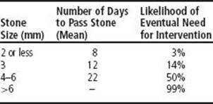
The ureter is the smallest diameter structure of the urinary tract. It is also the area most likely to be obstructed by a stone. Most stones <5 mm in diameter are likely to pass spontaneously with the likelihood of spontaneous stone passage decreasing with increased stone size. It is estimated that 2/3 of ureteral stones that pass spontaneously pass within 4 wk of the onset of symptoms.
REFERENCE
Kidney Stones. http://www.auanet.org/education/kidney-stones.cfm, Accessed April 5, 2014.
URETER, STRICTURE
DESCRIPTION Strictures are 1 of the main causes for ureteral obstruction that might lead to hydronephrosis and renal function impairment. Ureteral strictures may present with an insidious onset of irreversibly damaged renal parenchymal due to slow development of silent hydronephrosis. Common signs of symptomatic stricture are flank pain, elevated creatinine level, or decreased urine output. Imaging studies with contrast are an essential part of the diagnostic workup. The location and length of obstruction are important parameters for treatment planning. (See also Section I: “Ureter, Obstruction.” and (Image ))
CAUSES
• Congenital
• Extrinsic trauma
• Iatrogenic (gynecologic or other pelvic surgeries)
• Inflammatory and infectious factors (Crohn disease)
• Instrumentation (ureteroscopy)
• Malignant (intrinsic or extrinsic)
• Postchemotherapy and radiation therapy
• Sclerosing retroperitoneal fibrosis
TREATMENT
• Relieve the obstruction with concomitant antibiotic coverage, if needed.
• Surgical correction with endoscopic, laparoscopic, robotic, or open surgery.
• Reimplantation with excision for distal strictures.
REFERENCE
Ogan K, Abbott JT, Wilmot C, et al. Laparoscopic ureteral reimplant for distal ureteral strictures. JSLS. 2008;12(1):13–17.
URETER, VALVES
DESCRIPTION Ureteral valves obstruct the forward flow of fluid, causing a proximal hydronephrosis. About 35% of ureteral valves occur at the UPJ; 60% occur near the ureterovesical junction, and 5% in the upper ureter. Valves are 3 times more common in males. They are usually unilateral, although they may be bilateral, and, if so, usually lead to renal failure. 75% of valves occur on the left side. Treatment depends on the location of the valve and may include ureteral reimplantation (distal ureter), endoscopic ablation (midproximal ureter), or UPJ repair.
REFERENCE
Amis ES, Newhouse JH, eds. Essentials of Uroradiology. 1st ed. Boston, MA: Little Brown; 1991:63.
URETERAL JETS
DESCRIPTION Ureteral jets have been studied to diagnose urinary tract obstruction. Ureteral jets are visualized on color Doppler ultrasound as ureteral urine passes into the bladder. Sensitivity of 100% and specificity of 90.9% in detecting ureteral obstruction has been reported based on the absence of the ureteral jet on a given side. Patients must be well hydrated and often a prolonged imaging interval is needed to document the presence or absence of the ureteral jet (Image ).
REFERENCE
Deyoe LA, Cronan JJ, Breslaw BH, et al: New techniques of ultrasound and color doppler in the prospective evaluation of acute renal obstruction: Do they replace the intravenous urogram? Abdom Imaging. 1995;20:58–63.
URETERAL STRICTURE FOLLOWING URINARY DIVERSION
DESCRIPTION Ureteral strictures are seen in 2–10% of patients who receive a cystectomy and urinary diversion for bladder cancer. The stricture is most commonly located at the ureteroenteric anastomosis and more commonly involves the left ureter. Considered a late complication, strictures usually present in the 6–12 mo postoperative time period. Ureteral recurrence of urothelial carcinoma should be ruled out. Treatment includes percutaneous nephrostomy, indwelling ureteral stent, balloon dilatation, or reanastomosis.
REFERENCE
Tal R, Sivan B, Kedar D, et al. Management of benign ureteral strictures following radical cystectomy and urinary diversion for bladder cancer. J Urol. 2007;178:538–542.
URETERITIS
DESCRIPTION A generic characterization given to describe inflammation of the ureter which can be further qualified into subtype descriptions based loosely on its etiologic factors. For example, postoperative ureteritis, infective ureteritis, and noninfective ureteritis. Noninfective causes of ureteritis include ureteral amyloidosis, eosinophilic ureteritis, IgG-4 associated ureteral inflammation, and idiopathic segmental ureteritis.
REFERENCE
Atsuta T, Shimizu Y, Masuda N, et al. First report of idiopathic segmental ureteritis successfully treated by steroid therapy. Int J Urol. 2012;19:583–586.
URETERITIS CYSTICA
DESCRIPTION A benign and rare condition, ureteritis cystica is characterized by multiple cysts and space-filling defects in urothelium. Usually asymptomatic, it may present with hematuria, and if obstruction occurs, may lead to stone formation, UTIs, and renal compromise. The etiology is unknown, but associated with chronic UTI. Space-filling defects seen on retrograde pyelography or excretory urography may appears as smooth, round or oval filling defects of varying sizes that protrude into the lumen. It can mimic other conditions such as bladder cancer, blood clots, air bubbles, radiolucent stones, fibroepithelial polyps, and sloughed renal papillae. It manifests as cystic areas of glandular metaplasia associated with chronic urothelial inflammation; this is more commonly seen in the bladder, called cystitis cystica. Treatment is ureteroscopy and the mechanical disruption of cysts or instillation of chemicals such as silver nitrate to relieve obstruction.
REFERENCE
Rothschild JG, Wu G. Ureteritis Cystica: A Radiologic Pathologic Correlation. J Clin Imaging Sci. 2011;1:23.
URETERONEOCYSTOSTOMY, TECHNIQUES AND INDICATIONS
DESCRIPTION Ureteral reimplantation, or ureteroneocystostomy, is used when there is an abnormality at the ureterovesical junction. Abnormalities include an obstructing ureterocele, megaureter, ureteral reflux, distal ureteral stricture, or iatrogenic injuries. The surgical treatment options can be classified on the basis of the ureteral approach to the bladder as intravesical, extravesical, or combined or on the relationship of the submucosal tunnel to the site of the original ureteral hiatus as suprahiatal or infrahiatal.
REFERENCE
Khoury AE, Bägli DJ. Vesicoureteral reflux. In: Wein AJ, et al., eds. Campbell-Walsh Urology. 10th ed. Philadelphia, PA: Saunders; 2011.
URETHRA, ADENOCARCINOMA OF ACCESSORY GLANDS
DESCRIPTION In males, the urethral accessory glands can develop rare, aggressive neoplasms that are difficult to diagnose because of the local destructive nature of the lesions (Cowper and Littré glands). Cowper gland cancers are found in the bulbous urethra, while Littré gland lesions can arise along the entire urethra, but tend to arise distally. In females, the Skene glands can develop adenocarcinoma as well. Patients typically present with hematuria, dysuria, and progressive urinary obstruction. Management is similar to that for urethral adenocarcinoma. (See also Section II: “Cowper Gland Carcinoma”, “Skene (Paraurethral) Gland, Adenocarcinoma.”)
SYNONYMS
• Adenocarcinoma of Cowper gland
• Adenocarcinoma of Littré glands
• Adenocarcinoma of Skene (periurethral) glands
REFERENCE
Reuter VE. Urethra, Chapter 11. In: Bostwick DG, ed. Urologic Surgical Pathology. 2nd ed. Philadelphia, PA: Mosby Elsevier; 2008:608.
URETHRA, ADENOMATOUS POLYPS
DESCRIPTION Congenital, benign papillary-appearing lesions that occur most frequently in the prostatic urethra and contain benign prostatic epithelium. These have been reported in the anterior urethra. Cores of the papillary projections contain prostatic stroma and glands. The lesions typically present in the 1st decade of life, but can appear at any age. Hematuria, enuresis, and obstruction are common. Cystourethroscopy is usually diagnostic.
SYNONYMS
• Villous polyp of the urethra
• Ectopic prostatic tissue in the urethra
TREATMENT
Transurethral or suprapubic resection is curative.
REFERENCE
Chan JK, Chow TC, Tsui MS. Prostatic-type polyps of the lower urinary tract: Three histogenetic types? Histopathology. 2007;11(8):789–801.
URETHRA, BLEEDING (BLOOD AT MEATUS)
DESCRIPTION Usually associated with GU trauma, blood at the urethral meatus is the single most important sign of urethral injury. Patients often complain of abdominal pain or inability to urinate, and report a history of crush injury to the pelvis. Clinically, this finding is an absolute contraindication to immediate urethral catheter placement. Instead, urethrography should be performed (see below). This is distinct from idiopathic urethrorrhagia, which is bleeding from the urethra or blood spotting on the undergarments in preadolescents. Urethrorrhagia is a benign lesion and self-limited in most cases. (See also Section I: “Urethra, Trauma [Anterior and Posterior]”; Section II: “Urethrorrhagia, Idiopathic.”)
CAUSES
• Posterior urethral injury (prostatic and membranous urethra, proximal to urogenital diaphragm) associated with pelvic fracture and deceleration/shear injury
• Anterior urethral injury (bulbous and pendulous urethra, distal to urogenital diaphragm) associated with straddle injury and iatrogenic laceration
• Traumatic urethral catheterization; more common in men
• Nontraumatic causes: Idiopathic urethrorrhagia, malignancy (urethral, prostate, bladder), urolithiasis, urethral condyloma, urethral diverticulum, urethral stricture, benign prostatic bleeding, urethral hemangioma
TREATMENT
• In cases of urethral trauma, standard trauma management; shock and hemorrhage control
• Avoid urethral catheterization
• Retrograde urethrogram (12-Fr catheter in fossa navicularis with 3 cc in balloon; retrograde injection of 20–30 cc of water-soluble dye) to evaluate for extravasation beyond the urethra:
– Positive extravasation: Immediate open bladder exploration with placement of suprapubic cystostomy tube; delayed urethral repair (3 mo after injury) with silicone urethral catheter placement concomitant with primary anastomosis
– Negative extravasation: Careful urethral catheter placement; cystography
REFERENCE
Smith DR, et al. Smith’s General Urology. New York, NY: McGraw-Hill; 2004:291–304.
URETHRA, CALCULI
DESCRIPTION Urethral calculi comprise <2% of all urinary stone disease in the Western world, with a greater incidence in men than women, given increased urethral length. They are classified as migrant, from the proximal GU system, or native, developing in the urethra itself. Stones <10 mm should pass spontaneously. Although native stones tend to be asymptomatic, urethral calculi may present with irritative voiding symptoms, hematuria, a palpable mass, and/or urethral discharge.
CAUSES
• Migrant calculi: See Section I: “Urolithiasis, Adult, General and Bladder Calculi”
• Native calculi: Urinary stasis (urethral strictures, diverticula, foreign-body entrapment, hair-bearing graft following urethroplasty), chronic infections (UTIs, schistosomiasis)
TREATMENT
• Depends on location and size of stone
• Acute retention: Suprapubic tube placement (allows for definitive planning)
• Trial of spontaneous expulsion with 2% intraurethral lidocaine
• Forceps extraction with or without meatotomy
• Antegrade massage/milking of calculus
• Retrograde manipulation with intravesical lithotripsy
• Intraurethral/endoscopic lithotripsy and fragment extraction
• Open urethrotomy (2-layer closure)
REFERENCE
Koga S, Arakaki Y, Matsuoka M, et al. Urethral calculi. Br J Urol. 1990;65(3):288–289.
URETHRA, CONDYLOMA (WARTS)
DESCRIPTION Condyloma (condylomata acuminatum, venereal warts) are a common finding in the lower genital tract, but a rare finding in the urinary tract. Condyloma of the urethra or bladder is often associated with immunosuppression. It is estimated that 0.5–5.0% of patients with condylomata of the genitalia may also have urethral involvement. Clinically, urethral involvement is suspected when pyuria or urethral discharge appears in a patient with genital verrucae. The cause is human papilloma virus (HPV), and primary treatment is ablative with cryotherapy, laser, or surgical excision. Urethral instillation via urethral syringe of fluorouracil injection mixed with lidocaine gel has also been reported. (See also Section I: “Condylomata Acuminata [Venereal Warts]”) and (Image ).
REFERENCES
Frenkl TL. Sexually transmitted infections. Urol Clin N Am. 2008;35(1):33–46.
Gammon DC, Reed KA, Patel M, et al. Intraurethral fluorouracil and lidocaine for intraurethral condyloma acuminata. Am J Health Syst Pharm. 2008;65(19):1830–1833.
URETHRA, DIVERTICULAR CARCINOMA
DESCRIPTION Carcinoma of the urethral diverticulum is a rare pathologic entity commonly found in females, with an average age at presentation of 52. Reported symptoms include urethral bleeding (most common), dysuria, vaginal mass, and urethral obstruction. Adenocarcinoma occurs more frequently than transitional and squamous cell cancers combined and carries a more favorable diagnosis. Female urethral diverticula can be diagnosed through MR imaging with a high index of suspicion in patients with recurrent UTIs, dysuria, urgency, and postvoid dribbling. (See Section I: “Urethra Diverticula, Female”; “Urethra, Carcinoma, General.”)
TREATMENT
• Surgical: Radical cystourethrectomy with pelvic node dissection is recommended by most authors.
• Diverticulectomy has been suggested for low-stage adenocarcinoma, if close follow-up is assured.
REFERENCES
Clayton M, Siami P, Guinan P. Urethral diverticular carcinoma. Cancer. 1992;70:665.
Pathi SD, Rahn DD, Sailors JL, et al. Utility of clinical parameters, cystourethroscopy, and magnetic resonance imaging in the preoperative diagnosis of urethral diverticula. Int Urogynecol J. 2013;24:319–323.
URETHRA, DIVERTICULUM, MALE
DESCRIPTION Urethral diverticulum are usually found in the ventral anterior urethra but have been reported from the bulbous to the mid pendulous. These can be acquired (often iatrogenic from the treatment of urethral pathology) and very rarely, congenital. Congenital urethral diverticula may be either saccular or tubular. The saccular type has a true neck and may cause urinary obstruction when the cavity fills at the beginning of micturition. The tubular or diffuse type is located proximal to the urethral bulb, where urinary stasis can cause infection and/or calculous formation. Most are asymptomatic unless infection or obstruction develops. A mass can be palpated in the ventral aspect of the anterior urethra, which empties with compression. The diagnosis is made by cystoscopy, urethrography, and voiding urethrography. Management can be endoscopic unroofing for small diverticulum with open repair reserved for larger lesions.
REFERENCE
Ballesteros Sampol JJ, Cortadellas Angel R, Juanpere Rodero N. Acquired male urethra diverticula. Report of seven cases. Bibliographic review. Arch Esp Urol. 2008;61(1):1–6.
URETHRA, DUPLICATION
DESCRIPTION Duplication of the urethra is rare and afflicts mainly boys. Duplication of the urethra may be complete, extending from the bladder to the dorsum of the penis, or partial, extending from the dorsal surface or, less commonly, the ventral surface of the penis and ending blindly. In only 15% of cases of duplicated urethra, whether complete or partial, is there a connection with the functional urethra. Urethral duplication is often associated with GU and GI abnormalities that maybe be severe. Most cases are asymptomatic, but the most common complication is infection. Patients may have urinary obstruction caused by compression of the functional urethra by a mass of material in the blind, accessory urethra. In other cases, patients may complain of incontinence or double urinary streams. Complete resection of the nonfunctioning urethra, if symptomatic, is curative.
REFERENCE
Arena S, Arena C, Scuderi MG, et al. Urethral duplication in males: Our experience in ten cases. Pediatr Surg Int. 2007;23(8):789–794.
URETHRA, FOREIGN BODY
DESCRIPTION Cases of self-inflicted foreign bodies in male urethra have been reported, including objects such as fishhooks, bones, screws, safety pins, and light bulbs. Cause for inserting foreign bodies varies, including psychiatric disorder, intoxication, and erotic stimulation. Endoscopic retrieval is usually successful using modern instruments. Open surgery may also be considered. IV perioperative antibiotics followed by PO antibiotics for 1 wk has been recommended. Delayed complications include stricture disease, therefore close urologic follow-up is recommended (Image ).
REFERENCE
Rahman NU, Elliott SP, McAninch JW. Self-inflicted male urethral foreign body insertion: Endoscopic management and complications. BJU Int. 2004;94(7):1051–1053.
URETHRA, HEMANGIOMA
DESCRIPTION Urethral hemangiomas are extremely rare tumors. The lesion is believed to be congenital, arising from the embryonic rest of unipotent angioblastic cells that fail to develop into normal blood vessels. The clinical presentation is bloody urethral discharge or frank urethral bleeding. These lesions are benign in nature. They are treated by local resection or ablation with electrocoagulation or laser. (See also Section II: “Urethra, Bleeding [Blood at Meatus].”)
REFERENCE
Parshad S, Yadav SP, Arora B, et al. Urethral hemangioma. An unusual cause of hematuria. Urol Int. 2001;66(1):43–45.
URETHRA, LEIOMYOMA
DESCRIPTION Rare benign neoplasm arising from smooth muscle. The majority occur in females, with a peak age of 30–40 yr. It usually presents as an asymptomatic mass, or with dysuria, UTI or obstruction, and dyspareunia. The proximal urethra is most commonly involved. No etiology is known, but it is hormonally associated and many of these tumors enlarge in pregnancy. Treatment is local excision, and prognosis is excellent as no malignant transformation or local recurrence has been reported in the literature. (See also Section I: “Urethral Mass.”)
REFERENCE
Pahwa M, Saifee Y, Pahwa AR, et al. Leiomyoma of the female urethra – a rare tumor: Case report and review of the literature. Case Rep Urology. 2012:1–2.
URETHRA, LEIOMYOSARCOMA
DESCRIPTION Leiomyosarcoma is a smooth muscle tumor that often exhibits necrosis, hemorrhage, and cystic degeneration. Leiomyosarcomas are extremely rare tumors that are more common in females than in males. Patients present with hematuria, pain, or mass. The prognosis is poor, and the treatment is radical excision with consideration of adjuvant radiation. (See also Section I: “Urethral Mass.”)
REFERENCE
Ahalla Y, Tazi MF, Khallouk A, et al. Primary leiomyosarcoma of the male urethra: a case report. Cases J. 2009;2:207.
URETHRA, LEUKOPLAKIA
DESCRIPTION The term leukoplakia (also called squamous metaplasia) refers to the presence of grossly discernible white patches commonly seen on the mucosal surfaces of areas of squamous metaplasia. There seems to be an increased incidence in patients with diabetes, as well as in those with chronic irritation or infection. Generally believed to be a premalignant lesion caused by chronic infection or irritation, it may progress to squamous cell carcinoma (SCC). It is treated by biopsy and ablation.
REFERENCE
Benson RC, et al. Relationship of urethral leukoplakia to urothelial malignancy. J Urol. 1984;13:507–511.
URETHRA, LYMPHOMA
DESCRIPTION Primary malignant lymphoma rarely affects the lower urinary tract. When it does, it generally affects the bladder. Initial presentation within the urethra is extremely rare. Concurrent or subsequent regional or systemic lymphoma is generally the rule. Only 11 cases of lymphoma presenting in the urethra have been documented, and 10 were in women. (See Section II: “Lymphoma, Urologic Considerations.”)
TREATMENT
• The high probability of regional or systemic extension is an argument against radiotherapy as a primary treatment.
• Chemotherapy is an excellent treatment, and the prognosis is good.
REFERENCE
Hatcher PA, Wilson DD. Primary lymphoma of the male urethra. Urology. 1997;49(1):142–144.
URETHRA, MALACOPLAKIA
DESCRIPTION This designation refers to a peculiar pattern of inflammatory reaction, characterized macroscopically by soft, yellow, slightly raised mucosal plaques (classic intracytoplasmic Michaelis–Gutmann bodies and von Hansemann cells). The disease shows a predilection for involving the bladder, ureter, renal pelvis, UPJ, and urethra. The disease predominates in females by a 4:1 ratio, and the peak age is in the 6th decade. Apart from symptoms associated with UTIs, the clinical manifestations are usually unremarkable. Most often, the bladder is involved, and symptoms of bladder irritability or hematuria may be present. Malacoplakia occurs with increased frequency in immunosuppressed transplant recipients. Pathogenesis is unknown, but an altered host response is suspected. There is an association with diabetes mellitus, alcoholic liver disease, sarcoidosis, and mycobacterial infection. When the lower urinary tract is involved, long-term antibiotics are successful.
REFERENCE
Karaiossifidi H, Kouri E. Malacoplakia of the urethra: A case of unique localization with follow-up. J Urol. 1992;148(6):1903–1904.
URETHRA, MALIGNANT MELANOMA
DESCRIPTION Primary urethral malignant melanoma is rare, with <100 cases reported in the literature. 90% of patients are diagnosed in the 6th–7th decades. 80% of cases were reported to be in the fossa navicularis and the meatus. The most common presentations are dysuria, hematuria, deviated urinary stream, or urinary obstruction. Endoscopically, a pigmented nodular mucosal mass or masses, which may be ulcerated, may be seen. Local recurrence is common. Metastasis is usually to inguinal and pelvic lymph nodes. Hematogenous spread to liver, lung, and brain is also common. Staging for urethral melanoma has not yet been standardized. Prognosis depends on the thickness of the lesion.
TREATMENT
• Surgical: Urethrectomy or penectomy with regional lymph node dissection
• The role of radiotherapy, immunotherapy, or chemotherapy is yet to be defined
REFERENCES
Kokatas NS, Kallis EG, Fokitis PJ. Primary malignant melanoma of male urethra. Urology. 1982;18:392.
Ramos JA, et al. Melanoma of the female urethra. Indian J Urol. 2011;27(4):448–450.
URETHRA, MEATUS, NORMAL CALIBER
DESCRIPTION Normal limits of male urethral calibration are as follows:
• 6 wk–3 yr: 15% <8 Fr; 85% 10 Fr
• 4–10 yr: 8% tight at 8 Fr; 76% 12 Fr
• 11–12 yr: 5% <10 Fr; 75% 14 Fr
Normal limits for female urethra are as follows:
• 2–4 yr: 14 Fr
• 6–10 yr: 16 Fr
• 12 yr: 20 Fr
• >14 yr: 24 Fr
REFERENCE
Elder JS. Congenital abnormalities of the genitalia. In: Walsh PC, et al., eds. Campbell’s Urology. 7th ed. Philadelphia, PA: Saunders; 1998:2128, 2137.
URETHRA, METASTASIS TO
DESCRIPTION Primary urethral tumors are rare, accounting for <1% of urologic cancers in the United States with metastatic urethral involvement even more uncommon. This involvement is mainly a result of local spread from the surrounding organs. Metastatic lesions to the urethra usually originate from the prostate, bladder, and rectum, although origin from more distant sites such as testicle has been reported. In a patient with a known malignancy, pain, hematuria, and/or urethral obstruction may suggest the diagnosis.
REFERENCES
Roberts TW, Melicow MM. Pathology and natural history of urethral tumors in females. Urology. 1977;10:583.
Ararwal V, Wah T, Chilka S, et al. Urethral metastasis from nonseminomatous germ cell tumor: A case report. J Med Case Rep. 2011;5:12.
URETHRA, NEPHROGENIC METAPLASIA (ADENOMA)
DESCRIPTION Nephrogenic metaplasia is a rare metaplastic lesion of urethral epithelium, with a classic triad of tubular, cystic, and papillary–polypoid patterns microscopically. Occurs at all ages, with a 3:1 male predominance. 15% of nephrogenic metaplasia is found in the urethra. Etiology is unknown, but resembles distal renal tubules and is associated with surgical trauma, calculi, indwelling catheter, chronic infections, and immunosuppression. This condition must be differentiated from prostatic carcinoma. Presenting symptoms include irritative voiding symptoms and hematuria, or the patient may be asymptomatic. Diagnosed by cystoscopy and biopsy, the clinical course is usually benign, although the problem may persist or recur. Rarely, a metaplastic lesion can cause carcinoma.
SYNONYMS
• Adenomatoid metaplasia
• Adenomatoid tumor
• Adenomatous metaplasia
• Hamartoma
• Tubular metaplasia
• Nephrogenic adenoma
TREATMENT
• Regular cystoscopic exam
• Removal of underlying cause
• Transurethral excision
REFERENCE
Xiao GQ, Burstein DE, Miller LK, et al. Nephrogenic adenoma: Immunohistochemical evaluation for its etiology and differentiation from prostatic adenocarcinoma. Arch Pathol Lab Med. 2006;130(6):805–810.
URETHRA, OBSTRUCTION
DESCRIPTION Urethral obstruction can occur anywhere from the meatus to bladder neck, and can be congenital or acquired. Posterior urethral valves are the most common cause of obstructive uropathy in boys. The incidence of posterior urethral valves is 0.25–0.5:10,000 births and anterior urethral valves occur 10 times less frequently than posterior valves. Other causes of urethral obstruction in both sexes include meatal stenosis, stricture, foreign body, phimosis (males), urethral calculus, urethral abscess, urethral diverticulum (acquired or congenital), or urethral neoplasm. (See also Section I: “Urethra, Mass.”)
REFERENCE
Narasimhan KL, Choudhary SK, Kaur B. Anterior urethral valves. Indian Pediatr. 2005;42:708–710.
URETHRA, POLYPS (FIBROEPITHELIAL, ADENOMATOUS, INFLAMMATORY)
DESCRIPTION Uncommon benign polypoid of papillary lesions of the urethra, these are usually limited to male patients and occur most often in children. Polyps vary in microscopic features, which result in their classification into fibroepithelial, adenomatous, or inflammatory. Adenomatous polyps are thought to represent prostatic glandular material from a congenital developmental error. Fibroepithelial polyps consist of stromal elements. Inflammatory polyps have a distinct inflammatory infiltrate. Presenting symptoms can include hematuria, hematospermia, obstruction, or UTI. Cystourethroscopy with biopsy is the test of choice. Transurethral resection with fulguration is the treatment of choice, along with removal of the source of inflammation (eg, catheter or stone removal) if present. (See Section II: “Cystitis, Polypoid and Papillary;” “Urethritis, Polypoid.”)
REFERENCE
Walsh IP, et al. Benign urethral polyps. Br J Urol. 1993;72:937–938.
URETHRA, PROLAPSE (FEMALE)
DESCRIPTION Prolapse of the urethra is a rare condition, described as complete eversion of urethral mucosa through the external urethral orifice; the etiology is unknown. It is primarily associated with prepubertal girls and postmenopausal women with African American girls more commonly afflicted. Vaginal bleeding is often the presenting symptom, followed by urinary complications such as dysuria. Associated factors involve increased abdominal pressure, such as coughing or constipation, and trauma or infections of the vagina or urinary tract. Management ranges from conservative medical treatment to a variety of surgical corrective procedures, such as excision and urethroplasty. Medical treatment includes local hygiene, sitz bath, topical antibiotics, steroid, or topical estrogen creams (adults). Surgical intervention is indicated for more severe cases: significant bleeding, thrombosis, or gangrenous changes or if topical estrogen is contraindicated. Surgical treatment is usually accomplished with the modified Kelly–Burnam operation. The prolapsed mucosa is excised, and the mucocutaneous junction is reapproximated with absorbable sutures.
REFERENCES
Fernandes ET, Dekermacher S, Sabadin MA, et al. Urethral prolapse in children. Urology. 1993;41(3):240–242.
Olumide A, Kayode Olusegun A, Babatola B. Urethral mucosa prolapse in an 18-year-old adolescent. Case Rep Obstet Gynecol. 2013;2013:231709.
URETHRA, VILLOUS ADENOMA
DESCRIPTION An adenomatous lesion of the urethra, usually polypoid in nature, covered by mucinous material. Masses as large as 2–4 cm in the urinary tract have been described. Etiology is possibly due to an embryologic origin similar to that of the rectosigmoid. Urinary obstruction and/or hematuria can be presenting symptoms. Best treated by complete removal, due to the premalignant changes seen in some lesions and malignant potential seen in adenomas of the colon. These lesions are most commonly encountered in the male prostatic urethra. Histogenesis has been suggested to be secondary to residual cloacal epithelium in the prostatic urethra.
REFERENCES
Ulgaba F, Matias-Guiu X, Badia F, et al. Villous adenoma of the prostatic urethra. Eur Urol. 1988;14:255–257.
Zarineh A, et al. Recurrent villous adenoma with high grade dysplasia arising in a urethral diverticulum. Case Rep Med. 2009;1–3.
URETHRAL HYPERMOBILITY
DESCRIPTION Also called type II stress urinary incontinence (SUI), urethral hypermobility is caused by weak support of pelvic floor supporting structures, in which increased intra-abdominal pressure causes the descent of the bladder neck and proximal urethra. Women with hypermobility present with SUI, although some continent women have it as well. The degree of hypermobility is measured by the Q-tip test, in which a well-lubricated sterile cotton-tipped applicator is placed into the bladder and then withdrawn to the point of resistance. The patient is then asked to strain and the motion of the Q-tip is observed. Hypermobility is defined as a resting or straining angle 30% from horizontal. Treatment is periurethral collagen injection or pubovaginal sling procedure.
REFERENCE
Bakas P, Liapis A, Creatsas G. Q-tip test and tension free vaginal tape in management of female patients with genuine stress incontinence. Gynecol Obstet Invest. 2002;53(3):170–173.
URETHRAL PRESSURE PROFILE (UPP)
DESCRIPTION The UPP is a graphic representation of the intraluminal pressure along the length of the urethra. This static study provides no assessment of physiologic urethral function during voiding. The micturitional urethral pressure profile, however, is a dynamic study that can be performed by withdrawing a catheter from the urethra during micturition. The study can define the site of urethral obstruction by demonstrating a drop in urethral pressure immediately distal to the obstructive lesion in the urethra. Controversy exists on its use in clinical practice due to the variability in the reproduction of measurements and lack of standardization.
REFERENCES
Sullivan MP, Comiter CV, Yalla SV. Micturitional urethral pressure profilometry. Urol Clin N Am. 1996;23(2):263–278.
Valentini FA, Robain G, Marti BG. Is a sequence of tests during urethral pressure profilometry correlated with symptoms assessment in women? Int Brazil J Urol. 2012;38(6):809–817.
URETHRAL SLING, INDICATIONS AND ANATOMIC POSITIONS
DESCRIPTION A urethral sling is a surgically placed to support pelvic structures or lift the urethra to enhance the bladder and pelvic floor’s ability to retain urine in patients with stress urinary incontinence (SUI). Slings can be made from autologous, allograft, xenograft, or synthetic tissues that provide strength. They can be placed at the proximal urethra (pubovaginal slings) or mid urethra (tension-free transvaginal tape [TVT] and transobturator [TOT] and other mid urethral slings). The urethral sling is a very effective treatment for SUI, with cure rates of 80–90% vs. other options such as the periurethral injection of bulking agents. Complications include erosion into surrounding structures such as vagina, urethra, and bladder, as well as bladder perforation, urinary tract infection, and new onset irritative voiding symptoms. (See also Section I: “Incontinence, Urinary, Adult Female”; Section II: “Sling Materials.”)
REFERENCES
Schulz JA. Midurethral minimally invasive sling procedures for stress urinary incontinence. J Obstet Gynaecol Can. 2008;30(8):728–740.
Rehman H, Bezerra CC, Bruschini H, et al. Traditional suburethral sling operations for urinary incontinence in women. Cochrane Database Syst Rev. 2011;(1):CD001754.
URETHRA, STENOSIS/STRICTURE, FEMALE
DESCRIPTION A decrease in the caliber of the urethra, uncommon in females compared with males. Causes can include recurrent UTIs, previous endoscopic instrumentation, surgical management of urethral pathology or diverticular repair, trauma (including childbirth), neoplasia, or pelvic radiation, or it can be idiopathic. The patient usually presents with recurrent UTI or obstructive urinary symptoms (weak stream, straining to urinate, incomplete emptying). Female urethral stricture has been formally defined as a fixed anatomic narrowing between the bladder neck and distal urethra of <14 Fr preventing catheterization, with the diagnosis confirmed by cystourethroscopy, and/or videourodynamics. Intermittent catheterization has been used successfully, with internal urethrotomy or urethroplasty also as options.
REFERENCES
Smith AL, Ferlise VJ, Rovner ES. Female urethral strictures: Successful management with long-term clean intermittent catheterization after urethral dilatation. BJU Int. 2006;98(1):96–99.
Tsivian A. Dorsal graft urethroplasty for female urethral stricture. J Urol. 2008;176(2):8-611–613.
URETHRAL SYNDROME
DESCRIPTION A nonspecific term used in the past to describe symptoms such as urinary frequency, urgency, dysuria, and pelvic/perineal discomfort having no obvious cause. Because this term is so nonspecific, it is not meaningful for diagnosis or treatment planning. A more effective approach is to delineate each of the patient’s specific symptoms (eg, frequent voiding), then pursue the differential diagnosis and treatment options for each symptom. The concept of chronic or acute urethral syndrome is now essentially historical, and is no longer used in modern medical literature.
REFERENCE
Hanno PM. Painful bladder syndrome/interstitial cystitis and related disorders. In: Wein AJ, et al., eds. Campbell-Walsh Urology, 9th ed. Philadelphia, PA: Saunders; 2007.
URETHRITIS, ACUTE
DESCRIPTION Syndrome of urethral inflammation marked by painful urination, urethral pruritus, and discharge. Usually caused by a STD/STI, but other causes are not uncommon. Untreated cases may gradually resolve, but complications, such as urethral stricture in males or pelvic inflammatory disease (PID) in women, may ensue. Cause is predominantly N. gonorrhoeae and C. trachomatis infection; often together. Less common infectious agents include Ureaplasma urealyticum, Trichomonas vaginalis, herpesvirus, and Mycoplasma genitalium. Rare noninfectious causes include foreign bodies, soaps, shampoos, douches, spermicides, and urethral instrumentation. Gram stain of discharge with >5 WBC/HPF strongly suggests urethritis. Intracellular gram-negative diplococci are strongly indicative of gonorrhea. Cultures may be difficult to obtain, but are important for antimicrobial sensitivity testing and should be performed in all symptomatic patients. Routine urine analysis may be normal in simple urethritis. st-void urine is often positive for leukocyte esterase and should show ≥10 WBC/HPF in acute urethritis. NAAT utilizing PCR assay on urine is very sensitive and specific, but costly. Wet prep of discharge may reveal Trichomonas; this is usually reserved in males who fail adequate treatment for gonorrhea and chlamydia. Syphilis, HIV, and hepatitis B serology is performed as indicated to rule out concomitant STDs.
TREATMENT
• The United States Preventative Services Task Force recommends screening all sexually active women ≤25 yo and all other women at increased risk of infection.
• All sexual partners who came in contact with the patient within 60 days should be evaluated, tested, and treated for gonorrhea and chlamydia.
• Gonorrhea: Ceftriaxone 250 mg in a single IM PLUS Azithromycin 1 g orally in a single dose or doxycycline 100 mg orally twice daily for 7 days
• Chlamydia: Azithromycin: 1 g PO single dose or doxycycline: 100 mg PO BID for 7 days
• Trichomoniasis: Metronidazole 2 g PO single dose or 250 mg TID for 7 days or tinidazole 2 g PO in a single dose
REFERENCE
Centers for Disease Control and Prevention. Sexually Transmitted Diseases Treatment Guidelines, 2010. MMWR. 2010;59(No. RR-12).
URETHRITIS, CHRONIC, FEMALE
DESCRIPTION Chronic urethritis is a common urologic problems of females, as the distal urethra normally harbors pathogens. Infection may be increased by wearing contaminated diapers, by insertion of an indwelling catheter, by spread from cervical or vaginal infections, or by intercourse with an infected partner. Urethral inflammation may also occur from the trauma of intercourse or childbirth, particularly if urethral stenosis, either congenital or following childbirth, is present. The urethral mucosa is reddened, sensitive, and often stenotic. Granular areas are often seen, and polypoid masses are common. The symptoms resemble those of cystitis, although the urine is not infected. Complaints include dysuria, frequency, and nocturia. Discomfort in the urethra may be felt, particularly when walking. Urethral dilations may help if stenosis is found (dilate to 36 Fr). Empiric doxycycline or azithromycin can be tried.
REFERENCE
Tanagho EA, et al. Disorders of the female urethra. In: Tanagho EA, McAninch JW, eds., Smith’s General Urology. 17 ed. New York, NY: McGraw-Hill; 2008.
URETHRITIS, POLYPOID
DESCRIPTION A urethral counterpart of polypoid cystitis, it occurs as single or multiple polypoid/papillary lesions. A nonneoplastic inflammatory lesion that is usually found in the prostatic urethra near the verumontanum. The lesions are edematous stroma with distended blood vessels and chronic inflammatory infiltrate. Usually resolves after removal of the inflammatory stimulus; if necessary, resection of the lesions usually leads to a cure. If the lesions persist, malignancy should be ruled out.
REFERENCE
Reuter VE. Urethra Chapter 11 in Bostwick DG, ed Urologic Surgical Pathology. 2nd ed Philadelphia, PA: Mosby Elsevier, 2008:598.
URETHRITIS, SENILE
DESCRIPTION After physiologic (or surgical) menopause, hypoestrogenism occurs and retrogressive (senile) changes take place in the vaginal (atrophy) and the urethral walls. Some eversion of the mucosa at the urethral orifice, from atrophy of the vaginal wall, is usually seen and can be misdiagnosed as a caruncle. Many postmenopausal women have symptoms of vesical irritability (burning, frequency, urgency) and stress incontinence. Dysuria may occur due to urine contact with the inflamed atrophic tissues themselves or because of the increased incidence of UTIs in these women. Best treated symptomatically or with DES vaginal suppositories 0.1 mg nightly for 3 wk. The patient may also benefit from other topical agents such as estradiol vaginal tablets or from conjugated estrogen cream or slow release intravaginal therapy such as the Estring.
REFERENCES
Manonai J, Theppisai U, Suthutvoravut S, et al. The effect of estradiol vaginal tablet and conjugated estrogen cream on urogenital symptoms in postmenopausal women: A comparative study 2001:27:255–269.
Tanagho EA, et al. Disorders of the female urethra. In: Tanagho EA, McAninch JW, eds., Smith’s General Urology. 17 ed. New York, NY: McGraw-Hill; 2008.
URETHROCELE
DESCRIPTION Urethrocele is a form of pelvic prolapse in which the urethra protrudes into the anterior wall of the vagina, due to loss of the normal urethral support from damage such as childbirth. Cystocele is also commonly present. In women, a urethrocele can cause voiding difficulty, some degree of incontinence, UTI, and dyspareunia. The condition can also develop in children after urethroplasty, with distal obstruction causing proximal dilation of the neourethra; it is very rarely congenital. (See also Section I: “Pelvic Prolapse [Cystocele and Enterocele].”)
REFERENCE
Lentz G. Anatomic defects of the abdominal wall and pelvic floor: abdominal and inguinal hernias, cystocele, urthrocele, enterocele, rectocele, uterine and vaginal prolapse: diagnosis and management. In: Lentz GM, Lobo RA, Gershenson DM, Katz VL, eds., Lentz: Comprehensive Gynecology. 6th ed. Philadelphia, PA: Mosby; 2012.
URETHRORRHAGIA, IDIOPATHIC
DESCRIPTION Bleeding from the urethra or blood spotting on the undergarments in preadolescents average age (around 10 yr); this is a benign condition, self-limited in most cases. The etiology is unknown. Routine radiographic, lab, and endoscopic evaluation is unnecessary for evaluating urethrorrhagia. Watchful waiting is indicated, as the condition resolves in 71% and 91.7% of patients at 1 and 2 yr, respectively. Evaluation should be considered in patients with prolonged urethrorrhagia because urethral stricture may be identified. (See also Section II: “Urethral, Bleeding [Blood at Meatus].”)
REFERENCE
Walker BR, Ellison ED, Snow BW, et al. The natural history of idiopathic urethrorrhagia in boys. J Urol. 2001;166(1):231–232.
URGE INCONTINENCE/URGE URINARY INCONTINENCE (UUI)
DESCRIPTION Urge urinary incontinence (UUI) is the involuntary leakage of urine immediately preceded by a sense of urgency; it is caused by detrusor muscle overactivity. Etiology is categorized into idiopathic; neurogenic, as from stroke or multiple sclerosis (MS); or nonneurogenic, including infection, bladder stones, and cancer. Initial workup includes a good history and physical, evaluation for other associated urinary symptoms, check of postvoid residual, urine analysis. Cystoscopy should be performed in the setting of simultaneous hematuria. Urodynamic evaluation can be considered in medically refractory patients. (See also Section I: “Incontinence, Female”; and “Incontinence, Male.”)
TREATMENT
• Symptomatic control mainstay of treatment.
• Dietary and behavioral modifications: Limiting caffeinated substances fluid intake, artificial sweeteners, and timed voiding should be the initial treatment.
• Biofeedback-assisted pelvic muscle training
• 1st-line medical therapy: Anticholinergics, β3-agonists
• 2nd-line therapy: Intravesical Botulinum toxin, sacral neuromodulation, and augmentation cystoplasty.
REFERENCES
Greer JA, Smith AL, Arya LA, et al. Pelvic floor muscle training for urgency urinary incontinence in women: a systematic review. Int Urogyneol J. 2012;23(6):687–697.
Holroyd-Leduc JM. What type of urinary incontinence does this woman have? JAMA. 2008;299(12):1446–1456.
URGENCY PERCEPTION SCORE (UPS)
DESCRIPTION The UPS is a system based on the grade of sensation of bladder fullness at each micturition. This system proposes that urgency is always abnormal, that it lies on a continuum, and that it can be graded on a scale of 0–4 where 0 is no urge; 1, mild urge; 2, moderate can hold (>10–60 min); 3 severe can hold (<10 min); and 4, desperate urge must go (immediately). The UPS can be a useful tool in a bladder diary, and it becomes a treatment outcome tool because improvements can be shown as decreases in grade, and in number or urgency of voids.
REFERENCE
Blaivas JG, Panagopoulos G, Weiss JP, et al. The Urgency Perception Score. J Urol. 2007;177(1):199–202.
URIC ACID NEPHROPATHY
DESCRIPTION Uric acid nephropathy can be both acute and chronic. Acute uric acid nephropathy is caused by intrarenal precipitation of uric acid crystals in the distal nephron (collecting tubules) due to its acidic environment. This can lead to obstruction of nephrons and development of ARF. Acute acid nephropathy more likely occurs in patients with leukemia and lymphomas undergoing chemotherapy (plasma urate concentration generally above 15 mg/dL or 893 μmol/L). Chronic uric acid nephropathy or gouty nephropathy occurs in individuals with more protracted forms of hyperuricemia. Monosodium urate crystals deposit in distal tubules, collecting ducts and the renal interstitium. Deposits induce a tophus from a fibrotic reaction. Tubular obstruction leads to cortical atrophy and scarring. Typically this form is a subtle disease with slow progression.
REFERENCES
Conger JD. Acute uric acid nephropathy. Med Clin North Am. 1990;74(4):859–871.
Alpers CE. Urate Nephropathy. In: Kumar V, Abbas AK, Fausto N, Aster JC, eds, Kumar: Robbins and Cotran Pathologic Basis of Disease, Professional Edition. 8th ed. Philadelphia, PA: Saunders; 2010.
URINARY ASCITES (UROPERITONEUM)
DESCRIPTION Urinary ascites is usually seen in infants, because of the relative lack of dilation of the newborn collecting system, when compared to that in adults. The condition most often occurs in neonates due to intraperitoneal bladder or upper tract perforation as a result of distal urinary obstruction. It is rare in adults. The most common cause is posterior urethral valves, accounting for 70% of cases. Persistent cloaca may allow the reflux of urine into the peritoneal cavity without perforation. Mortality rate is as high as 70%. Signs and symptoms may include abdominal distention, acidosis, electrolyte abnormalities, and respiratory compromise from increased abdominal pressure. In older patients, hyponatremia and increased serum creatinine can be observed. Diagnosis includes imaging (CT or US), voiding cystourethrography, and paracentesis to check creatinine levels in ascitic fluid.
A newer cause of urinary ascites relates to urinary extravasation following laparoscopic or robotically assisted radical prostatectomy (RP). Since the peritoneum is opened as part of the procedure urinary extravasation becomes urinary ascites. It can lead to ileus.
TREATMENT
• Relieve obstruction (ie, ablation of posterior urethral valves).
• Catheter bladder drainage in posterior urethral valves and upper tract drainage may be necessary.
• Correct fluid balance and electrolyte abnormalities.
• Direct repair at the perforation site is usually not indicated (Image ).
REFERENCES
Adams MC, Ludlow J, Brock JW, et al. Prenatal urinary ascites and persistent cloaca: Risk factors for poor drainage of urine or meconium. J Urol. 1998;(1606 Pt 1):2179–2181.
Hu JC, Nelson RA, Wilson TG, et al. Perioperative complications of laparoscopic and robotic assisted laparoscopic radical prostatectomy. J Urol. 2006;175(2):541–546; discussion 546.
URINARY DIVERSION, ELECTROLYTE, AND OTHER ABNORMALITIES
DESCRIPTION Fluid and electrolyte complications can arise from solute transfer from urine across a bowel segment used for urinary diversion. The specific segment of bowel used, the amount and time of contact of urine with bowel mucosa, the age of the conduit, and renal function are all factors that can affect fluid and electrolyte balances. In addition to the metabolic disturbances listed below, bowel segment absorption can be associated with abnormal drug metabolism. For example, methotrexate toxicity in patients with ileal conduits is well recognized, and patients with continent diversion who receive chemotherapy should be monitored closely and stay well hydrated; the reservoir is drained during treatment. Other drugs reported to be absorbed from intestinal segments in the urinary tract include phenytoin, theophylline, and antibiotics. Diabetics have enhanced ability to absorb glucose from intestinal reservoirs so screening with urine tests may be inaccurate. Glucose blood testing is recommended.
• Ileal and colonic conduits can produce hyperchloremic metabolic acidosis. The mechanism is the absorption of ammonium chloride (a weak acid) in exchange for carbonic acid (CO2 and water). Treatment, if necessary, consists of urinary alkalinization (sodium bicarbonate, Bicitra, Polycitra) or blockade of chloride transport (chlorpromazine 25–50 mg TID or nicotinic acid 400 mg TID)
• Jejunum is least attractive for use in urinary diversions, due to its high absorptive capacity, and it is associated with hyponatremic, hyperkalemic metabolic acidosis with azotemia.
• Gastric segments cause a hypochloremic, hypokalemic metabolic alkalosis. This is not normally a significant problem unless renal failure develops and the segment needs to be taken down.
• Distal ileum resection may result in macrocytic anemia due to B12 deficiency over long periods and may require supplementation.
REFERENCE
Gerharz EW, Hautmann RE, et al. Urinary diversion. Urology. 2007;69(Suppl 1A):17–49.
URINARY DIVERSION, RISK OF MALIGNANCY
DESCRIPTION Segments of bowel used for urinary diversion have an increased risk of malignant transformation. Some studies have shown an increase from 5% up to as high as 40% 10–20 yr after a urinary diversion. The etiology is unknown; adenocarcinomas, adenomatous polyps, sarcomas, TCCs, signet ring carcinomas, and SCCs have been identified. Many investigators now recommend annual screening in patients who have intestinal segments in contact with urine beginning 10 yr after the initial surgery. Patients who have previously undergone a ureterosigmoidostomy are at particularly increased risk for development of adenocarcinoma of the sigmoid colon along the anastamotic line. It is recommended that these patients undergo periodic colonoscopy to accurately define any morbidities from their previous diversion.
REFERENCES
North AC, Lakshmanan Y. Malignancy associated with the use of intestinal segments in the urinary tract. Urol Oncol. 2007;25(2):165–167.
Kalble T, Hofmann I, Riedmiller H, et al. Tumor growth in urinary diversion: A multicenter analysis. Eur Urol. 2011;60(5):1081–1086.
URINARY FLOW RATE (UROFLOWMETRY)
DESCRIPTION Uroflowmetry is the study of urinary flow rate. Urinary flow rate is defined as the product of detrusor contractility against bladder outlet resistance. Deviations from normal urinary flow rate may represent abnormalities of either process. It should not be used alone, but in combination with a determination of bladder residual volume and symptoms to determine the presence of bladder outlet obstruction. To interpret a uroflow, a voided volume of at least 125–150 mL is required for an adequate study. Some normal values are listed here, although clinical scenarios vary widely, with no given cutoff value to document the appropriateness of therapy:
• Males: <40 yr: >22 mL/s; 40–60 yr: >18 mL/s; >60 yr: >13 mL/s
• Female: <50 yr: >25 mL/s; >50 yr: >18 mL/s
A Qmax of <15 mL/s does not differentiate between obstruction and bladder hypocontractility. Men with >15 mL/s Qmax seem to have a poorer outcome with bladder outlet procedures such as prostatectomy. The study consists of a graphical flow rate pattern, along with values for maximum flow rate, also called peak flow rate (Qmax), average flow rate (Qave), maximum flow time, and total flow time. Various nomograms have been published to aid in the interpretation of uroflow data (Siroky, Abrhams, and Griffiths). A normal graphical flow rate pattern represents a bell-shaped curve. (See also Section II: “Pressure–Flow Studies.”)
REFERENCE
Smith JC. The measurement and significance of the urinary flow rate. BJUI. 2008;38(6):701–706.
URINARY RESIDUAL VOLUME (POSTVOID RESIDUAL)
DESCRIPTION Urinary residual volume is the amount of urine present in the urinary bladder immediately after a complete voiding. Also known as postvoid residual, it can assist in differentiating between disorders of emptying and disorders of storage in urinary incontinence. It provides clinical quantitative information on the degree of obstruction in certain conditions, such as BPH, or efficiency of bladder emptying in neurogenic bladder, for example. Chronic high urinary residual volumes can predispose a patient to bladder hypertrophy, ureterovesical reflux, increased intravesical pressure, incontinence, or loss of detrusor muscle tone. Residual volume is measured by US or catheterization and is usually interpreted in the context of uroflowmetry. Treatment of high urinary residual volumes is to treat the underlying cause and to provide bladder drainage.
REFERENCE
Simforoosh N, Dadkhah F, Hosseini SY, et al. Accuracy of residual urine measurement in men: Comparison between real-time US and catheterization. J Urol. 1997;158:59–61.
URINARY RETENTION FOLLOWING BRACHYTHERAPY
DESCRIPTION Urinary retention occurs in up to 22% of patients who undergo brachytherapy. Risk factors for postbrachytherapy retention include a large prostate (>36 g) and elevated I-PSS scores prior to treatment. Medical therapy that may aid with retention include corticosteroids, celecoxib, and α-blockers. Clean intermittent catheterization (CIC) is done to allow drainage of urine. Patients who fail medical therapy can safely undergo a cautious transurethral resection of the prostate. A temporary Spanner urethral stent has also ben used.
REFERENCE
Mabjeesh NJ, Chen J, Stenger A, et al. Preimplant predictive factors of urinary retention after iodine 125 prostate brachytherapy. Urology. 2007;(703):548–553.
URINARY RETENTION, POSTOPERATIVE
DESCRIPTION Postoperative urinary retention occurs in 4% of surgical patients, although abnormal voiding is seen up to 80% of postop patients. Subclinical obstructive uropathy, overdistention of bladder during operation, sympathomimetic and anticholinergic medication use, and inability to stand after surgery are common causes. Treatment with decompression of the bladder using an indwelling Foley catheter or with clean intermittent catherization should also include early ambulation and a bowel regimen to prevent constipation, as well as limited use of narcotic pain meds.
REFERENCE
Burchko BL, Robison LE. An evidence-based approach to decrease early post-operative urinary retention following urogynecologic surgery. Urol Nurs. 2012;(325):260–264, 273.
URINARY TRACT INFECTION (UTI), CATHETER-RELATED (CAUTI, CA-UTI)
DESCRIPTION UTI is the most common hospital-acquired infection in the United States and a major focus to improve patient outcomes. Any passage of a urethral catheter can introduce bacteria into the bladder. Once the catheter is in place, bacteria enter the bladder around the catheter (extraluminal infection) and from intraluminal infection failure of closed drainage or contamination of the urine collection bag. The incidence of bacteriuria in patients with indwelling bladder catheters is directly related to the duration of catheterization. Even with optimal bladder care, 3–10% develop significant bacteriuria daily. Of these patients, 10–25% develop symptomatic UTI (catheter associated UTI or abbreviated CAUTI), and 4% may develop bacteremia. Avoiding unnecessary catheterization, removing the catheter as soon as possible, and appropriate catheter management (closed catheter drainage) are the most effective methods to reduce these infections. Silver-coated urinary catheters have been shown to reduce infections. Patients who require long-term bladder drainage should be managed with intermittent catheterization, if possible. This approach is associated with a lower rate of bacteriuria and symptomatic UTI than management with long-term indwelling catheterization. Prophylactic antibiotics are not consistently of proven benefit. Catheter-associated urinary tract infection definitions by the CDC have recently been narrowed to exclude asymptomatic bacteriuria. (See alco Section I: “Urinary Tract Infection [UTI], Catheter Associated [CAUTI, CA-UTI].”)
REFERENCES
Press MJ, Metlay JP. Catheter-associated urinary tract infection: does changing the definition change quality? Infect Control Hosp Ediomiol. 2013;34(3):313–315.
Saint S, Kowalski CP, Kaufman SR, et al. Preventing hospital-acquired urinary tract infection in the US: A national study. Clin Infect Dis. 2008;46(2):243–250.
URINE, ABNORMAL COLOR
DESCRIPTION Normal urine is clear and pale to dark yellow due to the presence of urochrome. Foods, medications, metabolic products, and infection can cause abnormal urine colors. (See also Section IV: “Urine Studies.”):
• Cloudy: Phosphaturia precipitated phosphate crystals in alkaline (urine), pyuria (UTI), fungal infection, chyluria, lipiduria, hyperoxaluria, diet high in purine-rich foods (hyperuricosuria)
• Colorless: Diabetes insipidus (DI), diuretics, excess fluid intake
• Brown: Nitrofurantoin, metronidazole, aloe, porphyria, bile pigments, myoglobin
• Brown-black: Bile pigments, melanin, acute intermittent porphyria, methemoglobin, medications: Cascara, chloroquine, ferrous salts/ iron dextran, levodopa, methocarbamol, methyldopa, metronidazole, nitrates, nitrofurantoin, quinine, senna, sulfonamides
• Deep yellow: Very concentrated urine, carrots, cascara
• Green-blue: Pseudomonal UTI; Medications: Indigo carmine, methylene blue, amitriptyline
, indomethacin , doxorubicin, triamterene, methcarbamol
• Muddy: Pyuria, phosphaturia, chyluria
• Yellow-brown: Medications: Bismuth, chloroquine, cascara, metronidazole, nitrofurantoin, primaquine, senna, sulfonamides
• Orange-yellow: Bile pigments. Medications: Phenothiazines, phenazopyridine, laxatives, vitamin B, rifampin, warfarin, heparin, chlorzoxazone, sulfasalazine
• Pink or red: Hematuria, hemoglobinuria, myoglobinuria, porphyria, anthocyanin in beets, blackberries, rhubarb. Medications: phenolphthalein, rifampin, daunorubicin, doxorubicin, heparin, ibuprofen, methyldopa, phenothiazines, phenytoin, phenylbutazone, rifampin, salicylates, senna
• Tea-colored: Old blood
REFERENCES
Hanno PM, et al. Clinical Manual of Urology. 3d ed. New York, NY: McGraw-Hill; 2001:75.
Simerville JA, Maxted WC, Pahira JJ. Urinalysis: A comprehensive review. Am Fam Physician. 2006;74(7):1096.
URINE, CYTOLOGY
DESCRIPTION Urine cytology is the microscopic evaluation of shed urothelial cells in the urine. A positive reading suggests the existence of urothelial malignancy. It is highly specific (94% for high-grade tumors), but has low sensitivity (40–60%) especially for low-grade tumors (11%). It is combined with cystoscopy and upper tract imaging for workup of urothelial malignancy. The urine is stained by a multichromatic staining process called Papanicolaou stain (Pap stain). It is used to differentiate different cell types.
REFERENCES
Brown FM. Urine Cytology. It is still the gold standard for screening? Urol Clin North Am. 2000;27(1):25–37.
Jones JS. DNA-based molecular cytology for bladder cancer surveillance. Urology. 2006;673(Suppl 1):35–45.
URINE, FOAMING
DESCRIPTION Foaming urine is a clinical finding often associated with proteinuria and kidney disease and its observation dates back to Hippocrates. Increasing degrees of proteinuria produce decreasing degrees of surface tension. In addition, the ellipsoid shape of protein molecules at the air–water interface produces increased surface activity. These factors establish an environment in which molecules are unable to reorient in a monolayer due to electrostatic repulsion; this produces foam. Occasionally, foaming is transient and caused by a forceful urination into water in a toilet. (See also Section IV: “Urinalysis and Urine Studies.”)
TREATMENT
• Treat the underlying cause of proteinuria
• Qualitative dipstick analysis, 24-hr urine collection, renal workup
REFERENCE
Diskin CJ, Stokes TJ, Dansby LM, et al. Surface tension, proteinuria, and the urine bubbles of Hippocrates. Lancet. 2000;355:901–902.
URINE, ODOR
DESCRIPTION Urine odor is related to the volume, concentration, and composition of a variety of excreted chemicals and physiologic contributions from the urinary system. Normally, dilute urine ranges from odorless to mildly aromatic, often described as nutty or “urinous.” Changes in odor are often temporary and carry little prognostic indication; however, medical conditions can occasionally present with distinct urinary odors, as described below. (See also Section IV: “Urine Studies.”)

REFERENCES
Rezvani I. Metabolic diseases. In: Kliegman R, et al., eds. Nelson Textbook of Pediatrics. 18th ed. Philadelphia, PA: Saunders; 2007.
Robertson J, Shikofski N. Inborn errors of metabolism. In: Gunn VL, et al., eds. The Harriet Lane Handbook. 17th ed. Elsevier Mosby. Philadelphia, PA: Elsevier Mosby; 2005.
URINE, PARTICLES IN
DESCRIPTION In the gross observation of particulate matter in the urine, differential diagnoses include infection (bacterial, fungal), enterovesical fistula (mucus, feces, or undigested food particles), blood clots from hematuria of any cause, papillary necrosis, ATN, chyluria, urolithiasis, crystalluria, bilharzia (schistosomiasis), urothelial or other carcinoma with sloughing tissue, clumping of excessive urinary cast or protein, postoperative (eg, suture material), or vaginal contamination. The condition can be a normal finding with any form of urinary diversion that uses a bowel segment (usually mucous or sloughed epithelium). Note that a urine sample left standing at room temperature may cause precipitation of phosphate salts. Urine samples that will not be analyzed immediately (in <2 hr) should be refrigerated. Clearing of the specimen after addition of a small amount of acid indicates that precipitation of salts is the probable cause. (See also Section II: “Fecaluria.”)
REFERENCE
McPherson RA, et al. Chapter 28: Basic Examination of Urine. In: McPherson, Pincus, eds. Henry’s Clinical Diagnosis and Management by Laboratory Methods. Philadelphia, PA: Elsevier; 2012.
URINOMA (PERINEPHRIC PSEUDOCYST)
DESCRIPTION A collection of urine outside the urinary tract (extravasation), commonly seen from rupture of the collecting system (usually calyceal rupture) due to high pressures from obstruction (stones, posterior urethral valves, strictures, others), postoperative surgical leak (ureteral anastomosis, collecting system closure, urethral anastomosis, etc.), or traumatic disruption of the urinary tract (iatrogenic, penetrating, or blunt). Leakage of urine into the perirenal or periureteral tissues in excess of an amount that can be absorbed results in urinoma formation. The urine causes lipolysis of surrounding fat, creating a fibrous sac around the extravasated urine.Perinephric pseudocyst is the term sometimes used for an urinoma that surrounds the kidney. The urinoma fluid’s creatinine level is typically elevated, indicating urine as the source. Urinoma must be distinguished from hematoma, abscess, or lymphocele. Classically, the urinoma is thin-walled and smooth, and the walls tend to enhance secondary to inflammatory neovascularity. In contrast, the walls of abscesses and hematomas tend to be thick and more irregular with even more prominent vascularity. Typically, urinomas demonstrate homogeneous Hounsfield units between –10 and +30 HU, and hematomas and abscesses demonstrate heterogeneous Hounsfield units. Treatment is directed at correcting the cause (relief of obstruction, stenting, or repair of leak). Small urinomas will reabsorb spontaneously, and drainage is not necessary. If the urinoma is large, CT- or US-guided drainage or aspiration can be performed. In perirenal urinomas due to trauma to the kidney, viability of the parenchyma must be assessed by contrast-enhanced CT. If nonviable tissue components extend to the collecting system (suggestive of necrosis), there is an increased risk for continued urine leak and debridement is necessary. (See also Section I: “Renal Trauma, Adult” and (Image ).)
REFERENCE
Heikkilä J, Taskinen S, Rintala R. Urinomas associated with posterior urethral valves. Urology. 2008;180(4):1476–1478.
URINOTHORAX
DESCRIPTION Urinothorax is rare and refers to the presence of urine in the pleural space. It usually occurs secondary to obstructive uropathy, from the leakage of urine into a retroperitoneal urinoma and then its passage into the pleural space directly or via lymphatics. Cases have been reported in the setting of interventions including percutaneous nephrolithotomy, ESWL, and others. It is classified as a transudate. The diagnosis can be confirmed by finding a pleural fluid-to-serum creatinine ratio >1 (often >10). Relief of obstruction causing the persistent urine leak and thoracentesis or chest tube placement is therapeutic.
REFERENCE
Mora, Silvente CM, Nieto JM, et al. Urinothorax: presentation of a new case as pleural exudate. South M J. 2010;103(9):931–933.
URODYNAMICS, INDICATIONS AND NORMAL VALUES
DESCRIPTION A series of investigational tests to assess lower urinary tract function. Usual components include uroflowmetry, cystometry, abdominal pressure monitoring, electromyography, and voiding pressure–flow studies. Through simultaneous measurement of bladder and abdominal pressures, the detrusor pressure can be inferred and used to interpret neuromuscular events during voiding. UDS can be used to identify contributing factors to voiding dysfunction and assess their relevance, predict consequences of that dysfunction on the upper tracts, to predict the consequences and outcomes of therapeutic intervention, to confirm or understand the effects of interventional techniques, and to investigate the reasons for treatment failure. See table for Normal UDS values in Adults (Image )
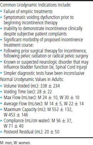
REFERENCES
Lenherr SM, Clemens JQ Urodynamics : with a focus on appropriate indications. Urol Clin North Am. 2013;40(4):545–557.
Wyndaele JJ. Normality in urodynamics studied in healthy adults. J Urol. 1999;161:899–902.
Winters JC, Dmochowski RR, Goldman HB, et al. Urodynamic studies in adults: AUA/SUFU guideline. J Urol. 2012;188(6 Suppl):2464–2472.
UROGENITAL DISTRESS INVENTORY (UDI-6)
DESCRIPTION The UDI-6 is a 6-item questionnaire that assesses LUTS, including incontinence, in women. It is a short form of an original 19-question inventory. All questions are relatively easily understood by patients, and individual responses have been shown to correlate with different types of urinary incontinence (#3 >2 = SUI; #1, #2 >2 = detrusor overactivity; #5 > all other scores = bladder outlet obstruction) (see table on previous page).
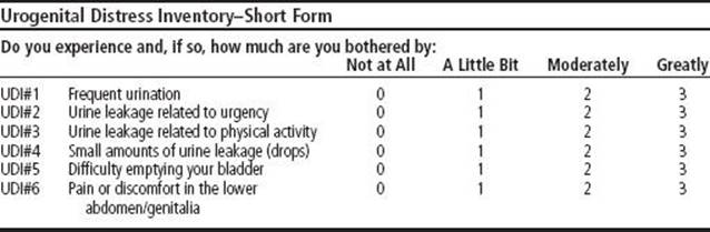
REFERENCE
Lemack GE, Zimmern PE. Predictability of urodynamic findings based on the Urogenital Distress Inventory-6 Questionnaire. Urology. 1999;533:461–466.
UROLITHIASIS, DRUG INDUCED
DEFINITION
Drug-induced renal calculi represent about 1–2% of all renal calculi. Drugs used for the treatment of HIV have become the most frequent cause of drug-containing urinary calculi. Protease inhibitors used in the management of HIV are documented to induce kidney stones, especially indinavir and atazanavir, and more recently darunavir. Triamterene, topiramate and zonisamide (anticonvulsants), methotrexate, guaifenesin/ephedrine, ceftriaxone, trimethoprim-sulfamethoxazole, vitamin D, and calcium supplements are also responsible for some cases of drug-induced urolithiasis. Patients with triamterene-containing stones are more likely to have a prior history of stone disease, suggesting that metabolic predisposition is an important risk factor. Renal calculi have been noted in up to 64% of low–birth-weight infants receiving furosemide loop diuretic therapy. Laxative abuse should be suspected in patients who form ammonium acid urate stones in the absence of bowel disease or urinary tract infection. Loop diuretics, carbonic anhydrase inhibitors, and abused laxatives can cause metabolic abnormalities that facilitate the formation of stones.
REFERENCES
Izzedine H, et al. HIV medication-based urolithiasis. Clin Kidney J. 2014;7(2):121–126.
Matlaga BR, Shah OD, Assimos DG. Drug-induced urinary calculi. Rev Urol. 2003;5(4):227–231.
UROLITHIASIS, INDINAVIR AND OTHER PROTEASE INHIBITORS
DESCRIPTION Drug-induced renal calculi represent 1–2% of all renal calculi. In the last decade, drugs used for the treatment of HIV-infected patients have become the most frequent cause of drug-containing urinary calculi. Among these agents, protease inhibitors (PIs) are well known to induce kidney stones, especially indinavir and atazanavir, and more recently darunavir. A spectrum of asymptomatic crystalluria and renal colic secondary to urolithiasis, with or without dysuria or urgency, seen in patients who are HIV infected and being treated with indinavir and other PI’s. Indinavir stones are not radiopaque nor are they always identified on unenhanced CT imaging. A high index of suspicion is necessary to make the diagnosis and stones may appear as a filling defect on contrast enhanced imaging. (See Section I: “HIV/AIDS, urologic considerations” and Section II: “HIV nephropathy.”)
REFERENCE
Izzedine H, et al. HIV medication-based urolithiasis Clin Kidney J. 2014;7(2):121–126.
UROLITHIASIS, INFECTIOUS (STRUVITE)
DESCRIPTION Composed of magnesium, ammonium, and phophate mixed with carbonate; struvite stones directly correlate with the presence of urease-producing bacteria and active UTI. Associated with a urinary pH of >7.2, which causes struvite crystallization. They usually undergo rapid growth and may result in replacement of the entire pelvis with a stone. (See also Section I: “Urolithiasis, Staghorn” and Section II: “Struvite.”)
CAUSES
• Foreign body in the urinary tract
• Neurogenic bladder
• Urinary diversion
• LUTS
• Indwelling catheter
REFERENCE
Vella M, Karydi M, Coraci G, et al. Pathophysiology and clinical aspects of urinary lithiasis. Urol Int. 2007;79(Suppl I):26–31.
UROLITHIASIS, MATRIX
DESCRIPTION Also called matrix stone, matrix nephrolithiasis, or matrix calculus in the literature, this rare renal calculus has been described as being composed of coagulated mucoids with little crystalline component. Found mostly in individuals with infection due to urease-producing organisms such as Proteus, matrix calculi can be confused with uric acid calculi because they are radiolucent. Matrix calculi, however, are usually associated with alkaline urine from a UTI, whereas uric acid calculi usually form in acidic sterile urine. Standard treatment techniques such as ureteroscopic lithotripsy are used. (See also Section I: “Urolithiasis, Adult, General.”)
REFERENCE
Kim SH, Lee SE, Park IA. CT and ultrasound features of renal matrix stones with calcified center. J Comput Assist Tomogr. 1996;20(3):404–406.
UROLITHIASIS, MELAMINE
DESCRIPTION An increased incidence of kidney stones and renal failure recently reported in China are believed to be associated with ingestion of infant formula contaminated with melamine. Melamine (cyanuric acid, ammelide, ammeline) has industrial use as a resin or adhesive, and has been deliberately added to raw milk to boost its protein content. Although the mechanism is not clear, melamine is almost completely excreted by the kidney and appears to interact with cyanuric acid (a by-product or associated impurity) to form crystals. Low solubility promotes precipitation in renal tubules and causes progressive blockage and significant renal degeneration.
TREATMENT
• Immediately discontinue use of melamine-containing food products.
• Medically monitor renal function, fluid balance, and electrolyte status.
• Alkalize the urine.
• Treat acute kideny injury (AKI) if indicated; use blood or peritoneal dialysis.
• Consider surgical pyelolithotomy in refractory cases.
REFERENCES
World Health Organization. Melamine and cyanuric acid: Toxicity, preliminary risk assessment and guidance on levels in food 25 Sept 2008). Available online at: www.who.int/foodsafety/fs_management/melamine.pdf.
Yang L, Wen JG, Wen JJ, et al. Four years follow-up of 101 children with melamine-related urinary stones. Urolithiasis. 2013;41(3):265–266.
UROLITHIASIS, METHOTREXATE
DESCRIPTION Methotrexate is an anti-folate used to treat a wide variety of diseases including: Cancer, rheumatoid arthritis, lupus, ectopic pregnancy, and psoriasis. More than 90% of the drug is cleared in the urine. Drug and its metabolites are poorly soluble in acidic pH and can precipitate crystalline formation and subsequent tubular obstruction. Medication can also lead to an increase in uric acid levels in the body. Extremely rare with only a few reports of renal calculus to the FDA from using this medication. (See also Section I: “Urolithiasis, Adult, General” and Section II: “Urolithiasis, Drug Induced.”)
TREATMENT
• Urine alkalinization
• Hydration to induce high urine flow rates
REFERENCE
Yarlagada SG, Perazella MA. Drug-Induced Crystal Nephropathy: an Update. Expert Opin Drug Saf. 2008;7(2):147–158.
UROLITHIASIS, TRIAMTERENE
DESCRIPTION Renal calculus consisting either completely or partially of triamterene, a potassium-sparing diuretic often used with hydrochlorothiazide in the treatment of hypertension. Promotion of nucleation and growth of renal calculi, especially calcium oxalate monohydrate, has been shown to occur from triamterene and its metabolites. They are usually radiopaque. Although rare, they usually occur in a patient with a history of urolithiasis. (See also Section II: “Urolithiasis, Adult, General.”)
TREATMENT
• Avoid use of triamterene in patients with a history of urolithiasis.
• Discontinue use of triamterene in patients with triamterene urolithiasis.
REFERENCE
Carr MC, Prien EL Jr, Babayan RK. Triamterene nephrolithiasis: Renewed attention is warranted. J Urol. 1990;1446:1339–1440.
UROLITHIASIS, XANTHINE
DESCRIPTION Renal calculus composed of xanthine are usually associated with hereditary xanthinuria, an autosomal recessively inherited inborn error of metabolism characterized by a deficiency of xanthine oxidase. Other causes include allopurinol use in patients with Lesch–Nyhan syndrome, APRT deficiency, or endogenous uric acid overproduction. Xanthine calculi can be confused with uric acid calculi because they are both radiolucent. Xanthine calculi, however, are associated with low serum uric acid levels. Treat with high fluid intake and standard lithotripsy or stone removal techniques. (See also Section II: “Urolithiasis, Adult, General.”)
REFERENCE
Cameron JS, Moro F, Simmonds HA. Gout, uric acid, and purine metabolism in paediatric nephrology. Pediatr Nephrol. 1993;7:105–118.
URORADIOLOGY SIGNS
DESCRIPTION Uroradiology is rich with descriptions of imaging findings, often metaphorical, which have found common usage in the day-to-day practice of genitourinary radiology. (See Section VII: “Reference Tables: Uroradiology Signs.”)
REFERENCE
Dyer RB, Chen MY, Zagoria RJ. Classic Signs in Uroradiology. RadioGraphics. 2004;24:S247–S280.
UROTHELIAL DYSPLASIA
DESCRIPTION Urothelial dysplasia represents an early morphologic manifestation of progressive alterations between normal urothelium and carcinoma in situ (CIS). Changes in cellular architecture restricted to the intermediate and basal cell layers. A flat lesion with changes including: Loss of nuclear polarity, nuclear enlargement, nucleolar prominence, nuclear membrane irregularity, and nuclear hyperchromasia. Should only be diagnosed when changes cannot be attributed to inflammation and there is not marked cytologic atypia of CIS. Urothelial dysplasia has the possibility of progressing to carcinoma.
REFERENCE
Hodges KB, Lopez-Beltran A, Davidson DD, et al. Urothelial dysplasia and other flat lesions of the urinary bladder: Clinicopathologic and molecular features. Human Pathology. 2010;41:155–162.