
Q. A 60-year-old patient who is an ex-smoker presents with a large lump on his glans penis and a palpable lump in the left groin. The lump is shown in Figure 3.5. What is the likely diagnosis?

Figure 3.5
A. Figure 3.5: Penile tumour. This is an extensive carcinoma of the penis affecting the glans penis.
Q. Which is the most common type of cancer affecting the penis?
A. Squamous cell carcinomas (SCCs) account for over 95% of tumours.
Q. Which region of the penis does SCC most commonly affect?
A. SCC is commonly found on the glans penis or foreskin. The primary tumour is localised to the glans penis in approximately 48% of cases.
Q. What are the risk factors for SCC of the penis?
A. These include smoking, increasing age, previous HPV infection (type 16 and 18), uncircumcised males, poor penile hygiene and retained smegma. There is an association with exposure to psoralen and ultraviolet A photo chemotherapy (PUVA) and lichen sclerosus. (Premalignant lesions are mentioned in a previous question.)
Q. Which HPV subtypes are commonly implicated in penile SCC?
A. High-risk HPV subtypes 16 and 18 are most commonly associated with SCC.
Q. With this particular case what would you do next?
A. I would perform a biopsy of the lesion in order to confirm the diagnosis and organise further imaging to stage the tumour. As this is highly likely to be a penile cancer I would also ensure that the patient receives the appropriate information related to penile cancer and has access to a specialist nurse. The patient needs to be discussed at a specialist penile cancer multidisciplinary meeting with the results of the biopsy and the imaging.
Q. How would you stage the tumour?
A. The penile lesion can be imaged using penile magnetic resonance imaging (MRI) which requires an injection of intracavernosal prostaglandin (to induce an artificial erection) in order to assess whether the lesion invades the corpus cavernosum or the urethra. A computed tomography (CT) scan of the chest, abdomen and pelvis will identify enlarged or abnormal lymph nodes.
Q. Does CT imaging pick up all pathological lymph nodes?
A. No. The CT criteria for metastatic lymph nodes rely mainly on the size of the lymph nodes and abnormal features of the hilum or the shape of the lymph nodes. It will not detect small foci of metastatic disease.
Q. Describe the image presented in Figure 3.6.

Figure 3.6
A. Figure 3.6: This is an MRI of the penis demonstrating a penile tumour on the glans penis extending into the corpus spongiosum and corpus cavernosum.
Q. What is the likely T stage of this tumour?
A. The tumour is extending into the corpus spongiosum and cavernosum and There fore it is at least a T3 lesion according to the 2017 AJCC TNM classification (Table 3.1). (Previously it would have been classified as T2 on the AJCC 7th Edition classification.)
Table 3.1 TNM classification of penile cancer 2017 (8th edition)
|
TX |
Primary tumour cannot be assessed |
|
T0 |
No evidence of primary tumour |
|
Tis |
Carcinoma in situ (penile intraepithelial neoplasia - PeIN) |
|
Ta |
Non-invasive localised squamous cell carcinoma |
|
T1 |
Tumour invades subepithelial connective tissue (lamina propria) |
|
T1a |
Tumour invades subepithelial connective tissue without perineural or lymphovascular invasion and is not poorly differentiated or undifferentiated (TIGI-2) |
|
T1b |
Tumour invades subepithelial connective tissue with perineural or lymphovascular invasion or is poorly differentiated or undifferentiated (TIG3-4) |
|
T2 |
Tumour invades corpus spongiosum with or without invasion of the urethra |
|
T3 |
Tumour invades corpus cavernosum (including tunica albuginea) with or without invasion of the urethra |
|
T4 |
Tumour invades other adjacent structures (scrotum, prostate, bone) |
|
NX |
Regional lymph nodes cannot be assessed |
|
N0 |
No lymph node metastasis |
|
N1 |
Metastasis in one or two unilateral inguinal lymph nodes, but without extra-nodal extension |
|
N2 |
Metastasis in three or more unilateral inguinal lymph nodes without extra-nodal extension, or, any number of bilateral positive inguinal lymph nodes without extra-nodal extension |
|
N3 |
Metastasis to any number of inguinal lymph nodes with extra-nodal extension, or; metastasis to pelvic lymph nodes |
|
M0 |
No distant metastases |
|
M1 |
Distant metastases |
Q. How accurate is MRI in predicting invasion into the corpus cavernosum?
A. Penile MRI is very accurate and has been shown to predict corpus cavernosum/spongiosum invasion in all cases analysed [2].
Q. What would be the surgical treatment option in this case?
A. As the tumour is located distally, the tumour can be excised with clear margins by performing a partial penectomy.
Q. Would you advise any other surgical treatment if the tumour had involved the glans without extension into the corpus cavernosum?
A. Tumours which are limited to the glans penis can be managed by performing a glansectomy and reconstruction of the neoglans using a split skin graft. Spongiosal invasion of the glans or urethra is now classified as T2 according to the AJCC TNM 8th Edition.
Q. How would you perform a partial penectomy?
A. I would ensure that the patient is fully consented for the risks of surgery which include bleeding, infection, penile shortening, erectile dysfunction, spraying of urine, and graft loss if a split skin graft is being used as part of the reconstruction. The patient is placed in a supine position and the tumour is covered with a finger glove or condom. I would then mark and incise the penile shaft skin proximal to the tumour and deglove the penis.
The next step involves mobilising and ligating the neurovascular bundle followed by mobilisation of the urethra. The penis is then transected. There are several techniques described for the procedure e.g. straight transection or ‘fish mouth’ or urethral centralisation technique (be familiar with at least one). I would send the proximal shavings of the corpus cavernosum and urethra for frozen section analysis to ensure that they are tumour free. The corpora and Bucks fascia are oversewn with absorbable sutures. After spatulating the urethra, the neoglans can be covered either with the penile shaft skin or a split skin graft can be used to reconstruct the neoglans. I would leave a catheter in situ for approximately 1 week.
Q. How would you follow up a patient who has undergone penile preserving surgery with no inguinal lymph node disease?
A. Most recurrences will occur within the first 2-3 years following surgery. The EAU guidelines recommend that follow-up should be on a three-monthly basis for the first 2 years then six- monthly after that until 5 years depending on the inguinal lymph node status. At each follow up I would examine the penis and inguinal lymph nodes for any local or distant recurrence. Patients should also be encouraged to self-examine and request an urgent follow-up if any new lesion is palpable.
Q. What is the local recurrence rate following a partial or total penectomy?
A. This varies according to the initial stage of the tumour. Studies have generally reported a local recurrence rate of between 0% and 8%.
Q. How does this compare with the recurrence rates following penile preserving surgery?
A. Although early studies reported a local recurrence rate of up to 40%, more recent studies have shown a local recurrence rate of approximately 11%.
Q. How would you manage the palpable lump in the groin?
A. This is likely to be an enlarged inguinal lymph node. Bearing in mind the size and the stage of the primary penile tumour, it is likely that this is a metastatic lymph node since penile cancer will metastasise to the inguinal lymph nodes and then to the ipsilateral pelvic lymph nodes in a stepwise pattern. I would There fore confirm the diagnosis by arranging for an ultrasound of the groin combined with fine needle aspiration cytology (FNAC) of the enlarged lymph node.
Q. If the FNAC confirms SCC in the palpable inguinal node, what would be the next step in the management of this patient?
A. I would advise the patient that there is confirmed metastatic disease in the palpable lymph node and a possibility of micrometastatic disease within the other inguinal lymph nodes on the same side and recommend that he undergoes a radical inguinal lymphadenectomy.
Q. Following the radical inguinal lymphadenectomy, a single node is found to be involved with metastatic SCC in this patient, what is the N stage according to the 2017 AJCC TNM classification (8th Edition)?
A. As only one (unilateral) lymph node is involved, the nodal staging is N1.
Q. Assuming that this patient was found to have a single metastatic lymph node in both the right and left groin, what would be the N stage according to the 2017 AJCC TNM classification?
A. For inguinal nodes, regardless of the number involved, bilateral nodal involvement is classified as N2.
Q. What percentage of palpable inguinal lymph nodes is involved with metastatic disease?
A. This is controversial. In previous studies, mainly from South America, it was suggested that only 50% of palpable inguinal lymph nodes were involved with tumour and the remaining 50% were enlarged as a result of associated sexually transmitted or other infections. There fore traditionally a short course of antibiotics was given in an attempt to differentiate between the two with the infected ones likely to reduce in size. However, recent data have suggested that over 90% of palpable inguinal nodes are involved with metastatic disease and There fore require a radical inguinal lymphadenectomy. There fore patients should be assumed to have metastatic disease in the palpable lymph nodes and further imaging for staging using CT and ultrasound should not be delayed.
Q. What is this surgical procedure?
A. Figure 3.7 shows an open inguinal lymphadenectomy. The saphenous vein has been preserved (demonstrated with the loop).

Figure 3.7
Q. What are the anatomical boundaries of the femoral triangle?
A. The femoral triangle is bordered by the inguinal ligament superiorly, medial border of the sartorius muscle laterally, lateral border of the adductor longus and adductor longus tendon medially and the floor comprises the pectineus muscle medially and iliopsoas muscle laterally together with the femoral artery and vein.
Q. What is the difference between a superficial modified and a radical inguinal lymph node dissection?
A. The modified superficial lymph node dissection uses a much smaller incision and the boundaries are reduced compared to a radical lymph node dissection. The saphenous vein is preserved and the femoral vessels do not have to be skeletonised deep to the fascia lata.
Note that if any nodes are found to be positive in a modified superficial inguinal node dissection then one proceeds to a radical inguinal node dissection. Also, if two or more inguinal lymph nodes are found to be involved on histology or there is a single metastatic node with extracapsular spread, the patient should proceed to a pelvic lymph node dissection on the ipsilateral side.
Q. What are the complications of a radical inguinal lymph node dissection?
A. The morbidity of this procedure is reported to be between 30% and 50%. The complications include early haemorrhage, wound infection, flap necrosis, lymphoedema of the lower limb, lymphocele, prolonged lymph drainage and patchy sensory loss of the thigh.
Q. What is the advantage of performing a superficial modified inguinal node dissection compared to a radical inguinal node dissection?
A. The superficial inguinal node dissection can only be performed for patients with impalpable lymph nodes and is a technique which reduces the morbidity of the inguinal lymphadenectomy procedure by utilising a smaller inguinal incision with less extensive dissection required to develop the skin flaps. The saphenous vein is also preserved in order to try and reduce the degree of lower limb lymphoedema. The procedure also avoids the need to use a sartorius transposition flap to cover the exposed femoral vessels.
Q. Does superficial modified lymphadenectomy reduce the lymphoedema rate?
A. Yes, it does. Approximately 20% of patients will suffer from lymphoedema with a very small proportion developing persistent lower limb lymphoedema.
Q. Can you describe what has happened here? (Figure 3.8)
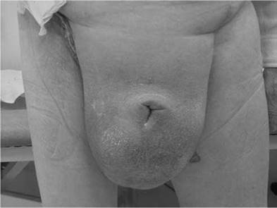
Figure 3.8
A. Figure 3.8: Extensive scrotal lymphoedema. This patient has had surgery to the penis combined with bilateral inguinal lymphadenectomy as indicated by the surgical scars in the inguinal region. This has resulted in extensive genital lymphoedema.
Q. How would you manage the post-operative complication of genital lymphoedema?
A. Initially conservative management includes the use of supportive underwear or specially designed compressive garments which also cover the lower limbs. Patients should ensure that they avoid trauma to the skin in areas affected by lymphoedema, e.g. scratches, pressure sores, and insect bites. Once the patient is mobile, the lymphoedema may start to reduce. However, for troublesome and persistent scrotal lymphoedema where the penis is buried, surgical excision is required which includes a scrotectomy combined with a scrotoplasty and unburying of the penis.
Q. In patients with impalpable inguinal lymph nodes, what is the risk of lymph node metastases?
A. The risk is approximately 15%-25%.
Q. Would you offer a radical inguinal lymphadenectomy to every patient diagnosed with penile cancer?
A. As the aim of radical inguinal lymphadenectomy is to remove metastatic lymph nodes from the inguinal region, subjecting everyone with impalpable inguinal nodes would result in overtreatment in approximately 75%-85% of patients. As the procedure is associated with significant morbidity including lymphoedema, wound complications, seromas and lymph fistulae, alternative techniques should be offered to minimise the complications.
Q. Do you know of any other techniques available to reduce the number of individuals undergoing inguinal lymphadenectomy in those patients with impalpable inguinal nodes?
A. In patients who have clinically impalpable inguinal lymph nodes, dynamic sentinel
lymph node biopsy can be offered which will target the first draining lymph node(s) only.
In centres where a dynamic sentinel lymph node biopsy programme has not been established, an alternative option would be to use a laparoscopic assisted or robot assisted inguinal lymphadenectomy. This is referred to as video endoscopic inguinal lymphadenectomy (VEIL).
Q. Which radioisotope is used for dynamic sentinel lymph node biopsy?
A. 99mTc nanocolloid.
Q. What is the false-negative rate of dynamic sentinel lymph node biopsy for penile cancer?
A. Approximately 4%-7% [3,4].
Q. Do you know of any modifications to the original technique of dynamic sentinel lymph node biopsy which has reduced the false-negative rate to 5%?
A. The false-negative rate has been reduced by incorporating pre-operative inguinal ultrasonography combined with FNAC of suspicious nodes. The rationale for this is to locate morphologically abnormal lymph nodes as well as those which are filled with extensive tumour and There fore unlikely to have normal lymphatic drainage and may not take up the 99mTc nanocolloid. Additionally the use of a blue dye (Patent Blue V) has also helped in localising the lymph nodes during inguinal explorations.
Q. Which pathological features correlate with lymph node metastases and prognosis?
A. T stage (>T2), lymphovascular and perineural invasion, histological grade of tumour, histological subtype, e.g. basaloid or sarcomatoid features. Other factors include depth of invasion of the primary tumour, positive margins following resection and the presence of corpus cavernosum invasion. Of all of these factors the most important appear to be perineural invasion, vascular invasion and high-grade tumours.
Q. An 83-year-old patient with a 2 cm primary SCC on the glans penis opts for radiotherapy to this lesion as opposed to surgical excision as he is worried about the perioperative risks and complications. What would you quote as the response rate and recurrence rate of external beam radiotherapy?
A. Radiotherapy is no longer considered a primary treatment option as the response rate is approximately 56% and the local failure rate is 40%.
Q. What are the main complications of external beam radiotherapy for penile SCC?
A. There is a risk of meatal stenosis and urethral stricture (up to 30%), telangiectasia (90%) and even radionecrosis. (Note that the patient must be circumcised prior to radiotherapy otherwise the prepuce will become fused to the glans.)
Q. Is there a role for radiotherapy for clinically impalpable nodes?
A. Currently there is no evidence to suggest that radiotherapy prevents the development of metastatic inguinal lymph nodes. However, there is a possible role in patients who have undergone inguinal lymphadenectomy for metastatic inguinal lymph nodes which show extracapsular spread.