
Q. What are the histological types of primary bladder carcinoma?
A. Almost 95% of patients with bladder cancer have a transitional cell carcinoma (TCC). The remaining patients have squamous cell carcinoma (4%) or rarely adenocarcinoma of the bladder.
Q. What proportion of patients present with non-muscle-invasive disease?
A. Over 80% of patients with TCC present with non-muscle-invasive Ta or T1 disease and the remaining patients present with muscle-invasive malignancy (T2 to T4).
Q. A 67-year-old man has been assessed in the haematuria clinic. Flexible cystoscopy is performed and reveals a 2 cm papillary lesion on the posterior wall of his bladder. A CTU has been performed which shows normal upper tracts. He is concerned about this diagnosis. How would you treat this man? How would the treatment differ if a solid/non- papillary lesion was found?
A. This man requires an examination under anaesthetic and transurethral resection of bladder tumour (TURBT) with a single intravesical instillation of mitomycin C. I would consent him for the risk of bleeding, infection and bladder perforation as well as further adjuvant treatment with the risk of recurrent disease. He will require catheterisation following his operation.
The European Association of Urology (EAU) guidelines [4] have outlined recommendations on performing TURBT systematically, which is performed either under general or spinal anaesthesia (Table 4.6). If the tumour is over the obturator nerve on the postero-lateral aspects of the bladder then a general anaesthetic is preferable to avoid obturator spasm and the almost inevitable risk of bladder perforation. Bimanual palpation under anaesthesia should be undertaken initially to assess for a bladder mass, which would suggest muscle-invasive disease.
The cystoscope is then inserted to examine the urethra and bladder. A 70° telescope allows a more complete examination of the urothelium. A continuous flow resectoscope can then be introduced to perform the operation. A monopolar (or bipolar) diathermy loop is used to resect the exophytic tumour in fractions (exophytic part, bladder tumour base with detrusor muscle and edges of resection) to obtain a histological diagnosis and fully stage the lesion. Rollerball monopolar diathermy is then utilised to provide haemostasis and fulgurate the edge of the lesion to destroy any potentially malignant urothelium. En bloc resection using monopolar or bipolar current, Thulium-YAG or Holmium-YAG laser is feasible in selected exophytic tumours. These provide high-quality resected specimens with the presence of detrusor muscle in 96%-100% of cases [4].
Table 4.6 Recommended steps for TURBT
|
Bimanual palpation under anaesthesia |
|
Insertion of resectoscope and inspection of urethra |
|
Inspection of whole urothelium of bladder |
|
Resection of tumour |
|
Prostatic urethra biopsy and cold cup biopsies if indicated |
Source: Adapted from European Association of Urology. http://uroweb.org/wp-content/uploads/EAU- Guidelines-on-Non-muscle-Invasive-BC_TaT1 - 2017.pdf
If any other abnormal areas of urothelium are seen then these sites need separate sampling with a cold cup biopsy and diathermy to detect CIS. Random biopsies of normal mucosa are rarely performed in Ta/T1 disease as the chance of detecting CIS is less than 2%. A three-way irrigating catheter can then be inserted to washout any malignant cells and blood following the procedure. Generally this can be removed the next day but with larger resections may have to wait 48 hours. Care must be taken when resecting deep lesions that a perforation of the bladder does not occur. Following the resection a further bimanual examination can then be performed to assess whether a bladder mass has resolved. A residual mass strongly suggests residual invasive disease (T3).
In the operation note, it is important to document tumour characteristics (size, location, number and appearance), all steps of the procedure undertaken and the extent and completeness of resection.
The EAU recommends additional biopsies from normal-looking mucosa when high- risk exophytic (i.e. solid/non-papillary) tumour is visualised. Prostatic urethra biopsies are indicated when a tumour arises from the bladder neck, abnormal areas are seen at the prostatic urethra or if bladder CIS is suspected.
Note: Conversely NICE guidelines recommend that no additional biopsies of normal-looking urothelium be taken unless there is a specific indication, such as unexplained positive cytology.
There is a significant risk of residual disease after TURBT with some studies reporting rates of up to 27% and 53% in those patients with Ta and T1 tumours, respectively. In those patients with T1 disease, upstaging to T2 disease occurs in approximately 21%-25% of patients following a second resection. Second resection is thus recommended 4-6 weeks later in all T1 and high-grade tumours (high-grade G2 and G3). Re-resection is also indicated after incomplete initial TURBT or if there is a lack of muscle in the initial resection (except in G1pTa tumours and primary CIS).
Post-operatively clinically superficial lesions should receive adjuvant intravesical mitomycin C. A meta-analysis by Sylvester et al. has shown a decrease in the relative risk of recurrence by 39% [11].
Q. What do you know about fluorescence cystoscopy?
A. Fluorescence cystoscopy, photodynamic diagnosis (PDD) or blue light cystoscopy relies on the molecular handling of 5-aminolaevulinic acid (5-ALA) by tumour cells. 5-ALA is instilled into the bladder at least 1 hour prior to cystoscopy and taken up by the urothelium. 5-ALA is converted to protoporphyrin, which is preferentially taken up by malignant cells. When blue light (375-440 nm) is used to illuminate the bladder, red fluorescence from abnormal mucosa is seen compared to the surrounding normal bladder mucosa.
In a systematic review by Kausch et al., PDD had a 20% higher detection rate in nonmuscle-invasive tumours and 39% increase diagnosis of CIS when compared to white light cystoscopy [12]. However, a higher detection rate in patients with positive cytology was not observed in a prospective randomised trial [13]. Although PDD has been shown to be more sensitive than white light cystoscopy, PDD has lower specificity.
Fluorescence cystoscopy, as compared to white light cystoscopy, has also been shown to reduce recurrence rates (<10% absolute reduction within 12 months) [14]. No benefit of PDD on progression rates or disease-specific survival has been demonstrated to date.
A prospective multicentre randomised trial comparing PDD guided bladder tumour resection with standard white light resection in patients with newly diagnosed non-muscle- invasive bladder cancer is currently under way with the objective of establishing the effect of PDD on time to recurrence and cost effectiveness (PHOTO trial).
Q. What factors predispose this man to non-muscle-invasive bladder cancer?
A. The main risk factors for bladder cancer are cigarethe smoking (increases risk by threefold particularly in slow hepatic acetylators), industrial carcinogens in rubber and paint industries (aniline dyes, a-naphthylamine), phenacetin, and cyclophosphamide (used in chemotherapy of many haematological cancers). Carcinogenesis in bladder cancer has been extensively reported. The main karyotypic changes involve aberrations of chromosome 9 (>50% tumours), chromosome 17 (p53 loci) and chromosome 13 (retinoblastoma gene loci). A loss of retinoblastoma gene expression, H-ras activation and overexpression of telomerase are early genetic changes in non-muscle-invasive bladder cancer with p53 mutations, FGF and VEGF overexpression leading to invasive or metastatic disease.
Two key theories govern the development of bladder cancer. The clonal theory argues that multifocal and recurrent tumours evolve from one single transformed cell from which all cells share identical genetic mutations. The field change or oligoclonal theory suggests a global change in the urothelium. Genetically pre-malignant cells then transform into clinically detectable tumours, which are genetically unrelated.
Q. Can you name any lesions that predispose a patient to bladder cancer?
A. Several clinically detected lesions can predispose to malignancy. These are summarised in Table 4.7.
Table 4.7 Lesions arising from the urothelium and their malignant potential
|
Keratinising squamous metaplasia |
This is seen in exstrophy, chronic bladder inflammation and schistosomiasis. It is pre-malignant. |
|
Non-keratinising squamous metaplasia |
Not pre-malignant. |
|
Urothelial dysplasia |
A flat non-invasive lesion typified by nuclear clustering. Can be seen with or without malignancy. |
|
Cystitis cystica |
Non pre-malignant enfolding of normal bladder lining cells. |
|
Cystitis glandularis |
Associated with adenocarcinoma. |
|
Papillary urothelial neoplasm of low malignant potential |
Difficult to define. Histology reveals exaggeration of normal glandular rests in bladder urothelium. |
|
Leucoplakia |
Clinically seen as thick and raised white plaques of squamous metaplasia on bladder surface. It is associated with chronic urinary infections. There is a weak association with malignancy. Histology shows keratinising squamous metaplasia. |
|
Malakoplakia |
Similar if not the same as leucoplakia. Malakoplakia histologically is characterised by Michaelis-Gutmann bodies with distinctive basophilic inclusions and foamy histiocytes. Clinically seen as yellow soft plaques on the urothelium. |
Q. This 67-year-old man mentioned earlier attended for a TURBT. Examination under anaesthesia (EUA) revealed no mass. A 2 cm papillary looking tumour was resected from the posterior wall of his bladder. Deep muscle samples were sent separately. He received a single dose of MMC intravesical chemotherapy post-operatively. The histology shows grade 1 disease invading sub-epithelial tissue. The deep muscle biopsies are clear of tumour. He attends clinic to have the histology explained to him. At what TNM stage is this man?
A. This man has G1 pT1 disease. The TNM bladder cancer staging system is reproduced in Table 4.8.
Q. Do you know of any risk stratification for bladder cancer?
A. Several authorities have proposed a risk stratification of non-muscle-invasive bladder cancer (NMIBC).
The NICE bladder cancer: Diagnosis and management guidelines 2015 risk classification outline three risk groups as detailed in Table 4.9. This risk stratification incorporates time to recurrence unlike the risk stratification proposed by the EAU.
The European Organisation for Research and Treatment of Cancer - Genito-urinary Cancer Group (EORTC-GUCG) have also published risk categories (Table 4.10) based on a scoring system utilising the following pathological features: number of tumours (i.e. multifocality), tumour diameter, prior recurrence rate, stage, concomitant CIS and grade. This scoring system is derived from seven trials in non-muscle-invasive bladder cancer organised by the EORTC [15]. It calculates a percentage risk of recurrence and progression at 1 and 5 years as well as defining scores for low-, intermediate- and high-risk disease (Tables 4.11 and 4.12).
Table 4.8 TNM classification of urinary bladder cancer 2017 (8th edition)
|
Tx |
Primary tumour cannot be assessed |
|
Ta |
Non-invasive papillary carcinoma |
|
Tis |
Carcinoma in situ |
|
Tl |
Tumour invades subepithelial connective tissue |
|
T2 |
T2a: Tumour invades superficial muscularis propria (inner half) |
|
T2b: Tumour invades deep muscularis propria (outer half) |
|
|
T3 |
T3a: Tumour invades perivesical tissue microscopically |
|
T3b: Tumour invades perivesical tissue macroscopically (extravesical mass) |
|
|
T4 |
T4a: Tumour invades prostate stroma, seminal vesicles, uterus or vagina |
|
T4b: Tumour invades pelvic wall or abdominal wall |
|
|
Nx |
Regional lymph nodes cannot be assessed |
|
N0 |
No regional lymph node metastasis |
|
N1 |
Metastasis in a single lymph node in true pelvis (hypogastric, obturator; external iliac or presacral) |
|
N2 |
Metastasis in multiple lymph nodes in true pelvis (hypogastric, obturator, external iliac or presacral) |
|
N3 |
Metastasis in common iliac lymph node(s) |
|
Mx |
Distant metastasis cannot be assessed |
|
M0 |
No distant metastasis |
|
Ml |
MIa: Non-regional lymph nodes |
|
MIb: Other distant metastases |
Table 4.9 NICE bladder cancer risk classification in NMIBC 2015
|
Low risk |
• Solitary GI/2 (low-grade) pTa tumour <3 cm • Any papillary urothelial neoplasm of low malignant potential (PUNLMP) |
|
Intermediate risk |
Urothelial cancer that is not low or high risk including: • Solitary GI/2 (low-grade) pTa tumour > 3 cm • Multifocal GI/2 (low-grade) pTa tumours • High-grade G2pTa tumour • Any low-risk NMIBC recurring within I2 months |
|
High risk |
• G3 tumour • G2/G3 pTI tumour • CIS • Aggressive variant tumour (e.g. micropapillary or nested variant) |
Table 4.10 EAU bladder cancer risk group stratification 2017 [4]
|
Low risk |
• Primary, solitary Ta, GI (PUNLMP/low grade), <3 cm, no CIS |
|
Intermediate risk |
• All tumours not defined in low- or high-risk groups |
|
High risk |
Any of the following: • TI tumour • G3 (high-grade) tumour • carcinoma in situ (CIS) • Multiple, recurrent and large (>3 cm) TaGIG2/low grade tumours (all features must be present) Subgroup of highest risk tumours: TIG3/high grade associated with concurrent bladder CIS, multiple and/or large TIG3/high grade and/or recurrent TIG3/high grade, TIG3/high grade with CIS in the prostatic urethra, some forms of variant histology of urothelial carcinoma, lymphovascular invasion. |
Source: Adapted from European Association of Urology, http://uroweb.org/wp-content/uploads/ EAU-Guidelines-on-Non-muscle-Invasive-BC_TaTI-20l7.pdf
Table 4.11 EORTC-GUCG scoring system
|
Factor |
Recurrence |
Progression |
|
Number of tumours |
||
|
Single |
0 |
0 |
|
2-7 |
3 |
3 |
|
>8 |
6 |
3 |
|
Tumour diameter |
||
|
<3 cm |
0 |
0 |
|
>3 cm |
3 |
3 |
|
Prior recurrence rate |
||
|
Primary |
0 |
0 |
|
< I recurrence/year |
2 |
2 |
|
> I recurrence/year |
4 |
2 |
|
Stage |
||
|
Ta |
0 |
0 |
|
TI |
I |
4 |
|
Concurrent CIS |
||
|
No |
0 |
0 |
|
Yes |
I |
6 |
|
Grade |
||
|
GI |
0 |
0 |
|
G2 |
I |
0 |
|
G3 |
2 |
5 |
|
Total score |
0-I7 |
0-23 |
Source: European Association of Urology.
Table 4.12 EORTC-GUCG probability of recurrence and disease progression
|
Recurrence score |
Probability of recurrence at 1 year |
Probability of recurrence at 5 years |
||
|
% |
(95% CI) |
% |
(95% CI) |
|
|
0 |
I5 |
(I0-I9) |
3I |
(24-37) |
|
I-4 |
24 |
(2I-26) |
46 |
(42-49) |
|
5-9 |
38 |
(35 41) |
62 |
(58-65) |
|
I0-I7 |
6I |
(55-67) |
78 |
(73-84) |
|
Progression score |
Probability of progression at 1 year |
Probability of progression at 5 years |
||
|
% |
(95% CI) |
% |
(95% CI) |
|
|
0 |
0.2 |
(0-0.7) |
0.8 |
(0-I.7) |
|
I-4 |
I |
(0.4-I.6) |
6 |
(5-8) |
|
5-9 |
5 |
(4-7) |
I7 |
(I4-20) |
|
I0-I7 |
I7 |
(I0-24) |
45 |
(35-55) |
Source: European Association of Urology.
G3pT1 disease is understaged at radical cystectomy in 30% of patients, with a very high risk of progression if concomitant CIS is found. CIS leads to invasive disease in 2 years if not treated. Progression will occur in <5% of patients with GlpTa disease, 10% of multifocal GlpTl, 30% of G3pT1, >50% for CIS and 50%-80% for G3 disease with CIS.
Q. Are you aware of the histological grading of non-muscle-invasive bladder cancer?
A. The 1973 World Health Organisation (WHO) classification of urothelial carcinomas, stratifies tumours histologically into groups of urothelial papilloma, grade 1 (well differentiated), grade 2 (moderately differentiated) and grade 3 (poorly differentiated).
In 2004, the WHO and International Society of Urological Pathology (ISUP) produced a new histological classification consisting of groups of urothelial papilloma (benign), papillary urothelial neoplasm of low malignant potential (PUNLMP), low-grade and high-grade lesions.
The 2004 WHO classification has been updated in 2016, which includes additional categories of urothelial proliferation of uncertain malignant potential and urothelial dysplasia. Current management guidelines are still based on the 2004 WHO classification.
Although the prognostic values of both 1973 and 2004 WHO classifications have been demonstrated, neither has been shown to be superior and are thus both used in current practice.
Q. What is the evidence for using mitomycin C intravesical chemotherapy in non-muscle-invasive bladder cancer and how do you consent a patient for and administer Mitomycin C?
A. Mitomycin C (MMC) is an anti-tumour antibiotic, which causes DNA cross-linking in bladder tumour cells. It has been extensively investigated in the treatment of Ta/T1 bladder cancer. Following on from work in the United Kingdom for the MRC by Parmar and Tolley in the late 1980s [16], a meta-analysis in 2004 by Sylvester et al., reported on seven trials studying the use of single-dose intravesical chemotherapy post TURBT. This showed a 39% decrease in the relative risk of recurrence with adjuvant treatment [11]. Its use is There fore ‘standard of care’ post TURBT. In the latest meta-analysis by Sylvester et al., benefit from a single instillation of chemotherapy after TURBT was evident only in patients with a prior recurrence rate <1 recurrence per year and those with an European Organisation for Research and Treatment of Cancer (EORTC) recurrence score <5 [17].
Sylvester et al. have published four meta-analyses for adjuvant treatment of bladder cancer, which are summarised in Table 4.13.
Table 4.13 Summary of the four meta-analyses published by Sylvester et al. for the use of adjuvant intravesical treatment following TURBT in NMIBC and bladder CIS
|
I. Meta-analysis comparing TURBT plus single-dose adjuvant chemotherapy versus TURBT [II] |
7 randomised controlled trials involving I476 patients with NMIBC. Resulting in an absolute risk reduction of recurrence of I2% (NNT 8.5) and relative risk reduction of recurrence of 39%. There fore single-dose MMC reduces the risk of recurrence. |
|
2. Meta-analysis comparing single instillation of chemotherapy after TURBT with TURBT alone [I7] |
II randomised controlled trials involving 2278 patients with NMIBC. Resulting in an absolute risk reduction of recurrence of I 3.7% and relative risk reduction of recurrence of 35%. NNT to prevent one recurrence within 5 years was 7. Only patients with a recurrence rate of <I recurrence per year or EORTC recurrence score <5 benefited. No effect on progression was observed. |
|
3. Meta-analysis ofTURBT plus BCG versus TURBT alone or another adjuvant treatment [ I8] |
24 randomised controlled trials involving 4863 patients with NMIBC. Resulting in an absolute risk reduction of progression of 4% and relative risk reduction of progression of 27%. There fore BCG reduces the risk of progression. |
|
4. Meta-analysis comparing intravesical chemotherapy and BCG in CIS of the bladder [ I9] |
9 randomised controlled trials involving 700 patients with NMIBC. Resulting in an absolute risk reduction in recurrence of 20.5% and relative risk reduction in recurrence of 59%. There fore BCG is more effective than intravesical chemotherapy in CIS. |
MMC is given at a dose of 40 mg in 40 mL of saline via a urinary catheter. The catheter is then clamped and the patient left with the solution in the bladder for 1 hour. The catheter is then removed or unclamped. The patient should be aware of the risks of extravasation postoperatively, irritative voiding symptoms and a chemical dermatitis of the palm of the hands.
Q. Tell me about BCG?
A. BCG or bacillus Calmette-Guérin was first used by Morales in 1976 [20]. It is live attenuated mycobacterium bovis. Connaught, OncoTice and RIVM are the three strains used. No difference in efficacy has been reported between these strains. The mechanism of action of BCG is still poorly understood. What is known is that it attaches to the urothelium via the fibronectin receptor and is internalised within the cell. Glycoproteins remain on the surface membrane of the cell and these antigens mediate the immune response by macrophage chemotaxis and cytokine production. Histologically BCG granulomas consist of epithelioid cells, Langerhans’ giant cells and lymphocytes.
In the setting of adjuvant intravesical treatment of bladder cancer, BCG has been shown to reduce the risk of recurrence and progression.
A meta-analysis by Sylvester et al. analysed data from 4863 patients enrolled in 24 RCTs observed an absolute risk reduction in progression of 4% (13.8% versus 9.8%) and a relative risk reduction in progression of 27% in patients with non-muscle invasive (stage Ta, T1 or carcinoma in situ) bladder cancer. It must be noted that the median follow-up was short (2.5 years) and most tumours recurring were of low or intermediate risk (only 7.6% were G3 recurrence) [18].
Malmstrom et al. conducted a meta-analysis of 2820 patients from nine RCTs comparing MMC and BCG, observed a 32% reduction in risk of recurrence in patients treated with maintenance BCG (compared to MMC). However, a 28% increase in recurrence risk occurred in patients treated with BCG without maintenance BCG [21]. Another RCT by Sylvester et al. comparing BCG to epirubicin demonstrated between overall and disease-specific survival in patients with intermediate- and high-risk Ta T1 disease treated with BCG [22].
Q. How do you consent a patient for and administer BCG intravesical immunotherapy?
A. Patients who receive intravesical BCG must be at least 2 weeks post TURBT or bladder biopsy. The instillation is performed via a catheter, which is immediately removed, and the BCG is held within the bladder for up to 2 hours. Precautions such as sitting down to void in order to avoid splashing, hand washing after voiding as well as rinsing the toilet with undiluted bleach, are recommended. Patients should be encouraged to increase their fluid intake and void regularly.
Common side effects include dysuria, frequency and malaise with a mild fever for up to 24 hours. More serious symptoms of a high fever for 48 hours, arthralgia, headaches, rash and increased malaise suggest BCG sepsis. This requires hospitalisation, resuscitation, blood cultures and commencement of anti-tuberculous treatment. Steroids should be considered. Expert help should be sought and medication continued for a period of at least 6 months. Many of the agents used are hepatotoxic, which requires regular monitoring.
Absolute contraindications of BCG instillation include recent TURBT within 2 weeks, visible haematuria, traumatic catheterisation and symptomatic urinary tract infection.
The treatment regime involves once weekly instillations for 6 weeks followed by a 6-week break and three further instillations once a week for 3 weeks. If maintenance treatment is instigated, once weekly instillations for 3 weeks every 6 months for up to 3 years is given. This is the basis of Lamm’s regime [23].
Thus the complete BCG treatment consists of
Initial post TUR - 6 week course
At 3 months - 3 week course
At 6 months - 3 week course
Then every 6 months thereafter a 3 week course up to 3 years
The complete course entails 27 doses of BCG, however poor compliance to complete the entire 3 year course was reported (only 16% achieved 27 doses). Quinolones have some antituberculous activity and should be avoided to maintain efficacy.
Instillation of a reduced dose of BCG has been proposed to reduce toxicity. However, a full dose of BCG is more effective in multifocal tumours. The CUETO study compared one- third dose (27 mg) to full dose BCG (81 mg) and demonstrated no difference in efficacy. The EORTC randomised study by Oddens et al., comparing one-third dose to full dose BCG, found no difference in toxicity but in high-risk patients, 3 year is associated with a reduction in recurrence only when given at full dose [24].
Q. Do you know any alternatives to intravesical BCG?
A. In patients who fail a course of BCG, intravesical immunotherapy combination with 50 mega-units of interferon alpha with one-third dose of BCG has shown an increased response rate of 50%. Electromotive MMC has also been used in combination with BCG and shown to improve recurrence-free interval and reduce progression rates. The inflammatory reaction induced by BCG is thought to increase the absorption of MMC into the transformed urothelium. The effect of hyperthermia and MMC is currently being studied in the HIVEC II trial, which is comparing hyperthermia and MMC with MMC alone in patients with intermediate-risk NMIBC.
Q. When do you offer patients re-resection and what is the evidence for this practice?
A. Residual disease after TURBT has been reported in T1 tumours (33%-55%) and G3Ta tumours (41.4%). Understaging of tumours by initial resection has been observed in 4%-25% of T1 tumours and up to 45% if no muscle was included in the initial resection.
In a meta-analysis by Brausi et al., of 2410 patients from seven EORTC trials, in patients with T1 disease, 15%-35% had residual tumour and disease upstaging occurred in 30% [25]. Herr et al. evaluated 710 patients with re-resection and reported only 39% had no evidence of disease on re-TUR. Upstaging of tumour was observed in high-grade Ta (9%) and G3T1 tumours (23%). 76% of patients with residual T1 tumour on re-resection were found to progress in stage or grade compared with 14% of patients who had other than T1 tumours on re-resection [26]. Re-resection There fore improves staging accuracy, which is critical as the treatment of T1 disease differs significantly from T2 disease.
Re-resection has also been demonstrated to increase recurrence-free survival. Grimm et al. conducted an observational study of patients undergoing re-resection and primary TUR alone and found lower recurrence rates at 3 years in the re-resection group (33% versus 61%) [27].
Based on the described evidence, recommendations for re-resection are outlined in EAU guidelines [4]. Re-resection should be performed in the following situations:
After incomplete initial TURBT
If there is no muscle in the specimen of the initial resection (with exception of G1Ta tumours and primary CIS)
In all T1 tumours
In all high-grade/G3 tumours (except primary CIS)
The NICE guidelines recommend re-resection within 6 weeks of initial resection for those patients with high-risk NMIBC.
Q. How do you manage G3pT1 disease?
A. High-grade T1 bladder cancer presents significant difficulties in management. For G3pT1 disease, 30% of patients will never have a recurrence, 30% will undergo deferred cystectomy and 30% will die of metastatic disease. Based on the EORTC-GUCG risk tables, G3 disease has a 25%-75% risk of disease progression. Aggressive management is thus warranted in G3pT1 disease.
Given the high risk of residual disease and 23% risk of upstaging of G3T1 tumours [26], re-resection is recommended in the EAU and NICE guidelines within 6 weeks of the first resection to accurately stage the tumour.
After discussion at the uro-oncology MDT, treatment options including adjuvant intravesical immunotherapy (BCG) or primary radical cystectomy (especially in the highest risk group) should be offered. Radiotherapy has shown no increase in progression-free interval, progression-free survival or overall survival. Patient and disease factors should be taken into account when deciding on treatment with intravesical immunotherapy or primary radical cystectomy (e.g. patient fitness, life expectancy, pathological variants and large tumours).
If intravesical strategies fail, consideration of radical cystectomy in carefully chosen individuals results in a 5-year survival rate of 90%, however, 5%-10% of patients will still have positive lymph nodes.
Follow-up should be tailored to the risk of recurrence. Table 4.14 summarises the investigation, treatment and follow-up of NMIBC from the NICE guidelines.
Table 4.1- NICE guidelines for the investigation, treatment and follow-up of NMIBC by risk group
|
Risk group |
Investigation |
Treatment |
Follow-up |
|
Low |
|||
|
• Solitary GI/2 (low grade) pTa tumour <3 cm • Any PUNLMP |
Cystoscopy Cytology or urinary biomarker test Histopathology |
Single dose of intravesical MMC at time of TURBT |
Cystoscopic follow-up at 3 and I2 months after diagnosis Discharge if no recurrence within I2 months |
|
Intermediate |
|||
|
Urothelial cancer that is not low or high risk including • Solitary GI/2 (low grade) pTa tumour > 3 cm • Multifocal GI/2 (low grade) pTa tumours • High-grade G2pTa tumour Any low-risk NMIBC recurring within I2 months |
Cystoscopy Histopathology Cytology |
Single dose of intravesical MMC at time of TURBT Offer a course of at least six doses of intravesical MMC |
Cystoscopic follow-up at 3, 9 and 18 months, and once a year thereafter Discharge after 5 years of disease free follow-up |
|
High |
|||
|
• G3 tumour • G2/G3 pTI tumour • CIS • Aggressive variant |
Cystoscopy Histopathology CTU (CT staging if radical treatment considered) Cytology Re-resection |
Single dose of intravesical MMC at time of TURBT Offer another resection within 6 weeks of initial TURBT Offer choice of intravesical BCG or radical cystectomy |
For patients treated with intravesical immunotherapy cystoscopic follow-up: • Every 3 months for the first 2 years then • Every 6 months for the next 2 years then • Once a year thereafter |
The EAU guidelines recommendations for follow-up in NMIBC patients do differ from the NICE guidelines in some respects:
Low-risk Ta tumours should undergo cystoscopy at 3 months, 12 months and then yearly for 5 years.
Surveillance of high-risk tumours should entail cystoscopy and cytology. Yearly upper tract imaging is recommended.
Random biopsies or PDD-directed biopsies should be considered after intravesical treatment in patient with CIS.
Q. How do you manage carcinoma in situ (CIS) of the bladder?
A. CIS is a histological diagnosis and is a flat, non-invasive (i.e. not crossing the basement membrane), high-grade malignancy of the bladder (Figure 4.1).
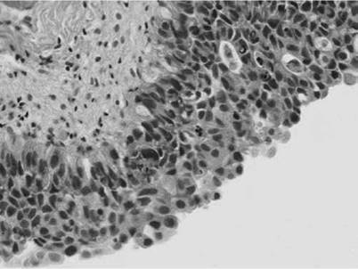
Figure 4.1 Haematoxylin and Eosin staining of carcinoma in situ of bladder displaying flat, non-invasive full thickness disordered proliferation of atypical cells with loss of cell polarity and cell cohesion (changes do not cross the basement membrane). (Thanks to Dr Rupali Arora, Consultant Histopathologist, University College Hospitals, London, UK, for providing this image.)
CIS leads to invasive disease in 2 years if not treated. Additionally, if untreated progression occurs in >50% of patients with CIS alone and in 50%-80% of patients of for G3 disease with CIS.
Primary CIS of the bladder is defined as CIS without any concomitant or previous TCC. Secondary CIS is defined as CIS with a TCC of the urothelium. Concurrent CIS is defined as CIS in the presence of any other urothelial bladder tumour.
Primary disease represents about 5% of CIS diagnosed. It occurs most commonly in men over the age of 50 years old. Secondary disease often occurs in association with high-grade invasive lesions. Urine cytology is very sensitive and specific for the detection of CIS (94% and 96%, respectively). Fluorescence cystoscopy has been used to detect carcinoma in situ but does have a 35% false-positive rate on biopsy. CIS cells show a high percentage of p53 mutations. The mainstay of treatment of CIS is with intravesical immunotherapy with BCG. A meta-analysis by Sylvester et al. shows that BCG is superior to MMC in the treatment of CIS [19]. With maintenance BCG up to 3 years, 75% of patients have an initial complete response with 50% remaining disease free at 5 years and 30% at 10 years. Response to intravesical BCG is a prognostic factor for progression and death with 10%-20% of complete responders progressing to muscle-invasive disease compared with 66% of non-responders.
For BCG refractory disease other novel approaches have been used, including BCG with electromotive MMC, combination BCG with interferon alpha and photodynamic therapy.
Upper tract CIS is a very dangerous condition and BCG treatment is associated with a higher degree of BCG-related complications due to increased systemic absorption.
Q. What do you know about squamous cell carcinoma of the bladder?
A. Squamous cell carcinoma of the bladder accounts for 3%-7% of all bladder tumours.
It is often associated with long-term inflammation and irritation of the bladder from catheterisation, stone disease or schistosomiasis (in sub-Saharan Africa). It generally presents with haematuria in advanced stages and carries a poor prognosis. Cystoscopy reveals an invasive ulcerative lesion often on the trigone or lateral walls of the bladder. Treatment is with cystectomy for loco-regional control. At presentation only 8%-10% of patients will have metastatic disease. Five-year survival is 50% in those whose aetiology is schistosomiasis.
Q. What do you know about adenocarcinoma of the bladder?
A. Adenocarcinoma of the bladder is rare and accounts for less than 1% of all bladder tumours. It is either primary (de novo carcinoma often on the trigone or posterior wall of the bladder) or secondary (metastatic or associated with an urachal remnant). ttere is an association with cystitis glandularis rather than with CIS. Adenocarcinoma also has an increased incidence in bladder exstrophy patients and bowel augmentation/bladder substitution into the urinary tract (after 10-20 years).
In urachal adenocarcinoma, the urachal remnant is patent in one-third of patients. These patients often present with both haematuria and a mucous discharge. Histology shows mucous-secreting cells in a glandular, colloid or signet ring pattern. Clinically an important consideration is to identify a colonic tumour extending into the bladder. The treatment is with radical cystectomy with excision of the urachus and umbilicus but with aggressive management the 5-year survival rate is only 40%. ttere is no proven role for chemotherapy in this rare tumour, which often presents with muscle-invasive disease at diagnosis.