
Q. A 62-year-old man is referred urgently from his GP with a raised PSA of 6.4 ng/mL.
He had the PSA blood test performed due to deterioration in his urinary symptoms.
A midstream specimen of urine (MSU) was negative and he is otherwise well. He is concerned and is anxious about further assessment. How would you assess this patient in clinic?
A. A full urological history should be taken including his age, race, LUTS, urinary tract infection, bone pain and family history of prostate cancer. A general examination would precede examination of his external genitalia and a DRE. If advanced disease is suspected one should also look for leg swelling, anorexia, weight loss, bone tenderness and neurological defects suggesting spinal cord compression. This relatively young patient has an abnormal PSA reading and There fore he should be counselled about further tests, either upfront TRUS prostate biopsy with an explanation that we are concerned about a diagnosis of a prostate cancer or, as is becoming more common, pre-biopsy multiparametric MRI of the prostate, if I have access to a high-quality service for this locally. I will explain the pros and cons of biopsy to him including the risks of severe bleeding (1%), severe infection (1%-2%), pain and urinary retention. Many men will experience haematospermia, rectal or urethral bleeding or both following this procedure. If his upfront biopsy results are negative, one would continue to follow him up with a further PSA blood test in 6 months’ time. If another biopsy is indicated, then I would consider an initial mpMRI or a transperineal template biopsy. If his biopsy is positive then he should be counselled regarding the available treatment options. He may need further investigation of his lower urinary tract symptoms prior to treatment.
Q. How do you perform a prostate biopsy?
A. Transrectal prostate biopsy is performed with a 7.5 MHz transrectal ultrasound probe.
Many are now bidirectional allowing multiplanar views. Antibiotic prophylaxis is required. Regimes vary between hospitals but commonly intravenous gentamicin, rectal metronidazole and three doses of a quinolone are prescribed. After informed consent, including the risks of bleeding, infection, pain and acute urinary retention, a digital rectal examination is first performed assessing the prostate for size and consistency. The ultrasound probe is then inserted into the rectum. The prostate is then scanned to detect any capsular breach or hypoechoic areas within the periphery of the prostate. Using the ellipsoid volume formula (height x breadth x width) x 0.52 the prostate volume can be calculated. The PSAD can then be calculated. Local anaesthetic peri-prostatic injections have been shown to reduce the pain of prostate biopsy in a RCT [22]. A total of 12 biopsy cores are now considered standard and is an acceptable compromise between increased cancer detection and the morbidity from the procedure. Targeting of the periphery of the prostate is essential due to the higher proportion of prostate cancers diagnosed in this anatomical location (approximately 85% of prostate cancers are found in the peripheral zone of the prostate and 15% in the transition zone).
Q. What are the chances of finding prostate cancer in further biopsies?
A. Djavan et al. looked at prostate biopsies in a series of 1051 men with a normal DRE and PSA between 4 and 10 ng/mL. This study reported that the chance of finding a prostate cancer on the first, second, third and fourth biopsy were 22%, 10%, 5% and 4%, respectively [23]. The more biopsies that are taken, the lower the Gleason score of the cancer is when it is detected. There fore a second biopsy is often considered in many patients with serial abnormal PSAs, but the more biopsies that are taken the more confident the clinician can be that the disease will be insignificant.
Q. Is there a role for pre-biopsy MRI in men with an elevated PSA?
A. ttere have been a number of studies, such as the UK PROMIS study [24], which have shown multiparametric MRI of the prostate can detect higher-risk disease with better sensitivity than TRUS biopsy. The important point about MRI of the prostate is that it has to be multiparametric (including T2-weighting, diffusion weighting, dynamic contrast enhancement), with a high-quality 1.5 Tesla or 3 Tesla scanner and reported by an expert uro-radiologist. ttere is still controversy about how to manage men with a negative or nonsuspicious mpMRI of the prostate as the false-negative rate can vary between 5% and up to 25%. This is often because of difference in expertise and experience between and within centres, different ways of comparing the mpMRI to histology and different definitions of what is high-risk cancer. Most urologists would still biopsy the prostate if the mpMRI is negative because of concern about missing some Gleason 7 tumours and concern about the inter-observer variability of reporters. In this case of a non-suspicious MRI, I would discuss with my patient the false-negative rate of mpMRI and TRUS biopsy, the risks and benefits of biopsy and the problems of missing important cancer if we do not biopsy and overall advise he undergo a systematic biopsy.
Q. The multiparametric MRI is as shown in Figure 1.1. What can you see?
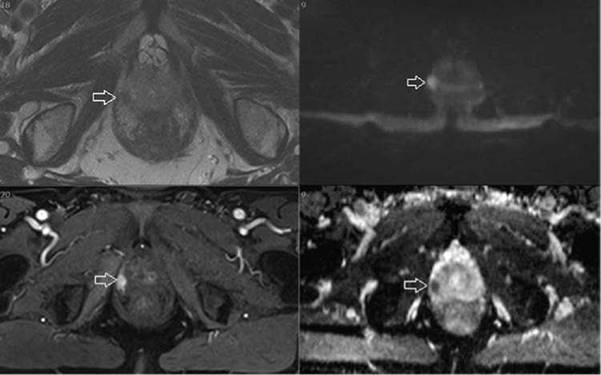
Figure 1.1
A. I would want to see the full series of scans (T2, diffusion weighted and dynamic contrast- enhanced images) using a dedicated DICOM software viewer or a PACS system. I would want this reported by a dedicated uro-radiologist.
However, I can see the following on these images provided:
Top left - This is an axial T2-weighted MRI scan of the prostate delineating the peripheral zone and the transition clearly. In the right peripheral zone I can see a hypointense/low signal area that is potentially suspicious.
Top right and bottom right - These are diffusion weighted images (DWIs).
Top right - This is the long b DWI image that shows restricted diffusion with high signal intensity in the area of concern in the right peripheral zone.
Bottom right - This is the ADC (apparent diffusion coefficient) map in which the lesion is seen as an area of low signal intensity.
Bottom left - This is a contrast-enhanced image (contrast used in mpMRI is gadolinium) in which the corresponding area is shown to have early intense contrast enhancement (tumours have early contrast enhancement and also demonstrate early washout of contrast).
In summary the lesion enhances early with contrast and has a diffusion signal so this lesion would be scored highly suspicious on the MRI and would need to be targeted with biopsies (in addition to taking systematic biopsies of the prostate).
Q. What is the PIRADS score on MRI?
A. PIRADS, Prostate Imaging Reporting and Data System, is a standardised way of reporting the level of suspicion that a radiologist has for there being clinically significant prostate cancer on a multiparametric MRI. There is an alternative scale called Likert that is similar. The scale means the following:
1 = Highly unlikely for the presence of clinically significant prostate cancer
2 = Unlikely for the presence of clinically significant prostate cancer
3 = Equivocal/uncertain
4 = Likely for the presence of clinically significant prostate cancer
5 = Highly likely for the presence of clinically significant prostate cancer the higher the score, the greater is the chance of finding high-risk prostate cancer.
Q. The highly suspicious lesion on mpMRI of the prostate in the previous questions is scored 5 on the Likert scale. What are the different ways you might target this?
A. The broad approach to targeting is whether one does this using a cognitive (or otherwise known as visually estimated) approach or using software and devices to help guide the needle. Most of the studies so far have shown no significant difference between cognitive and image-fusion targeting in expert hands. Fusion can be done using rigid or elastic methods, with elastic fusion taking into account the deformation that occurs when a TRUS probe is used. There is no definite evidence that this makes a difference in fusion. Targeting can also be done inside the MRI scanner although this can be expensive and take a significantly longer time. Targeting can be done using a transrectal approach or a transperineal approach with most of the transperineal cases being done under general anaesthetic. Recently, some groups have used local anaesthetic and sedation to do transperineal biopsies.
Q. How would you manage a patient with high-grade prostatic intraepithelial neoplasia (HGPIN) on biopsy?
A. HGPIN has been proposed as a precursor to prostate cancer but this remains unproven.
If HGPIN is found in one core then the patient has a 20% chance of prostate cancer on further biopsies. If more than one core identifies HGPIN then the next biopsy can be positive for prostate cancer in up to 70%. In 82% of radical prostate specimens HGPIN is shown, but only 40% of BPH specimens show HGPIN after transurethral resection of the prostate (TURP). HGPIN does not secrete PSA. With an overall positive biopsy rate of 24% on second biopsy, this is similar to the detection of prostate cancer in the PCPT trial. Clinically the European Association of Urology (EAU) guidelines recommend repeat biopsy if HGPIN is found in three or more prostate biopsies but the interval is unclear.
Q. How would you manage a patient with atypical small acinar proliferation (ASAP) on biopsy?
A. ASAP has a 40% detection of prostate cancer on repeat biopsy compared to 24% with HGPIN. The EAU guidelines recommend repeat biopsy but the interval is unclear. Due to the higher detection of prostate cancer on subsequent biopsy with ASAP a more aggressive approach to repeat biopsy is often undertaken. Recent studies in which MRI-targeted biopsies have found ASAP, the rate of Gleason 7 or more cancer on repeat biopsies is very low so men evaluated in this manner could just undergo PSA surveillance.
Q. What do you know about epidemiology, diet and chemoprevention in prostate cancer?
A. Prostate cancer affects 100 in 100,000 men in the Western world. Post-mortem series suggest that the prevalence is very high with 30% of 50-year-olds having the disease, increasing to 80% at 80 years. However, only a small proportion of these patients develop clinically significant, life-threatening cancer. In the United Kingdom in 2014, over 46,000 men developed prostate cancer making the disease a significant health problem.
Dietary factors have been shown to affect the risk of prostate cancer. The factors that increase the risk of prostate cancer include obesity (relative risk 1.1), meat (not much data) and dairy products (higher risk). A reduced risk of prostate cancer has been publicised for many components of a man’s diet with limited evidence. These include fruit and vegetables (no benefit), beta-carotene 50 mg (no benefit), lycopene (possible benefit), vitamin D (no benefit), vitamin E and selenium (both no benefit - in fact results from the Selenium and Vitamin E Cancer Prevention Trial, SELECT, demonstrated that men should avoid selenium or vitamin E supplementation at doses that exceed recommended dietary intakes as this resulted in an increased risk of being diagnosed with prostate cancer including high-risk cancers). In addition, the PCPT trial has shown the possible chemoprevention benefit of finasteride compared to placebo in decreasing the incidence of prostate cancer, although this may be at the expense of an increase in higher-grade cancers (see previous discussion).
Q. What do you know about hereditary prostate cancer?
A. Familial prostate cancer represents about 9% of prostate cancer patients and relates to a clustering of prostate cancer cases within a family. Hereditary prostate cancer by definition must have three generations affected with three first-degree relatives or three relatives under the age of 55 years. In addition, prostate cancer in a family infers an increased risk of prostate cancer. If one relative is affected then a 2-3 x risk is transferred, if two relatives are affected then a 5x risk is transferred and for three relatives an 11x risk is transferred. Familial breast cancer confers a greater risk of prostate cancer within a particular family. The BRCA 2 gene particularly has been implicated in this relationship.