
Thomas Johnston, Mark Rochester and Oliver Wiseman
Q. A 44-year-old woman presents with a 6-month history of recurrent urinary tract infections (UTIs) and occasional left loin ache. A recent midstream urine culture grew Proteus mirabilis 105 cfu/mL with >200 leucocytes. Her GP has requested an ultrasound of the renal tract and plain kidney, ureter and bladder (KUB) x-ray. What does the KUB x-ray in Figure 10.1 show?
A. Figure 10.1 shows a large left staghorn calculus and upper third right ureteric calculus.
Q. What further investigations are required?
A. This woman should have urine sent for culture and sensitivity in the first instance and blood for full blood count (FBC), urea and electrolytes (U+Es), calcium and urate and a urine spot test for cystine. Imaging to define the stone burden and calyceal anatomy, and also split renal function is then required prior to planning definitive treatment.
Q. Which imaging modalities would you use and why?
A. The choice of imaging to determine burden and anatomy is a non-contrast computed tomography of kidneys, ureters and bladder (CT-KUB) scan. Some endourologists would state that a contrast phase to look at the anatomy of the collecting system would be helpful to plan treatment, but this is not standard.
The function of the affected kidney is determined by renography, typically a 99mTc DMSA (99mTc dimercapto-succinic acid) renogram. DMSA is a protein which is actively extracted and bound by functioning renal tubules with very little filtered. It is the drug of choice for high-quality cortical imaging. The standard dose is 100 MBq. Images are taken 2-3 hours later with a gamma camera, or after a longer interval in the presence of renal failure.
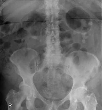
Figure 10.1
Posterior and posterior oblique views are taken.
Q. Should the left staghorn renal calculus be treated or be left alone? Justify your answer.
A. The case for a more aggressive approach to staghorn calculi was outlined in a paper by Blandy and Singh [1]. This paper was in three parts:
Part 1
The first part of this paper was a post-mortem study. The authors retrospectively studied 8,996 consecutive post-mortems. Only nine staghorns were discovered. Five of these had caused severe symptoms and were thought to have contributed to the death of the patients. The authors concluded that ‘the notion of an incidentally discovered silent staghorn is false.
Part 2
The second part of this paper concerned the conservatively managed staghorn calculus. Sixty staghorns were identified retrospectively over the period 1955-1975, in patients where stones were not removed. Twenty had early nephrectomy. Of 40 observed 16 went on to develop pyonephrosis and had drainage and difficult nephrectomies with a high mortality. Overall 17/60 (28%) died during follow-up (mostly from renal failure). All others were said to have had pain and infection.
Part 3
The final section of this paper described a case series of surgical removal of staghorn calculi. In this retrospective series 152 staghorns were found in 125 patients. The authors described stone ‘clearance’ in 80%, and ‘dust’ only remaining in a further 5%. The mortality was 7% during follow-up.
The authors concluded that there is no such clinical entity as a ‘silent staghorn’ based on the post-mortem study. Furthermore, they stated that long-term survival is better in those treated surgically (mortality 7%) than in those managed conservatively (mortality 28%).
Critical analysis of this paper allows the following points to be raised. It is a simple message which has been supported by subsequent data. Good data concerning the fate of residual fragments is presented. However, it could be criticised for its retrospective design, and no statistical analysis was performed. Case selection error is likely, and autopsy data may be incomplete. For example, five symptomatic staghorns are referred to in part 1, but 24 deaths in parts 2 and 3. Furthermore, almost half the staghorns found in the autopsy series were asymptomatic, perhaps not supporting the authors’ first conclusion. Complications of surgery were probably underestimated due to the retrospective design, and questions could be raised as to the method of assessment of renal function, which is not described.
Further guidance on the management of staghorn calculi can be taken from Teichman et al. [2], who analysed retrospectively 177 consecutive staghorn calculus patients to determine risk factors for ultimate renal deterioration and renal cause-specific death. Over a mean follow-up of 7.7 years, the overall rate of renal deterioration was 28%. This was associated more frequently with solitary kidneys (77% versus 21%), previous stone disease (39% versus 14%), hypertension, complete staghorn calculi and neurogenic bladder as well as those who refused treatment (100% versus 28%). With respect to mortality, no patient with complete clearance of fragments died of renal-related causes versus 3% of those without clearance of fragments and 67% of those who refused treatment, comparing favourably with the earlier findings of Blandy and Singh.
Q. The patient opts for percutaneous nephrolithotomy (PCNL). What are the indications and contraindications for this procedure?
A. Indications
1. Stone size
a. Stones >3 cm diameter.
b. Renal pelvis stones >2 cm.
c. Lower pole stones >1 cm.
d. Staghorn stones.
2. Obstruction
a. An anatomic abnormality is present that will prevent stone fragments from passing spontaneously, especially where extracorporeal shock wave lithotripsy (ESWL) is usually contraindicated.
3. Anatomical considerations
a. Abnormal renal anatomy such as horseshoe kidney or calyceal diverticular stones.
b. Abnormal patient anatomy such as kyphoscoliosis or obesity preventing ESWL.
4. Failed ESWL/ureteroscopy (URS)
5. Stones associated with a foreign body
6. Patient choice/desire for one treatment only
Contraindications
1. Absolute
a. Uncorrected bleeding disorder
b. Pregnancy
c. Sepsis
d. Poor kidney function (e.g. <15%), where nephrectomy would be indicated
e. Need for coincidental open procedure
2. Relative
a. Medical problems - Patient high risk for anaesthesia
b. Anterior calyceal diverticulum
Q. Describe how you would take informed consent for this procedure.
A. Informed consent must include a discussion of available alternative treatment options, as described above, the intended benefit of the proposed procedure, and the potential complications, which are listed as follows with approximate percentages in parentheses:
Complications related to access
Bleeding
Requiring transfusion (2%-3%)
Requiring embolisation (1%)
Requiring nephrectomy (rare)
Perforation of adjacent organs (bowel <1%, pneumothorax 0%-5%)
Access failure (up to 5%)
Complications related to stone removal
Infection (bacteriuria 77%; sepsis 0.25%-1.5%)
Transurethral resection (TUR) syndrome
Irrigant extravasation (30%)
Renal pelvis injury
Residual stones (>10%), dependent upon stone complexity
Others
Pleural effusion (10%)
Hypertension and fibrosis (late)
Mortality (0.3%)
Q. Are there any advantages of supine versus prone PCNL?
A. Patient positioning during supine PCNL is less time consuming and the potential risks of musculoskeletal injuries (e.g. cervical spine, brachial plexus), cardiovascular (increased cardiac output, venous stasis and thromboembolic events) and visual complications are decreased. The supine position also allows easy access to the urethral meatus for simultaneous retrograde procedures during PCNL, is more ergonomic for the surgeon during the procedure, may reduce radiation exposure, promotes spontaneous drainage of stone fragments due to the relatively downward direction of the tract and provides lower irrigating pressures. Prone PCNL provides more options and a wider surface area for puncture (upper pole/multiple punctures), may reduce the risk of damage to visceral organs (retro-renal colon and lateral rotation of spleen/liver away from puncture site), allows greater manipulation with the nephroscope and may reduce perirenal injury (excessive anterio-medial movement with supine increases risk of injury during dilation) [3].
Three meta-analyses have been carried out to date comparing outcomes of prone versus supine PCNL and have reported conflicting results [4-6]. Two of these reported similar stone-free rates (SFRs) (prone 81.6%-83.4% versus supine 83.5%-84.5%), length of stay and complication rates but shorter operative times with supine [4,5]. In contrast, Zhang et al. [6] found better SFRs with the prone position compared to supine (77.3% versus 72.9%).
Q. In this case what is this stone likely to consist of?
A. This is most likely to be a struvite stone, named after the nineteenth-century Russian diplomat Baron von Struve. They are also referred to as triple-phosphate stones (calcium, ammonium and magnesium, phosphate), infection stones or urease stones. The following conditions must coexist for crystallisation of struvite:
Alkaline urine pH > 7.2
Ammonia in urine
The driving force is UTI with urease-producing bacteria. Urease-producing bacteria hydrolyse urea to ammonia molecules and carbon dioxide (Figure 10.2).
High urine pH with high ammonia concentration, abundant phosphate and magnesium lead to crystallisation of magnesium ammonium phosphate and the subsequent formation of large branched staghorn stones.
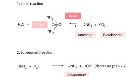
Figure 10.2 Urease-producing bacteria hydrolyse urea to ammonia molecules and carbon dioxide.
Q. Which bacteria produce urease?
A. Gram —ve
Proteus (mirabilis)
Providencia Klebsiella Pseudomonas Gram +ve
Staphylococcus
Mycoplasma
Ureaplasma urealyticum