
Q. A 20-year-old man attends casualty with a 7-hour history of a painful erection. How would you assess him?
A. A prolonged erection of this duration is due to priapism which is a urological emergency. I would see the patient immediately in the emergency department. I would ensure that he has adequate analgesia including a local anaesthetic penile block, intravenous access and then perform baseline blood tests which would include a full blood count (FBC) and sickle cell screen if indicated, renal function and electrolytes. I would then enquire as to whether this is the first or recurrent episode of the problem. An accurate history is required in order to establish as to whether this is an ischaemic or non-ischaemic priapism.
Q. What is important in the history?
A. In the history the important features are whether the erection is related to sexual stimulation or not (unlikely if it is 7 hours), the onset and duration of the erection and whether it is painful. It is then important to ask about any specific risk factors such as pelvic, genital or perineal trauma which may precede this episode by several weeks, any pharmacotherapy for erectile dysfunction, both oral and intracavernous injections, other medications particularly antipsychotics, a history of haematological disease such as sickle cell disease or leukaemia. This may be a first episode in which case document the pre-existing erectile function. However, in a proportion of patients a priapism episode is preceded by recurrent short selflimiting erections termed stuttering priapism.
Q. What is important in the examination?
A. It is important to ensure that analgesia is given before examining the patient. The baseline blood pressure and pulse must be recorded. A focused urological examination is performed to assess for any signs of trauma or which may have precipitated the event. The genitalia, perineum and abdomen should be carefully examined to assess for evidence of trauma and to ensure that there is no obvious intra-abdominal or pelvic lesion. Older patients should have a careful PR examination to exclude an underlying advanced pelvic malignancy. The degree of rigidity of the corpus cavernosum and glans may indicate the of priapism subtype.
Q. What types of priapism are you aware of?
A. The two common types of priapism are either ischaemic (low-flow) or non-ischaemic (high- low). A more uncommon subtype is stuttering priapism or recurrent priapism.
Q. How would you distinguish between the two types of priapism?
A. On the basis of the history, examination and with the aid of diagnostic investigations such as a cavernous blood analysis and radiological investigations such as penile Doppler ultrasound.
Q. How would you manage him in casualty?
A. After ensuring adequate analgesia and taking a focused history and examining the patient,
I would send off baseline blood tests including a full blood count and sickle cell screen or haemoglobin electrophoresis, renal function and electrolytes. I would then perform a penile block and aspirate blood from the corpus cavernosum using a large 19-gauge butterfly needle inserted through the glans penis and into the distal corporal tip or inserted into the side of the penis. Following aspiration of blood, a sample is sent for blood gas analysis and if possible glucose analysis. I would then continue aspirating blood (up to 100 mL) from the corpus cavernosum until the penis is flaccid.
Q. The blood gas analysis is shown as follows. The blood was dark when aspirated. What is the likely priapism subtype of priapism?
PO2 12 mm Hg PCO2 72 mm Hg pH 7.1
Glucose 0.2 mmol/L
A. The blood gas analysis is consistent with ischaemia as there is evidence of hypoxia and acidosis and glucopenia. There fore this is an ischaemic (low flow) priapism.
Q. What is the success rate of cavernosal aspiration in low flow priapism?
A. Reported success rates are 24%-36% but this is dependent on the duration of the priapism at presentation.
Q. When is aspiration alone successful in the management of priapism?
A. When it is done at an early stage generally within 12 hours.
Q. Are there any alternate ways of differentiating low and high flow priapism without cavernosal aspiration?
A. Yes, penile Doppler ultrasound may be utilised as an alternative to differentiate between ischaemic and non-ischaemic priapism. Patients with ischaemic priapism have little or no flow in the cavernosal arteries and corpus cavernosum on duplex ultrasound, whereas non ischaemic priapism shows high peak systolic velocities and a possible fistula.
Q. The priapism responds to cavernosal aspiration but then 2 hours later re-occurs. Now what would you do?
A. A step-wise approach is recommended. I would move the patient to an area where he can be monitored haemodynamically. I would repeat the aspiration using a large-gauge butterfly needle in 50 mL portions and also wash out the corpus cavernosum. If the priapism persists I would inject an a-agonist such as phenylephrine into the corpus cavernosum. I would also ensure that the Hb level is normal and that this is not a manifestation of an acute sickle cell crisis.
Q. How would you administer the phenylephrine?
A. Phenylephrine is available as 10 mg in 1 mL aliquots. I would dilute the phenylephrine in 19 mL of saline such that the concentration becomes 0.5 mg/mL. I would then inject 0.5 mL aliquots (250 g,g) every 10-15 minutes until detumescence occurs. Careful monitoring of the blood pressure (BP) and pulse is required.
Q. Assuming he presents with an ischaemic priapism beyond 24 hours’ duration which has not responded to corporal blood aspiration and a-agonists. Now what will you do?
A. I would then proceed to consenting the patient for shunt surgery.
Q. What types of shunts are you aware of?
A. Proximal and distal shunts.
Q. What types of distal shunts are you aware of?
A. I am aware of the Winter shunt (large biopsy needle) or Ebbehoj shunt (scalpel) where a fistula is created between the glans and corpus cavernosum through the glans. ttere is also a T-shunt described by Lue again using a scalpel through the glans and into the corporal tip followed by a 90° rotation.
Q. What is the shunt in Figure 13.10?
A. Figure 13.10 is a picture of an Al-Ghorab shunt where a piece of the tunica albuginea is excised at the tips of the corpora cavernosa via a dorsal transverse incision (on each side) distal to the coronal ridge.

Figure 13.10
Q. A Winter shunt is performed bilaterally. However, this again fails to resolve the priapism. What is your next step of management?
A. The easiest shunt to perform is the T-shunt. If this fails despite being performed on both sides, I would proceed to a tunnelling procedure using a narrow size 8 Hegar dilator and inserting this into the glans and corpus cavernosum and repeating this on the opposite side.
Q. What types of proximal shunt are you aware of?
A. I am aware of the Quackels (corporo-spongiosal) and the Grayhack (corporo-saphenous) procedures.
Q. What are the success rates of shunt procedures and are there long-term problems?
A. The literature suggests success rates of 73%-77% for the shunt procedures, but this depends on the duration of the priapism. Although they may succeed in detumescence there is a high rate of long-term erectile dysfunction (over 90%).
Q. Despite undergoing shunt procedures, the priapism persists. It is now 72 hours since the priapism began. What is your plan now?
A. I would again discuss the case with a specialist centre. It is likely that he will require transfer to a tertiary centre.
Q. He is transferred to a tertiary centre. What do you think they will plan for him and why?
A. It is likely that they will undertake a penile MRI and explore the patient surgically and undertake a cavernosal muscle biopsy to determine whether necrosis has already occurred. This would allow patients with prolonged ischemic priapism with non-viable tissue to be treated with the immediate insertion of a malleable penile prosthesis. This minimises penile shortening and deformity and allows adequate rigidity for sexual function.
Q. What is the role of penile prosthesis insertion?
A. Prolonged ischaemic priapism using conservative measures or shunt surgery may result in cavernosal fibrosis, penile induration and shortening. Unfortunately the resulting erectile dysfunction is usually severe and unresponsive to pharmacotherapy and the subsequent placement of a penile prosthesis into a fibrotic penis can be extremely difficult and is associated with a higher complication rate. It is now recommended that in cases of severe prolonged low-flow priapism, a (malleable) penile prosthesis is inserted early to maintain penile length and allow for an exchange to an inflatable prosthesis at a later date.
Q. Another 36-year-old patient attends casualty with a persistent erection for >8 hours. What is important in the history?
A. In the history the important features are whether the erection is related to sexual stimulation or not, the onset and duration of the erection and whether it is painful or not. It is then important to ask about any relevant risk factors such as pelvic, genital or perineal trauma, therapy for erectile dysfunction or other medications, a history of haematological disease such as sickle cell or leukaemia and any previous history of neurological disease. This may be the first or a recurrent episode. It is imperative to ask about the previous erectile function as priapism may result in erectile dysfunction.
Q. The patient is in pain but the pain is in his perineum. What is important in his examination?
A. It is important to ensure that analgesia is given before examining the patient. The baseline blood pressure and pulse must be recorded. A focused urological examination is performed to assess for any signs of trauma or infection which may have precipitated the event. In this case it is important to assess the genitalia, perineum and abdomen carefully for evidence of trauma. Again the degree of rigidity of the corpus cavernosum and glans may indicate the type of priapism present.
Q. He has severe bruising of his genitalia. He was involved in a motorbike accident earlier in the evening but did not attend casualty as he was not seriously injured. How would you manage him?
A. I would make sure he is comfortable. I would send off baseline blood tests and ensure intravenous access. It is important to exclude any other co-existing injuries. Once this has been done, I would ask a radiologist to perform a penile duplex ultrasound on him.
Q. Would you aspirate the corpus cavernosum?
A. As the history and examination are highly suggestive of a high-flow priapism I would aspirate a small amount of blood to confirm the diagnosis but only after the penile duplex has been performed to prevent aberrant blood flow.
Q. ’Шє duplex ultrasound confirmed high-flow priapism. How would you manage this patient?
A. This is not a urological emergency and can be managed conservatively. If a fistula is demonstrated on the Duplex ultrasound, compression with the probe can be applied.
Q. The patient is discharged home after undergoing investigations and confirming the diagnosis. His priapism persists and he returns after 1 week. He requests treatment. How would you manage him?
A. The site of the vascular injury may be diagnosed by pudendal angiography with superselective embolisation of any fistula that is seen.
Q. What type of material would you recommend he is embolised with?
A. Non-absorbable materials (39%) used during embolisation pose a greater risk of erectile dysfunction than absorbable (5%) materials. It is There fore recommended that autologous clots and absorbable gels are preferable to coils and permanent chemicals.
Q. How successful is embolisation for high-flow priapism?
A. The literature suggests the success rate is 74%-78% regardless of whether absorbable or nonabsorbable materials are used.
Q. Unfortunately the embolisation fails. He has had persisting priapism for 6 weeks. What would you do?
A. I would ask for a further colour duplex ultrasound and consider repeating the embolisation.
If imaging demonstrates a thick-walled cystic mass, I would counsel the patient for an open penile exploration and direct ligation. However, this only occurs rarely.
Q. What is the success rate and risk of complications?
A. The literature suggests that open exploration and ligation is successful in 63% of cases but has a very high risk of ED (up to 50%).
Q. You are asked to see an 86-year-old man in casualty. He has a long-term catheter in situ and is from a nursing home. You are told that he has scrotal swelling (Figure 13.11). What are you concerned about the most?
A. Figure 13.11 shows Fournier’s gangrene, which is a form of necrotizing fasciitis affecting the perineum and male genitalia.

Figure 13.11
Q. What are the important aspects in the history?
A. It is important to ascertain the age of the patient, whether this has appeared suddenly or insidiously and since when. It is also important to enquire about risk factors such as:
Any recent instrumentation of the urinary tract?
Any recent surgery in the ano-genital area or gynaecological procedures if a female?
Is a long-term catheter present?
Is there reduced mobility? In particular wheelchair bound/paraplegic/bed bound?
Is the patient normally continent?
Are there any co-morbidities which may result in immunosuppression, i.e. diabetes, alcohol abuse or steroid treatment?
Q. What is important in the clinical examination?
A. It is important to first resuscitate these patients aggressively as they are often very sick.
It is important to record the vital observations such as the temperature, BP, O2 saturations, pulse, peripheral circulation, sensorium and urine output to exclude signs of signs of shock.
A focused uro-genital examination is required including an evaluation of the perineum, perianal region and the genitals. Specifically one is looking for any areas of skin necrosis and the presence or absence of crepitus in the anterior abdominal wall. Perianal involvement signifies an anorectal source and the presence of skip lesions suggests more extensive involvement.
Q. Where does the infection normally arise from?
A. Infection most commonly arises from the skin, urethra or anorectal regions. There is an association between stricture disease and urethral instrumentation and the development of Fournier’s gangrene. Predisposing factors include diabetes mellitus, local trauma, paraphimosis, periurethral extravasation or urine, perirectal or perianal infections, and surgery such as circumcision or hernia repair.
Q. How are you going to manage him?
A. Prompt diagnosis is critical because of the rapidity with which the process can progress.
It may initially be difficult to differentiate the necrotizing fasciitis from cellulitis but the presence of marked systemic toxicity out of proportion to the local finding is often found.
I would then make sure he is transferred urgently to a urological ward or intensive therapy unit/high-dependency unit (ITU/HDU) depending on his clinical severity. There I would ensure he is resuscitated with adequate intravenous hydration and antimicrobial therapy so that he may be suitably prepared for surgical debridement.
Q. What investigations are required?
A. I would ensure baseline bloods tests are performed (FBC, urea and electrolytes [U&E], liver function test [LFT] Group and Save (G+S) and Glucose) as well as blood gases to exclude a metabolic acidosis. I would send cultures of the blood and urine as well as culture of any obvious pus from the affected region. In severe cases, I would ask the anaesthetist to insert appropriate arterial and intravenous lines for monitoring before surgery. If possible a computed tomography (CT) scan is useful pre-operatively to identify the possible source of infection.
Q. Which organisms are usually responsible?
A. There is a synergistic action so normally multiple organisms are present. The most common organism is Escherichia coli, however they are often mixed containing facultative organisms (E. coli, Klebsiella, enterococci) along with anaerobes (Bacteroides, Fusobacterium, Clostridium, microaerophilic streptococci).
Q. Which antibiotic do you plan to use?
A. I would use an antimicrobial regimen recommended following discussion with the microbiologist. It would commonly include triple therapy such as co-amoxiclav or a parenteral third-generation cephalosporin such as ceftriaxone, along with gentamicin, and metronidazole.
Q. How would you consent the patient?
A. The patients are often gravely ill and unable to give consent. It is There fore important to involve the family early and explain that the patient needs an operation as a matter of extreme urgency. I would explain the gravity of the situation and that more than one procedure is likely to be required. I would explain that we need to remove the subcutaneous gangrenous tissue and that a urinary diversion with a supra-pubic catheter is likely. In the longer term I would explain that large skin and subcutaneous tissue defects are likely which may require plastic surgery for functional and cosmetic results. I would explain that the patient would be required to stay in ITU/HDU depending on his clinical condition for optimal support.
Q. Explain what you will do in the theatre?
A. An incision should be made through the skin and subcutaneous tissues, going beyond the areas of involvement which show end arteritis until normal fascia is found and the subcutaneous tissue is bleeding. Necrotic fat and fascia should be excised, and the wound should be left open. A second procedure 24 hours later is always indicated. A suprapubic diversion should be performed in cases in which urethral trauma or extravasation is suspected. Colostomy should be performed if there is colonic or rectal perforation.
Q. Do these patients normally require an orchidectomy?
A. Orchidectomy is almost never required, because the testes have their own blood supply independent of the compromised fascial and cutaneous circulation to the scrotum.
Q. What is his long-term outlook?
A. The literature suggests that the mortality rate averages approximately 20%. Higher mortality rates are found in diabetics, alcoholics and those with colorectal sources of infection who often have a less typical presentation, greater delay in diagnosis, and more widespread extension.
Q. Are there any scoring systems which can predict mortality and outcome in these patients?
A. The mortality risk can be assessed using the Laor scoring system (Fournier’s gangrene severity index) which looks at parameters on admission including the temperature, heart rate, respiratory rate, sodium, potassium, creatinine, packed cell volume and whole blood cell count. A score of over 9 predicts mortality in 75%.
Q. Are there any adjunctive therapies which may be helpful in wound healing?
A. Using hyperbaric oxygen therapy in patients with Fournier’s gangrene has reported favourable results. Hyperbaric oxygen therapy has shown some promise in shortening hospital stays, increasing wound healing, and decreasing the gangrenous spread when used in conjunction with debridement and antimicrobials.
A small recent study suggests that vacuum-assisted closure (VAC) is equally effective in healing the wounds as compared to conventional management. However with the use of VAC, patients had fewer dressing changes, less pain, fewer skipped meals and greater mobility resulting in greater patient and physician satisfaction.
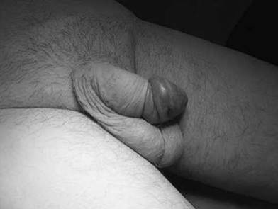
Figure 13.12
Q. You are asked to see a patient on the ward who had a TURP 2 days ago. He has been complaining of penile pain. The nurse is asked to remove the catheter. What is the diagnosis (Figure 13.12)?
A. Figure 13.12 shows a paraphimosis.
Q. How does it commonly occur?
A. It is often iatrogenic and frequently occurs after a well-meaning healthcare professional has examined the penis or inserted a urethral catheter and forgothen to replace the foreskin in its natural position. It develops when the tip of the foreskin retracts proximal to the coronal sulcus and becomes fixed in position and develops a constriction ring. Severe oedema of the foreskin occurs within several hours, depending on the tightness of the ring of the foreskin.
Q. What is your management plan?
A. In most cases, manual compression of the glans with placement of distal traction on the oedematous foreskin allows reduction of the paraphimotic ring.
Q. What is different about the dorsal band traction technique and how is it performed?
A. Most methods of reduction of paraphimosis focus on decreasing the oedema before reduction. This technique uses the basic surgical principles of traction and countertraction by applying a pair of Adson forceps directly to the band formed by the retracted preputial opening.
Q. What is the Dundee technique?
A. This is a technique in which the oedematous prepuce is first cleaned with an antiseptic cream and then a 26-gauge needle (outer diameter 0.45 mm) is used to make ~20 puncture holes in the oedematous prepuce. Using gentle but firm pressure, the oedema fluid is then expressed from the foreskin until it had been completely decompressed, allowing easy reduction of the prepuce.
Q. What will you do once the foreskin is reduced?
A. If the tip of the foreskin is tight, then there is a risk of recurrence. I would then list the patient for an elective circumcision. However as the tissue planes can be difficult it is advisable to wait until the oedema has settled completely. If all procedures fail it is possible to perform a dorsal slit under a local anaesthetic.
References
1. Mulcahy JJ. Penile implant infections: Prevention and treatment. Curr Urol Rep 2008; 9: 487-491.
2. Evers JL et al. Surgery or embolisation for varicocele in subfertile men. Cochrane Database Syst Rev 2004; (3): CD000479.
3. Hancock P, Woodward BJ, Muneer A, Kirkman-Brown JC. Laboratory guidelines for postvasectomy semen analysis: Association of Biomedical Andrologists, the British Andrology Society and the British Association of Urological Surgeons. J Clin Pathol 2016; 69(7): 655-660.
4. Belker AM et al. Results of 1,469 microsurgical vasectomy reversals by the Vasovasostomy Study Group. J Urol 1991; 145: 505-511.
5. Ralph DJ et al. The Nesbit operation for Peyronie’s disease: 16-year experience. J Urol 1995; 154: 1362-1363.
1. Mulcahy JJ. Penile implant infections: Prevention and treatment. Curr Urol Rep 2008; 9: 487-491.
2. Evers JL et al. Surgery or embolisation for varicocele in subfertile men. Cochrane Database Syst Rev 2004; (3): CD000479.
3. Hancock P, Woodward BJ, Muneer A, Kirkman-Brown JC. Laboratory guidelines for postvasectomy semen analysis: Association of Biomedical Andrologists, the British Andrology Society and the British Association of Urological Surgeons. J Clin Pathol 2016; 69(7): 655-660.
4. Belker AM et al. Results of 1,469 microsurgical vasectomy reversals by the Vasovasostomy Study Group. J Urol 1991; 145: 505-511.
5. Ralph DJ et al. The Nesbit operation for Peyronie’s disease: 16-year experience. J Urol 1995; 154: 1362-1363.