
Q. What do you understand by the term staging and why is it important in testicular cancer? What imaging modalities do we commonly use to stage testicular cancer?
A. Staging is a process by which clinically, radiologically and pathologically the extent of the disease is defined to prognosticate for relapse and survival for the patient, and the need for additional treatments.
The mainstay of radiological staging is abdominal CT to assess retro-peritoneal nodes and chest CT to assess the lungs and mediastinum. The EAU recommends a chest CT for both seminomas and NSGCTs. Occasionally there can be a role in special select cases for MRI in those with a contrast allergy for whom a CT cannot be used and PET scanning in re-staging residual masses following chemotherapy, specifically in seminoma. CT scans of the head for brain metastases and liver imaging are used where clinically indicated.
Q. What staging systems do you know for testis cancer?
A. There are clinical and pathological staging systems. We use primarily the American Joint Committee on Cancer (AJCC) staging classification of TNM and serum markers (Table 2.2).
The AJCC TNM stage groupings of testicular germ cell tumours (Table 2.3) are similar to the Royal Marsden Staging System (Table 2.4). Stage I disease is that confined to the testis, stage II has varying degrees of retroperitoneal nodal involvement depending on size (stage IIA, B and C) and stage III indicates supra-diaphragmatic and visceral metastatic disease with varying degrees of raised tumour markers. The use of tumour markers in the TNM staging is unique to testis cancer.
Other staging systems include the Boden/Gibbs classification and the previously mentioned Royal Marsden system (Table 2.4), which are similar to the AJCC TNM stage grouping classification (Table 2.3).
Table 2.2 TNM classification of testicular cancer
|
pT |
Primary tumour |
|
pTX |
Primary tumour cannot be assessed (T-Primary Tumour) |
|
pT0 |
No evidence of primary tumour (e.g. histological scar in testis) |
|
pTis |
Intratubular germ cell neoplasia (carcinoma in situ) |
|
pTI |
Tumour limited to testis and epididymis without vascular/lymphatic invasion: tumour may invade the tunica albuginea but not tunica vaginalis |
|
PT2 |
Tumour limited to testis and epididymis with vascular/lymphatic invasion, or tumour extending through tunica albuginea with involvement of tunica vaginalis |
|
pT3 |
Tumour invades spermatic cord with or without vascular/lymphatic invasion |
|
pT4 |
Tumour invades scrotum with or without vascular/lymphatic invasion |
|
N |
Regional lymph nodes clinical |
|
NX |
Regional lymph nodes cannot be assessed |
|
N0 |
No regional lymph node metastasis |
|
N1 |
Metastasis in a lymph node mass 2 cm or less in greatest dimension or multiple lymph nodes, none more than 2 cm in greatest dimension |
|
N2 |
Metastasis in a lymph node mass more than 2 cm but not more than 5 cm in greatest dimension or multiple lymph nodes, any one mass more than 2 cm but not more than 5 cm in greatest dimension |
|
N3 |
Metastasis in a lymph node mass more than 5 cm in greatest dimension |
(Continued)
Table 2.2 (Continued) TNM classification of testicular cancer
|
pN |
Pathological |
|
|
pNX |
Regional lymph nodes cannot be assessed |
|
|
pN0 |
No regional lymph node metastasis |
|
|
pNI |
Metastasis in a lymph node mass 2 cm or less in greatest dimension and five or fewer positive nodes, none more than 2 cm in greatest dimension |
|
|
pN2 |
Metastasis in a lymph node mass more than 2 cm but not more than 5 cm in greatest dimension and five or fewer positive nodes, none more than 2 cm in greatest dimension |
|
|
pN3 |
Metastasis in a lymph node mass more than 5 cm in greatest dimension |
|
|
M |
Distant metastasis |
|
|
MX |
Distant metastasis cannot be assessed |
|
|
M0 |
No distant metastasis |
|
|
MI |
Distant metastasis |
|
|
MIa |
Non-regional lymph node(s) or lung |
|
|
MIb |
Other sites |
|
|
S |
Serum tumour markers |
|
|
SX |
Serum tumour markers not available or not performed |
|
|
S0 |
Serum marker study levels within normal limits |
|
|
LDH (U/l) |
hCG (mLU/mL) |
AFP (ng/mL) |
|
SI < I.5 x N and |
<5000 and |
< I000 |
|
S2 < I.5-I0 x N or |
5000-50,000 or |
I000-I0,000 |
|
S3 > I0 x N or |
>50,000 or |
> I0,000 |
Note: N indicates the upper limit of normal for the LDH assay. LDH - lactate dehydrogenase; hCG - human chorionic gonadotropin; AFP - alpha-fetoprotein.
Table 2.3 AJCC stage groupings for testicular tumours
|
Stage |
TNM classification |
|
Stage I |
pTI-4, N0, M0, SX |
|
Stage II |
Any pT/Tx, N1-3, M0, SX |
|
IIA |
Any pT/Tx, NI, M0, SX Any pT/Tx, NI, M0, SI |
|
IIB |
Any pT/Tx, N2, M0, S0 Any pT/Tx, N2, M0, SI |
|
IIC |
Any pT/Tx, N3, M0, S0 Any pT/Tx, N3, M0, SI |
|
Stage III |
Any pT/Tx, any N, MI, SX |
|
IIIA |
Any pT/Tx, any NI, MIa, S0 Any pT/Tx, any NI, MIa, SI |
|
IIIB |
Any pT/Tx, NI-3, MIa, S0 Any pT/Tx, any N, MIa, S2 |
|
IIIC |
Any pT/Tx, NI-3, M0, S3 Any pT/Tx, any N, MIa, S3 Any pT/Tx, any N, MIb, any S |
Table 2.4 Comparison of several staging systems for testicular cancer
|
Royal Marsden system |
TNM system |
Description |
|
I |
Tx, N0, M0 |
Disease confined to testis and peri-testicular tissue |
|
II A <2 cm B 2-5 cm C >5-10 cm D >10 cm |
Tx, N1 or N2a, M0 Tx, N2b, M0 Tx, N3, M0 |
Fewer than six positive lymph nodes without extension into retroperitoneal fat; no node >2 cm (infradiaphragmatic) Six or more positive lymph nodes, well-encapsulated and/or retroperitoneal fat extension; any node >2 cm Any node >5 cm |
|
III |
Tx, Nx, Ml |
Supradiaphragmatic and infradiaphragmatic lymphadenopathy (no extralymphatic metastasis) |
|
IV |
Disseminated disease (lungs, liver; bone) |
Q. A patient has an orchidectomy and histologically this is a seminoma that is confined to the testicle. His staging CT of chest, abdomen and pelvis is normal and he has normal tumour markers. What stage disease does he have?
A. This is clinical stage I disease. Stage I is disease confined to the testicle and surrounding structures without nodal spread or metastatic deposits (T1-4, N0, M0).
Stage I disease can be broken down further into IA where the T stage is T1 (confined to testis with no involvement of tunica vaginalis and no vascular invasion, stage IB where the tumour stage is T2-T4 but normal markers and stage IS where any T stage is accompanied by elevated serum tumour markers.
Q. Does this man need follow-up and if so by whom? What would you do with this man with clinical stage I seminoma?
A. Such patients are normally followed up by the oncologist. The purpose of follow-up is to detect distant disease relapse especially in the retroperitoneum and initiate early treatment.
For clinical stage I seminoma, about 15%-20% of men have subclinical metastatic disease usually in the retroperitoneum and will subsequently relapse after orchidectomy alone [17]. To minimise this rate of relapse an informed discussion with patients about the need for adjuvant treatment is undertaken that reduces this risk balanced with the risks of treatment.
Q. What options does this young man have for clinical stage I seminoma following radical inguinal orchidectomy?
A. The options are surveillance, single-dose adjuvant chemotherapy (carboplatin) following orchidectomy or adjuvant radiotherapy to the retroperitoneum.
Q. This man is interested in surveillance given that you have indicated to him an approximately 80%-85% chance of being cured by orchidectomy alone. What would you tell him in light of the above information about surveillance?
A. I would tell him that patients who choose surveillance have an 80%-85% cure rate with orchidectomy alone but will need close regular follow-up with which they must be compliant to look for relapse. If relapse were to occur it can be treated with adjuvant treatment resulting in an overall cancer-specific survival of 97%-100%. Given that ~16% will relapse [18] over a 5-year period of follow-up an intensive programme of clinic visits and imaging of the retroperitoneum by yearly CT for the first 4 years along with 6 monthly CXR along with physical examination and tumour marker assessment every 3 months is advocated. Patients have to be committed to this extensive surveillance approach.
In addition to the above, I would warn him that while the majority of relapses occur in the first few years of follow-up about 20% of late relapses occur after 4 years [17], and hence there is a necessity for prolonged follow-up which in some cases can be lifelong, with its attendant anxiety and psychological stress that this may generate in some patients. Any relapse following a period of observation requires a more intensive treatment schedule of radiotherapy or chemotherapy.
Q. Are there any prognostic factors that you are aware of for clinical stage I seminoma that may guide you in counselling this patient?
A. Yes. Data from retrospective meta-analysis of surveillance studies suggest that the tumour size (>4 cm) and invasion of the rete testis represent a group of men that are at higher risk for future relapse for stage I seminoma. Together, the presence of both these factors can represent a relapse rate of 32% versus 16% for the presence of one risk factor and 12% when both these risk factors are absent [19]. For this reason the notion of a risk-adapted approach has developed where patients at higher risk of failure are encouraged to undergo adjuvant therapy and those at lower risk can safely choose surveillance as a reasonable option in the first instance. Early data of the risk-adapted approach suggest that this approach can safely be used in this setting.
Q. What other options are available to him? What would you tell him about the other options for stage I seminoma?
A. He can consider adjuvant radiotherapy or chemotherapy. Both radiotherapy and chemotherapy are equally effective and reduce the risk of relapse in the retroperitoneum down to 3%-4% (from the 15%-20% risk from surveillance alone).
Radiotherapy is administered in a hockey-stick fashion to the para-aortic field and ipsilateral iliac nodes to a total dose of 20 Gy over a 2-week period, and chemotherapy consists of a single dose of carboplatin (EORTC trial AUC 7, MRC trial TE 19). After a follow-up of 4 years both have been shown to be equally effective [20].
Q. What do the oncologists in your unit generally now advise?
A. Given the equal efficacy of single-dose chemotherapy (i.e. one cycle of carboplatin) to radiotherapy, there is a general trend in offering the former as the preferred adjuvant modality for its ease of administration, shorter time to delivery and the recognised late secondary pelvic malignancy rate with the use of radiotherapy (albeit small and unquantified).
Q. This patient decides to have adjuvant chemotherapy. How would you then follow-up the patient?
A. Three-monthly clinic appointments with physical examination, tumour markers and 6 monthly CXRs and yearly CT scans for the first 3 years eventually stretching out at year 5 to annual assessments. The follow-up regime is more intensive in the first 2 years as this is the time when relapses are more likely to occur.
Q. Why do you think that follow-up needs to be extended in such cases?
A. While most disease relapse occurs within the first few years, there remains with seminoma a 20% risk of relapse after 4 years with some cases presenting after the 10-year mark and so some would advocate a lifelong follow-up schedule.
Q. If instead, following his staging investigations he presented with a CT as shown in Figure 2.4, what do you think this shows?
A. Figure 2.4: This shows the presence of enlarged retroperitoneal nodes encasing the inferior vena cava highly suggestive of metastatic disease in the retroperitoneum. This indicates that the patient has stage II disease or nodal metastatic disease.
In general, metastatic disease can be classified into low-volume metastatic disease which includes stages IIA and IIB (stage II disease can be divided into A [<2 cm nodal mass] and B [2-5 cm nodal mass]) and advanced metastatic disease which encompasses stage IIC (nodal mass >5 cm) and stage III disease (supradiaphragmatic or visceral metastases).
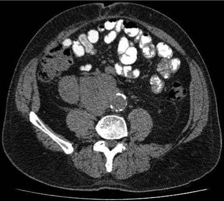
Figure 2.4
Q. What do you want to do with this patient now?
A. The mainstay of treatment for seminoma in this group (stage IIA/B) is three cycles of BEP chemotherapy (bleomycin, etoposide and cisplatin) or four cycles of EP (etoposide and cisplatin) for those in whom bleomycin should be avoided (age >40, smokers). There are, however, some centres that still would offer radiotherapy, 30-36 Gy, but this is not the norm in UK practice despite being popular in Europe, especially for stage IIA disease.
Treatment in metastatic germ cell tumours is based on the prognostic groups defined by the International Germ Cell Cancer Consensus Group (IGCCCG) which have validated a model for germ cell tumours that separates patients into good, intermediate and poor prognostic groups based on histology, location of primary tumour, location of metastases and post-orchidectomy tumour marker levels (Table 2.5). Treatment and specifically chemotherapy is tailored according to this classification allowing high cure rates with minimal toxicity for the good prognostic group and more aggressive chemotherapy reserved for the poor prognostic group.
Table 2.5 The International Germ Cell Cancer Collaborative Group (IGCCCG) prognostic-based staging system for metastatic seminoma
|
Good prognosis group |
|
|
(90% of cases) |
All of the following criteria |
|
5-year PFS 82% |
Any primary site |
|
5-year survival 86% |
No non-pulmonary visceral metastases |
|
Normal AFP |
|
|
Any hCG |
|
|
Any LDH |
|
|
Intermediate prognosis group |
|
|
(10% of cases) |
Any of the following criteria |
|
5-year PFS 67% |
Any primary site |
|
5-year survival 72% |
Non-pulmonary visceral metastases |
|
Normal AFP |
|
|
Any hCG |
|
|
Any LDH |
|
|
Seminoma |
|
|
No patients classified as poor prognosis |
|
Note: PFS - progression-free survival.
Q. Following from the above he has induction chemotherapy and has a follow-up CT scan 3 months later, which shows the persistence of a retroperitoneal mass. What do you do now?
A. I would check his tumour markers in the first instance. Seminomatous residual masses following chemotherapy or radiation therapy are not common and can be observed by serial imaging without the need to resort to immediate resection of the retroperitoneal mass irrespective of size. PET scanning has been found to be particularly useful in this group of patients in separating retroperitoneal masses that have active tumour in them. Generally it is agreed that residual masses >3 cm that do not regress after therapy should undergo this imaging modality to look for evidence of active tumour. A positive PET scan after a 4-6 week interval following the end of chemotherapy/radiotherapy is a very reliable predictor for viable tumour tissue in this group of patients. For such patients confirmation by way of biopsy may require further treatment such as retroperitoneal lymph node dissection (RPLND), salvage chemotherapy (or radiotherapy in those who did not receive this initially). In general a residual mass after primary treatment in seminoma is rare. Surgery after treatment for seminoma is generally more difficult and restricted to patients with ‘globular’ masses rather than a retro-peritoneal fibrosis type of picture.
Q. If the patient presented with advanced seminoma from the outset (stage IIC and greater) what would the mainstay of treatment have been?
A. The mainstay of treatment is three or four cycles of chemotherapy with BEP or EP if contraindications to bleomycin exist (i.e. smoker, >40 years of age, high burden of pulmonary metastases) which again is based on the IGCCCG classification grouping system (Table 2.5). The final regime that the patient receives is guided by the prognostic group he falls into. For the good prognosis group this is three cycles of BEP, for the intermediate group this is four cycles. There is no poor prognosis group for seminomatous disease.