
The abdomen, the region of the trunk between the thorax and the pelvis, contains the largest portion of the abdominopelvic cavity, a peritoneal-lined space that it shares with the pelvis. The abdomen houses the primary organs of the gastrointestinal and urinary systems, although some abdominal viscera (i.e., small intestine) typically overflow the boundaries of the abdomen to occupy pelvic spaces, and pelvic viscera, when distended (i.e., bladder and uterus), can extend superiorly into the abdomen (Fig. 7.1).
The abdominal wall, composed of skin, fascia, and muscles, is supported by its attachments to the ribs, lumbar vertebrae, and bony pelvis. It moves and stabilizes the trunk, supports the abdominal viscera, and creates intra-abdominal pressure that is crucial in digestion and respiration. The muscular abdominal wall provides little protection for underlying viscera, but much of the upper abdominal viscera lie under the dome of the diaphragm, where they are protected by the thoracic skeleton. The bony pelvis protects most viscera in the lower abdomen.
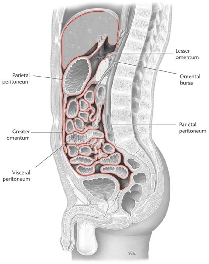
Fig. 7.1  Peritoneal relationships
Midsagittal section through male pelvis, viewed from the left side. Peritoneal cavity. The peritoneum is shown in red.
7.1 Regions and Planes of the Abdominal Wall
– In order to describe the location of abdominal viscera, we divide the abdomen into four quadrants or nine regions, using vertical reference lines and standard transverse planes (Fig. 7.2A and B).
– The transpyloric plane, a transverse plane measured halfway between the jugular notch and pubic crest, is a useful horizontal plane that provides orientation to the internal anatomy of the abdomen (Fig. 7.3; see also Fig. 1.4). The T12–L1 plane passes through (or very close to)
• the pylorus of the stomach,
• the ampulla of the duodenum,
• the celiac trunk,
• the superior mesenteric artery,
• the origin of the portal vein,
• the neck of the pancreas, and
• the left colic flexure of the large intestine.

Fig. 7.2 Criteria for dividing the abdomen into regions
A The abdomen is divided into four quadrants by two perpendicular lines that intersect at the umbilicus.
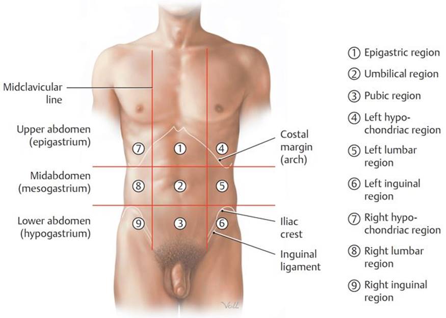
B Coordinate system composed of two vertical and two horizontal lines divide the abdomen into nine regions, each located in either the upper, middle, or lower abdomen. The two vertical lines are the left and right midclavicular lines. One of the two horizontal lines passes through the lowest point of the 10th ribs and the other through the summit of the two iliac crests.
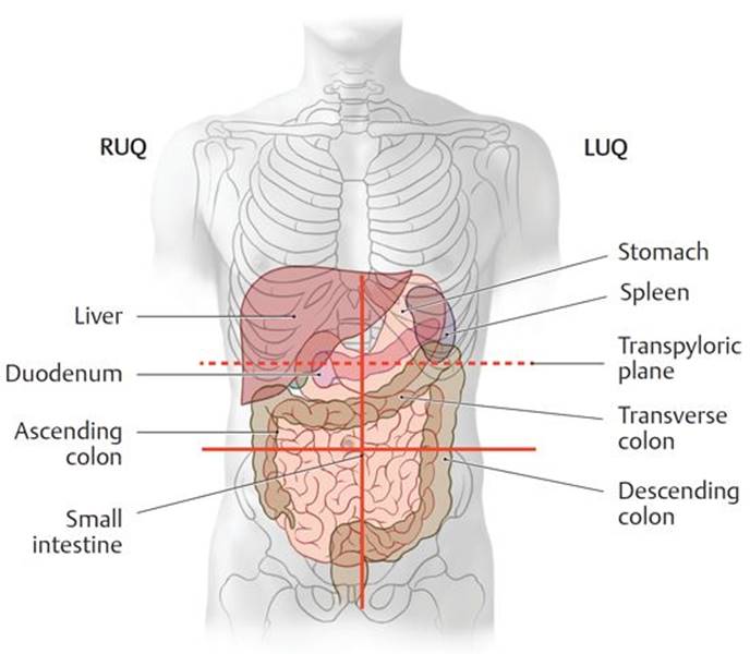
Fig. 7.3 Transpyloric plane (dashed red line) and its relationship to abdominal viscera
Anterior view. RUQ, right upper quadrant; LUQ, left upper quadrant.
7.2 Structure of the Abdominal Wall
7.1a Superficial Fascial Layer (see Fig. 7.5C)
– The superficial fascia lies deep to the skin and superficial to the muscular layer. It has two components:
• The superficial fatty layer (Camper’s fascia), a subcutaneous layer of fat whose thickness varies among individuals and that is continuous with the superficial fascia of the thorax, back, and lower limb
• The deep membranous layer (Scarpa’s fascia), a tough fibrous sheet that lies deep to the superficial fatty layer, covers the lower anterior abdominal wall, and extends inferiorly into the perineum, where it is continuous with the superficial perineal (Colles’) fascia.
7.1b Muscular Layer (Table 7.1)
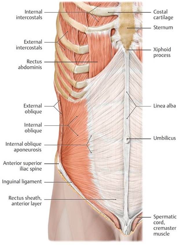
– Three flat muscles make up most of the muscular layer of the lateral and anterior walls of the abdomen: the external oblique, internal oblique, and transversus abdominis. Their large aponeuroses constitute the most anterior part of the abdominal wall (Fig. 7.4A, B, and C).
• The thickened inferior edge of the external oblique aponeurosis forms the inguinal ligament, which attaches laterally to the anterior superior iliac spine and medially to the pubic tubercle of the pubis (see Fig. 10.8A). Some fibers of the medial end of the ligament reflect downward as the lacunar ligament to attach to the superior edge of the pubis (see Table 7.2).
• Inferiorly, the aponeuroses of the internal oblique and transversus abdominis muscles join to form the conjoined tendon, where they attach to the pubis.
• In the anterior midline, the aponeuroses of the three muscles overlap with the contralateral muscles, forming the linea alba, a tendinous raphe (junction) that extends from the xiphoid process to the pubis. The umbilical ring, a remnant of the opening for the umbilical cord, interrupts the raphe at its midpoint.
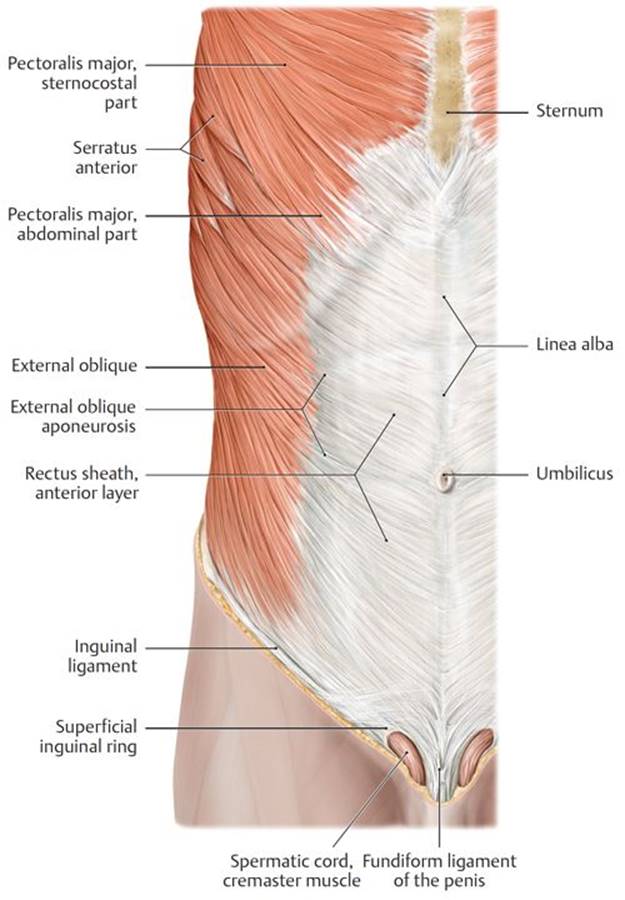
Fig. 7.4 Muscles of the anterior abdominal wall
Right side, anterior view.
A Superficial abdominal wall muscles.
B Removed: External oblique, pectoralis major, and serratus anterior.
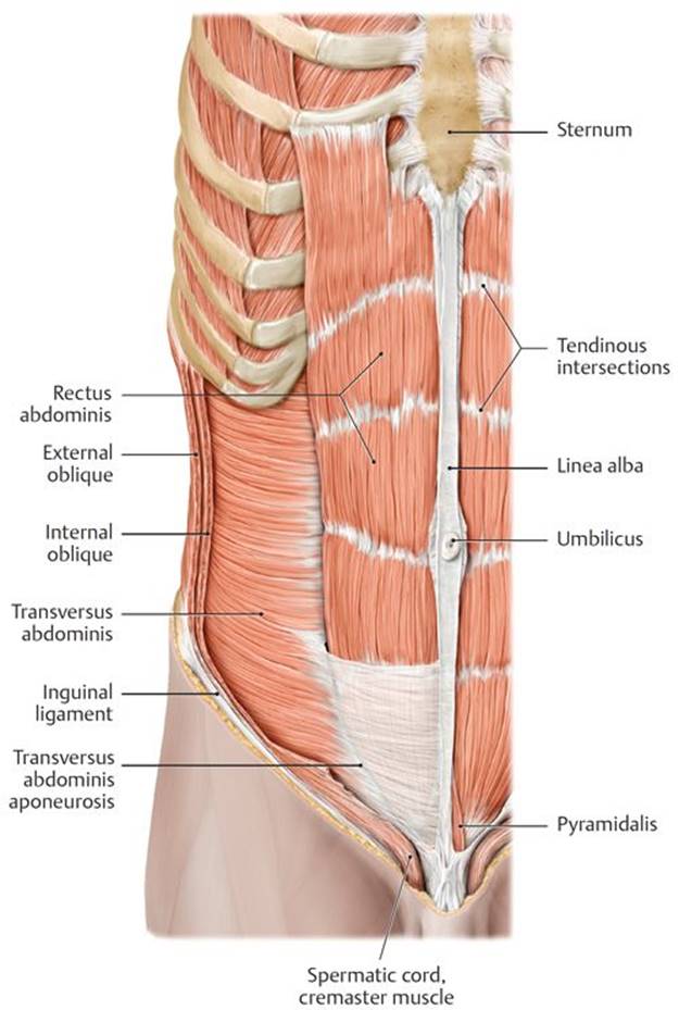
C Removed: Internal oblique.
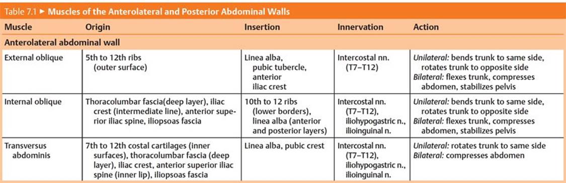

Fig. 7.5  Anterior abdominal wall and rectus sheath
A Posterior (internal) view of anterior abdominal wall.

B Section through the abdominal wall superior to the arcuate line.

C Section through the abdominal wall inferior to the arcuate line.
– A rectus sheath encloses the rectus abdominis and pyramidalis muscles on either side of the anterior midline (Fig. 7.5A, B, and C; see also Fig. 7.4C).
• The lateral edges of the sheath are visible externally as the semilunar lines.
• The sheath has anterior and posterior layers, formed by the aponeuroses of the anterolateral muscles as they split to pass around the rectus muscles.
• The arcuate line is the inferior end of the posterior layer of the rectus sheath. Inferior to this, the aponeuroses only pass anterior to the rectus muscles.
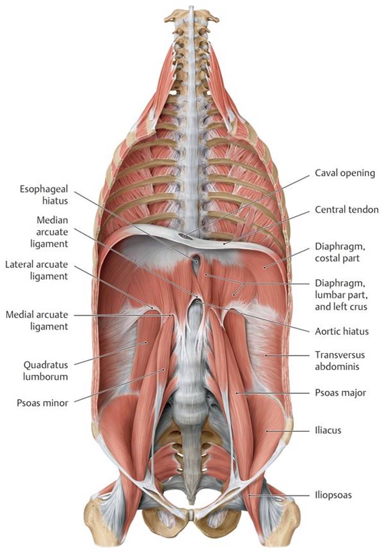
Fig. 7.6 Muscles of the posterior abdominal wall
Coronal section with the diaphragm in the intermediate position, anterior view.
– Five muscles form most of the posterior abdominal wall: the psoas major, psoas minor (sometimes absent), quadratus lumborum, iliacus, and diaphragm (Fig. 7.6).
• The psoas major and iliacus muscles unite to form the iliopsoas muscle, which passes into the thigh and acts on the hip joint.
• The thoracic diaphragm forms part of the superior portion of the posterior abdominal wall.
• The transversus abdominis muscle contributes to the lateral part of the posterior abdominal wall.
– Endoabdominal fascia is a deep fascial layer that lines the internal surface of the abdominal wall muscles. It lies superficial to (outside of) the parietal peritoneum and in most places is separated from it by a layer of fat called preperitoneal fat.
• Each part of the endoabdominal fascia is named for the muscle it lines: transversalis fascia (see Fig. 7.5B), diaphragmatic fascia, psoas fascia.
• In the inguinal region (groin), a thickened line of the transversalis fascia, the iliopubic tract, attaches to the inner edge of the inguinal ligament, where it supports the posterior wall of the inguinal canal (see Fig. 7.5A).
7.1c Internal Surface of the Anterior Abdominal Wall (Fig. 7.7)
The internal surface of the anterior abdominal wall is lined with transversalis fascia and parietal peritoneum, with a variable amount of intervening preperitoneal fat.
– Peritoneal folds form where structures tent the peritoneum as they course between it and the transversalis fascia. The folds include
• the median umbilical fold, a single midline fold created by the median umbilical ligament, a remnant of the urachus (a fetal connection between the bladder and umbilicus);
• the medial umbilical folds, paired folds created by the medial umbilical ligaments, remnants of the umbilical arteries in the fetus; and
• the lateral umbilical fold, paired folds created by the inferior epigastric vessels.
– Peritoneal fossae are formed between the peritoneal folds and are potential sites of herniation (protrusion of viscera through a wall or tissue). The fossae include
• the supravesical fossa between the median and medial umbilical folds;
• the medial inguinal fossa, commonly known as the inguinal triangle of Hesselbach (Hesselbach’s triangle) between the medial and lateral umbilical folds; and
• the lateral umbilical fossa, lateral to the lateral umbilical folds.
– The falciform ligament is a double-layered peritoneal reflection between the liver and the anterior abdominal wall that extends superiorly from the umbilicus to the roof of the abdominal cavity. It encloses the round ligament (remnant of the umbilical vein) and paraumbilical veins.
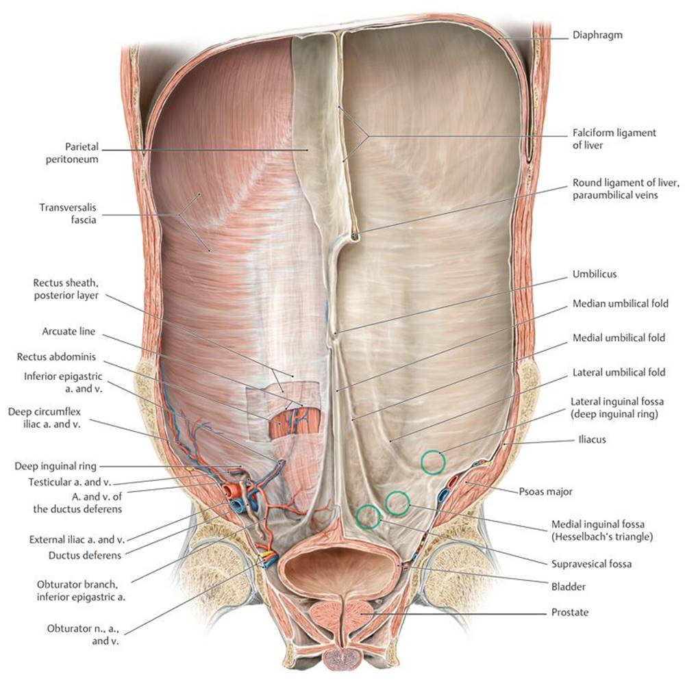
Fig. 7.7 Internal surface anatomy of the anterior abdominal wall in the male
Coronal section through the abdominal and pelvic cavity at the level of the hip joints, posterior view.
7.2 Neurovasculature of the Abdominal Wall (Fig. 7.8)
7.2a Arteries of the Abdominal Wall
Arteries of the abdominal wall, which anastomose extensively with one another, arise from the internal thoracic artery, the abdominal aorta, the external iliac artery, and the femoral artery.
– The branches of each internal thoracic artery are
• the musculophrenic artery and
• the superior epigastric artery, which descends within the rectus sheath posterior to the rectus abdominis muscle, where it anastomoses with the inferior epigastric artery.
– The paired segmental branches of the abdominal aorta are
• the intercostal, subcostal, and lumbar arteries.
– The branches of the external iliac artery are
• the inferior epigastric artery and deep circumflex iliac artery.
– The branches of the femoral artery in the thigh that supply the abdominal wall are
• the superficial epigastric artery and
• the superficial circumflex iliac artery.
7.2b Veins of the Abdominal Wall
– The deep veins of the abdominal wall accompany the arteries of similar name and drain to the superior and inferior venae cavae via the brachiocephalic, azygos, hemiazygos, and common iliac veins.
– An extensive subcutaneous venous network drains superiorly to the internal thoracic and lateral thoracic veins of the thorax and inferiorly to the inferior and superficial epigastric veins.
– Obstruction of the superior or inferior vena cava may alter the venous flow across the abdominal wall, resulting in the development or enlargement of a superficial anastomosis between the axillary and femoral veins through the thoracoepigastric vein (see Section 4.4).
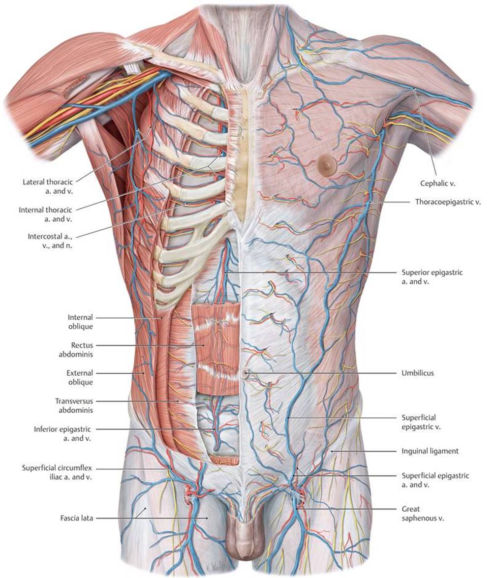
Fig. 7.8 Neurovascular structures of the anterior trunk wall
Anterior view. Left side: superficial dissection. Right side: deep dissection. Removed: pectoralis major and minor. Partially removed: external oblique, internal oblique, transversus abdominis, rectus abdominis, and intercostal muscles.
7.2c Lymphatic Drainage of the Abdominal Wall
– Lymphatic drainage of the abdominal wall is divided into upper and lower regions by a curved line (“watershed”) located between the umbilicus and costal margin (Fig. 7.9).
• From the upper region, lymph drains to axillary and parasternal nodes before draining to the right and left jugulosubclavian junctions (venous angles).
• From the lower region, lymph drains inferiorly to ipsilateral superficial inguinal nodes. These drain to external iliac and common iliac nodes and eventually to the thoracic duct.
7.2d Nerves of the Abdominal Wall
– Nerves of the abdominal wall arise from thoracic and lumbar spinal nerves and include (Fig. 7.10A and B)
• the lower intercostal nerves (T7–T11) and the subcostal nerve (T12) of the thorax, and
• the iliohypogastric nerve of the lumbar plexus.
– Dermatomes of the abdominal wall follow the slope of the ribs. Landmark dermatomes that correspond to visible surface features of the abdominal wall include T10 at the umbilicus and L1 at the inguinal ligament and top of the pubis.
7.3 The Inguinal Region
The inguinal region, or groin, includes the inferolateral region of the anterior abdominal wall (Fig. 7.11).

Fig. 7.9 Lymphatic pathways and regional lymph nodes of the anterior trunk wall
Anterior view. Arrows indicate direction of lymph flow.
7.3a Inguinal Canal
The inguinal canal is an oblique passage through the abdominal wall that allows structures to pass between the abdominal and pelvic cavities and the perineum. Deficiencies in the anterolateral abdominal muscles, their aponeuroses, and their deep fascia create the inguinal canal (Table 7.2).
– The boundaries of the canal are
• the anterior wall, formed by the aponeurosis of the external oblique muscle;
• the posterior wall, formed by transversalis fascia and conjoined tendon;
• the floor, formed by the inguinal ligament; and
• the roof, formed by the arching fibers of the aponeuroses of the internal oblique and transversus abdominis muscles.

Fig. 7.10 Cutaneous innervation of the anterior abdominal wall
A Sensory nerves of the anterior abdominal wall.
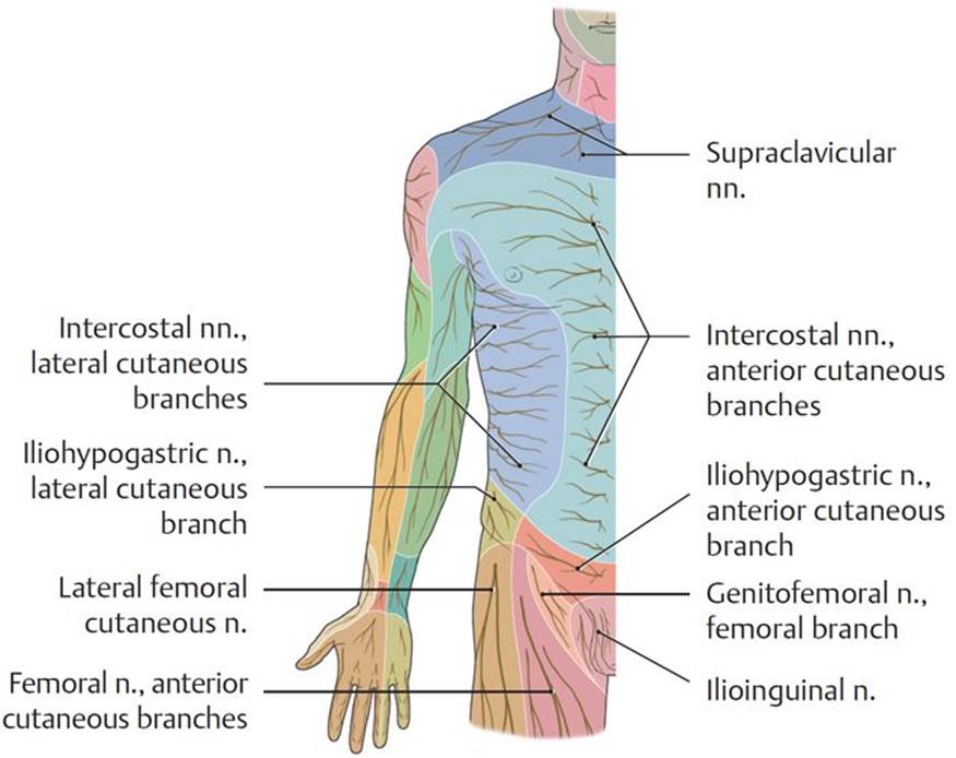
B Dermatomes of the anterior abdominal wall.
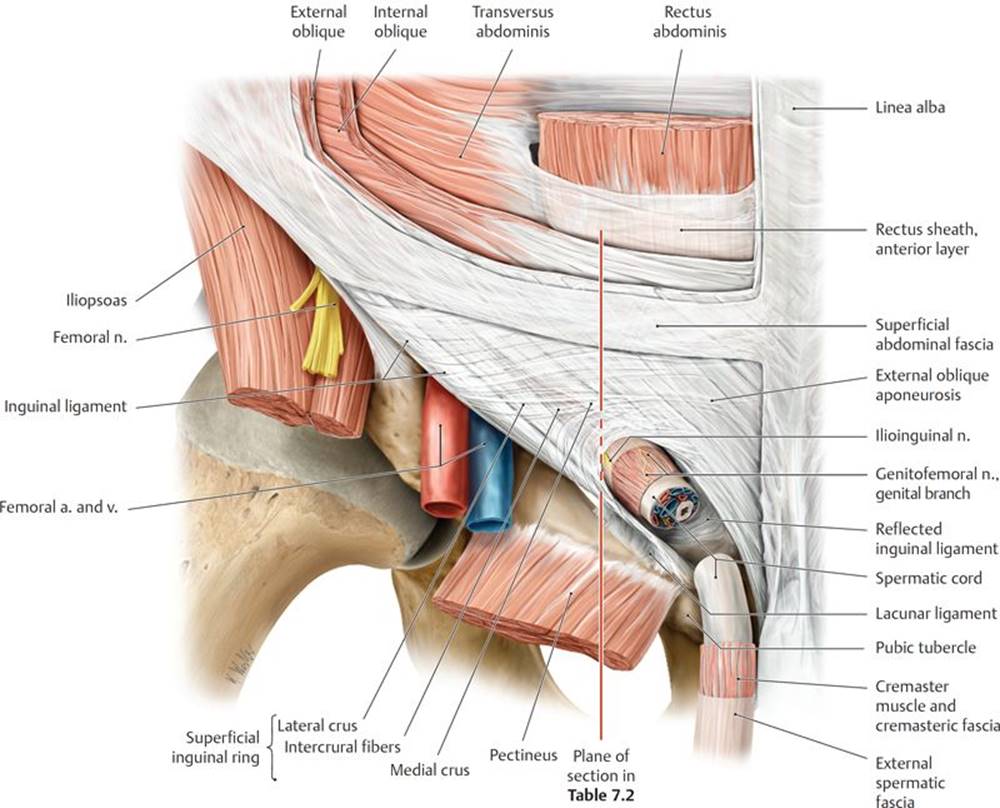
Fig. 7.11 Male inguinal region
Right side, anterior view.
TABLE 7.2  Structures of the Inguinal Canal
|
Structures |
Formed by |
|
|
Wall |
Anterior wall |
|
|
Roof |
|
|
|
|
||
|
Posterior wall |
|
|
|
|
||
|
Floor |
|
|
|
Openings |
Superficial inguinal ring |
Opening in external oblique aponeurosis; bounded by medial and lateral crus, intercrural fibers, and reflected inguinal ligament |
|
Deep inguinal ring |
Outpouching of the transversalis fascia lateral to the lateral umbilical fold (inferior epigastric vessels) |
|
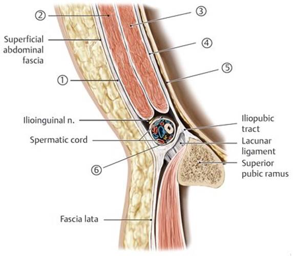
Inguinal region, sagittal section through plane in Fig. 7.11.
Inguinal hernia and hydrocele
Inguinal hernias account for the large majority of abdominal wall hernias, and of those, most occur in males. A hernia is the protrusion of a visceral structure into a space that it doesn’t normally occupy. Inguinal hernias involve the protrusion of parietal peritoneum, peritoneal fat, or the small intestine. Of the two types of inguinal hernias, the indirect hernia is the result of a congenital defect and is common in young males, whereas the direct hernia results from a weakening of the abdominal wall and generally occurs in middle-aged males.
During development, a tongue of peritoneum, the processus vaginalis, evaginates into the inguinal canal and accompanies the testis in its descent into the scrotum. Before birth most of the processes obliterate, closing the communication between it and the peritoneal cavity. If the processus vaginalis fails to obliterate, abdominal contents can herniate (indirect hernia) through its opening at the deep inguinal ring (lateral to the inferior epigastric vessels) and extend into the scrotum (or labia in females). Herniated viscera travel within the spermatic cord and are covered by the layers of the cord.
Weakening of the anterior abdominal wall in the inguinal triangle (medial to the inferior epigastric vessels) can allow viscera to protrude through the medial end of the canal and through an enlarged superficial ring (direct hernia). The herniated viscera travel outside the spermatic cord and are covered only by peritoneum and transversalis fascia of the abdominal wall.
The opening into a persistent processus vaginalis may be small enough to prevent herniation but large enough to form a hydrocele, the accumulation of excess peritoneal fluid. The hydrocele can be confined to the scrotum (hydrocele of the testis) or to the cord (hydrocele of the cord). Confirmation is by transillumination of the scrotum, which allows detection of the excess fluid.
– The canal has two openings:
• At the medial end of the canal, fibers of the external oblique aponeurosis split to create an opening known as the superficial inguinal ring. This ring lies in the anterior wall of the inguinal triangle of Hesselbach.
• At the lateral end of the inguinal canal, immediately lateral to the origin of the inferior epigastric vessels, the transversalis fascia evaginates into the canal and creates the deep inguinal ring. This ring lies in the lateral inguinal fossa (see Fig. 7.7).
– The contents of the inguinal canal include the spermatic cord in males and the round ligament of the uterus in females (Fig. 7.12; also see Section 11.2c).
7.3b The Spermatic Cord
The spermatic cord forms at the deep inguinal ring, traverses the inguinal canal, and exits through the superficial inguinal ring. It enters the scrotum and descends to the posterior surface of the testis (Fig. 7.13).
– The structures in the spermatic cord include
• the ductus deferens;
• the testicular artery and pampiniform plexus of veins, the artery of the ductus deferens, and the cremasteric artery;
• lymphatic vessels of the testis and spermatic cord; and
• sympathetic and parasympathetic fibers of the testicular plexus and the genital branch of the genitofemoral nerve.
– Derivatives of the muscles and fascia of the abdominal wall surround the contents of the spermatic cord as they pass through the inguinal canal. The layers formed by the muscles and fascia are the same as those surrounding the testis (Table 7.3):
• Internal spermatic fascia derived from the transversalis fascia
• Cremaster muscle and cremasteric fascia derived from the internal oblique muscle and fascia
• External spermatic fascia derived from the external oblique aponeurosis and fascia
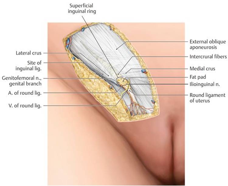
Fig. 7.12 Female inguinal region
Right side, anterior view.

Fig. 7.13 Spermatic cord
Male pelvis, anterior view. Opened: Inguinal canal and coverings of the spermatic cord.
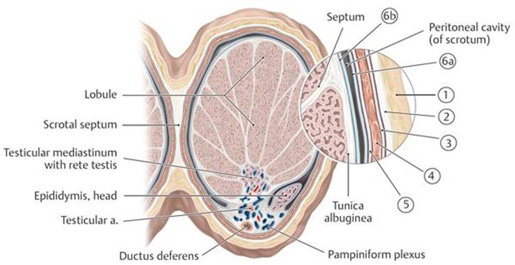
Transverse section through right testis, superior view.
TABLE 7.3 Coverings of the Testis
|
Covering of the Testis |
Abdominal Wall Derivative |
|
|
Abdominal skin |
|
|
Dartos fascia and muscle |
|
|
External oblique fascia |
|
|
Internal oblique muscle and fascia |
|
|
Transversalis fascia |
|
|
Peritoneum |
|
|
|
|
* The transversus abdominis has no contribution to the spermatic cord or covering of the testis. |
7.3c The Testes
The testes are paired ovoid reproductive organs, 4 to 5 cm long and 3 cm wide, located in separate compartments within the scrotum. They produce spermatozoa and secrete the male hormone testosterone (Fig. 7.14A and B; see also Table 7.3).
– An extension of the peritoneum known as the tunica vaginalis forms a closed sac that folds around the testis, surrounding it on all sides except on its posterior edge. The tunic has an outer parietal layer and an inner visceral layer that is adherent to the surface of the testis.
– Each testis is enveloped by the tunica albuginea, a tough capsule of connective tissue that thickens along the posterior border as the mediastinum of the testis and invaginates to divide the testis into over 200 lobules.
– Sperm are produced in the seminiferous tubules, highly coiled tubules within the lobules. They exit the testes through a ductal network, the rete testis in the mediastinum, and then pass through efferent ductules to the epididymis.
– The testicular artery, a branch of the abdominal aorta, supplies the testis. A rich collateral blood supply arises from anastomoses with the artery of the ductus deferens, the cremasteric artery, a branch of the inferior epigastric artery, and the external pudendal artery from the femoral artery (Fig. 7.15).
– The pampiniform plexus of veins drains the testis and converges to form the testicular vein. The testicular veins drain to the inferior vena cava on the right and to the renal vein on the left.

Fig. 7.14 Testis and epididymis
Left lateral view.
A Testis and epididymis in situ.
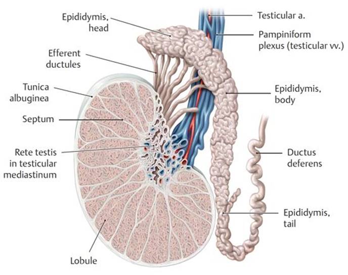
B Sagittal section.

Fig. 7.15 Blood vessels of the testis
Left lateral view.
Varicocele
The pampiniform plexus from each testis surrounds the testicular artery and converges to form a testicular vein. When the venous valves become incompetent, the plexus can become dilated and tortuous, forming a varicocele that is often reported to feel like “a bag of worms.” Varicoceles are predominantly on the left side. This is generally attributed to the abrupt termination of the left testicular vein at the left renal vein, which may slow venous return.
– The lymph vessels of the testes drain directly to lateral aortic and preaortic lymph nodes.
– The cremasteric reflex, initiated by stroking of the inner thigh, contracts the cremaster muscle and elevates the testis. The ilioinguinal nerve provides the sensory limb; the genital branch of the genitofemoral nerve provides the motor limb.
Absent or reduced cremasteric reflex
An absent or reduced cremasteric reflex that is accompanied by sudden testicular pain, inflammation and elevation of one testis, nausea, and vomiting may indicate testicular torsion (twisting of a testis). Prompt surgery for testicular torsion (to untwist the affected testis and anchor both testes) may prevent loss of a testis.
– The testicular nerve plexus arises from the aortic plexus and travels along with the testicular artery. It contains sympathetic fibers from the T7 spinal cord level, as well as visceral afferent and vagal parasympathetic fibers.
Testicular cancer
Testicular cancer is the most common cancer in males between 15 and 34 years of age. The vast majority of these cancers are seminomas or germ cell tumors that arise in the germ cells that produce immature sperm. Symptoms include a lump in the affected testis (usually only one testis is affected), a feeling of heaviness in the scrotum, pain in the affected testis or scrotum, a sudden collection of fluid in the scrotum, and the development of excess breast tissue (gynecomastia). Testicular cancer commonly metastasizes via lymph nodes to the lungs or via the bloodstream, commonly to the liver, lungs, brain, and spine.
7.3d The Epididymis and Ductus Deferens
The epididymis and ductus deferens are parts of the male ductal system that transport sperm from the testis to the genital structures in the pelvis (see Fig. 7.14A and B).
– The epididymis, a highly coiled tubule where sperm are stored and mature, hugs the posterior surface of the testis. Its expanded head contains the lobules with the efferent ductules, its body is made up of a long convoluted duct, and its tail is continuous with the ductus deferens.
– The ductus deferens is a muscular tube that transmits sperm from the scrotum to the pelvis.
• It begins at the tail of the epididymis and continues as part of the spermatic cord through the inguinal canal.
• At the deep inguinal ring, the ductus deferens descends into the pelvis posterior to the bladder, where, near its termination, it enlarges as the ampulla of the ductus deferens (see Fig. 11.2) .
• The ampulla joins with the duct of the seminal gland (vesicle) to form the ejaculatory duct (see Section 11.1a).
 External oblique aponeurosis
 Internal oblique muscles
 Transversus abdominis
 Transversalis fascia
 Parietal peritoneum
 In guinal ligament (densely interwoven fibers of the lower external oblique aponeurosis and adjacent fascia lata of thigh)
 Tunica vaginalis, parietal layer
 Tunica vaginalis, parietal layer