
The pelvic cavity contains the male or female genital organs, the pelvic urinary organs, and the rectum. These organs normally reside in the true pelvis, although when enlarged the bladder and uterus can extend into the abdominal cavity.
11.1 Male Genital Structures
The male gonad, the testis, is located in the inguinal region and is discussed in Chapter 7. The seminal glands (vesicles) and prostate gland are male accessory reproductive structures found in the pelvis (Fig. 11.1).
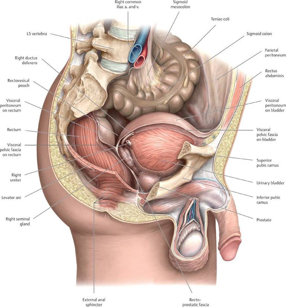
Fig. 11.1  Male pelvis
Parasagittal section, viewed from the right side.

Fig. 11.2 Accessory sex glands
The bladder, prostate, seminal glands, and bulbourethral glands, posterior view.
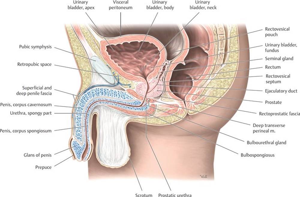
Fig. 11.3 Prostate in situ
Sagittal section through the male pelvis, left lateral view.
11.1a Seminal Glands (Vesicles)
The seminal glands are paired convoluted tubules that produce 70% of the seminal fluid (Figs. 11.2 and 11.3).
– They lie superior to the prostate, between the urinary bladder and the rectum.
– The seminal glands are subperitoneal, located immediately below the peritoneum of the rectovesical pouch.
– The duct of each seminal gland joins with the ampulla of the ductus deferens to form the ejaculatory ducts, which pierce the prostate and drain into the prostatic urethra.
– The middle rectal and inferior vesical arteries supply the seminal glands.
Veins of similar names accompany the arteries.
– Branches from the pelvic plexus innervate the seminal glands.
11.1b The Prostate
The prostate is an accessory reproductive gland that produces ~25% of the seminal fluid (Fig. 11.4A, B, and C; see also Figs. 11.2 and 11.3).
– The base, or superior surface, sits directly below the bladder. The apex points inferiorly and is in contact with the external urethral sphincter.
– It is posterior to the lower part of the pubic symphysis and anterior to the rectovesical septum, which separates it from the rectum.
– The prostate surrounds the proximal (prostatic) part of the urethra. Secretions from prostatic glands drain into the urethra through numerous prostatic ductules.
– A fibromuscular capsule surrounds the prostate. The prostatic capsule is separated from the outer prostatic sheath (derived from endopelvic fascia) by the prostatic venous plexus.
– Puboprostatic ligaments, anterior extensions of the tendinous arch of the pelvic fascia, attach the apex of the prostate (and neck of the bladder) to the pubis (see Fig. 11.14). Posterior extensions of the tendinous arch secure it to the sacrum.
– The anatomic lobes of the prostate include the following:
• A fibromuscular isthmus anterior to the urethra
• Right and left lateral lobes that are subdivided into lobules. The lower posterior lobule (sometimes referred to as the posterior lobe), which lies posterior to the urethra and inferior to the ejaculatory ducts, is palpable by digital exam
• A poorly defined middle lobe that sits above the lateral lobes between the urethra and ejaculatory ducts and is in close contact with the neck of the bladder
– For clinical purposes, the prostate is divided into three zones determined by their proximity to the urethra: periurethral, central (comparable to the anatomic middle lobe), and peripheral.
– Prostatic arteries are usually branches of the inferior vesical arteries. Middle rectal arteries also contribute to the prostatic blood supply (see Fig. 10.14A).
Benign prostatic hypertrophy (hyperplasia)
Benign prostatic hypertrophy (more accurately called hyperplasia) is the benign enlargement of the prostate gland that results from the proliferation of the epithelial and stomal tissues, particularly in the periurethral area. Although it is common in men of middle age, the incidence increases in later years and is thought to be due to the greater conversion of testosterone to the more active form, dihydrotestosterone (DHT), in the aging prostate gland. The hyperplasia constricts the internal urethral orifice, causing dysuria (difficult or painful urination), nocturia (excessive urination at night), and urgency (sudden desire to urinate). It is treated with a 5areductase inhibitor that blocks the conversion of testosterone to DHT by inhibiting the enzyme 5a-reductase. This may be used in combination with α-blockers that relax the muscles in the prostate and in the neck of the bladder, making urination easier. In severe cases, transurethral resection of the prostate may be necessary.

Fig. 11.4 Prostate
The prostate may be divided anatomically (top row) or clinically (bottom row).
A Coronal section, anterior view.
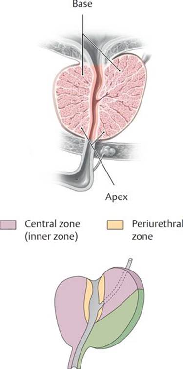
B Sagittal section, left lateral view.
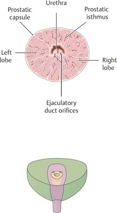
C Transverse section, superior view.
– The prostatic venous plexus, continuous with the vesical venous plexus of the bladder, drains to internal iliac veins. The prostatic plexus also communicates with the vertebral venous plexus (see Fig. 2.16).
– The lymph vessels of the prostate follow venous pathways to the internal iliac nodes.
– The prostatic nerve plexus is a derivative of the inferior hypogastric plexus. The role of parasympathetic innervation is unclear, but sympathetic nerves cause the smooth muscle of the gland to contract, expelling prostatic secretion into the prostatic urethra during ejaculation.
Carcinoma of the prostate
Carcinoma of the prostate is a slow-growing cancer affecting elderly men that commonly arises in the posterior lobe of the prostate. It metastasizes to the bony pelvis, vertebral column, skull, and brain via the vertebral venous plexus but also to the heart and lungs via drainage through the internal iliac veins and inferior vena cava. Symptoms include hesitancy (difficulty in beginning the flow of urination), frequency, and dribbling on urination (collectively known as prostatism). It can be diagnosed by digital examination of the rectum, ultrasonography, or prostate-specific antigen (PSA) levels. A PSA value greater than 4.0 ng/mL indicates disease activity.
Prostatectomy
Prostatectomy is the surgical removal of the prostate gland. Open radical prostatectomy involves removal of the prostate along with the seminal vesicles, ductus deferens, and pelvic lymph nodes via a retropubic or perineal incision. Transurethral resection of the prostate (TURP) is performed using a cystoscope that is advanced through the urethra to resect the prostate. Cavernous nerves carrying parasympathetic fibers that are responsible for penile erection run alongside the prostate and are particularly at risk during these procedures.
11.2 Female Genital Structures
Female genital structures, which include the ovaries, uterine tubes, uterus, and vagina, are located in the middle of the pelvis between the bladder anteriorly and the rectum posteriorly (Fig. 11.5).
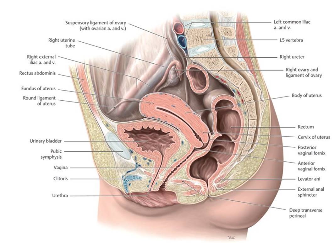
Fig. 11.5 Female pelvis
Sagittal section viewed from the left side.
11.2a The Ovary
The ovary is the female gonad, an ovoid structure that produces eggs and reproductive hormones and resides in the lateral wall of the pelvis (Fig. 11.6A and B).
– The ligament of the ovary attaches the ovary to the supralateral aspect of the uterus.
– The suspensory ligament of the ovary is a fold of peritoneum that encloses the ovarian vessels and nerves as they pass over the pelvic brim to the ovary.
– The mesovarium suspends the ovary from the posterior part of the broad ligament.
– The ovarian artery, a branch of the abdominal aorta at L2, supplies the ovary (see Fig. 10.17).
– A pampiniform plexus, which may converge to form a single ovarian vein, drains the ovary. The right ovarian vein is a direct tributary of the inferior vena cava; the left ovarian vein is a tributary of the left renal vein.
– Lymphatic vessels follow the ovarian vessels superiorly to the lateral aortic nodes.
– Both the ovarian nerve plexus, which follows the ovarian vessels, and the pelvic nerve plexus, which follows the uterine vessels, innervate the ovary.
11.2b The Uterine Tubes
The uterine tubes (fallopian tubes, oviducts), paired muscular tubes that extend laterally from the horns (supralateral corners) of the uterus, transmit ova from the ovary and sperm from the uterine cavity (see Fig. 11.6A and B).
– The uterine tubes are the normal sites of fertilization and are also the most common sites of ectopic pregnancies (implantation of a fertilized ovum outside the uterus).
– The uterine tube has four parts:
1. The uterine (intramural) part, the segment that passes through the wall of the uterus
2. The isthmus, the narrowest part
3. The ampulla, the longest and widest part and normally the site of fertilization
4. The infundibulum, the trumpet-shaped terminal part that is open to the peritoneal cavity, with fingerlike fimbriae that surround the ovary
– The uterine tube is ensheathed in the upper edge of the broad ligament, where it is supported by its mesosalpinx (see Section 11.2c).
– The uterine tube is supplied by the anastomosing ovarian and uterine arteries and drained by accompanying veins.
– Lymph vessels follow the ovarian veins to the lateral aortic nodes.
– The ovarian and uterine plexuses innervate the uterine tubes.
Ectopic pregnancy
Implantation of a fertilized ovum outside the uterus can occur anywhere, but the ampulla of the uterine tube is the most common site. Often the tube has been partially blocked by inflammation (salpingitis), preventing the blastocyst from completing its journey to the uterus. If not diagnosed early in the pregnancy, rupture of the uterine tube with consequent hemorrhage into the peritoneal cavity can result in a life-threatening situation for the mother. A ruptured ectopic pregnancy on the right side may be misdiagnosed as a ruptured appendix because both conditions irritate the parietal peritoneum and have similar presentations.
11.2c The Uterus
The uterus is a pear-shaped muscular organ located in the center of the pelvis, posterior to the bladder and anterior to the rectum (see Fig. 11.5). It is the site of implantation of the fertilized egg, subsequent development of the embryo, and parturition of the fetus.
– The uterus has two parts (see Fig. 11.6A and B).
1. The body is the superior two thirds of the uterus and includes
○ the fundus, the uppermost part above the openings of the uterine tubes, and
○ the uterine isthmus, a narrow inferior segment that extends into the cervix.
2. The cervix is the narrow inferior third of the uterus and its least mobile part.
○ A supravaginal part sits above the vagina.
○ A vaginal part protrudes into the upper vagina and is surrounded by the vaginal fornices (upper recesses).
– The uterine cavity, a narrow space within the uterine body,
• communicates with the lumen of the uterine tubes where they enter at the uterine horns, and
• extends inferiorly through the internal os (orifice) to the cervical canal and terminates where the external os opens into the vagina.
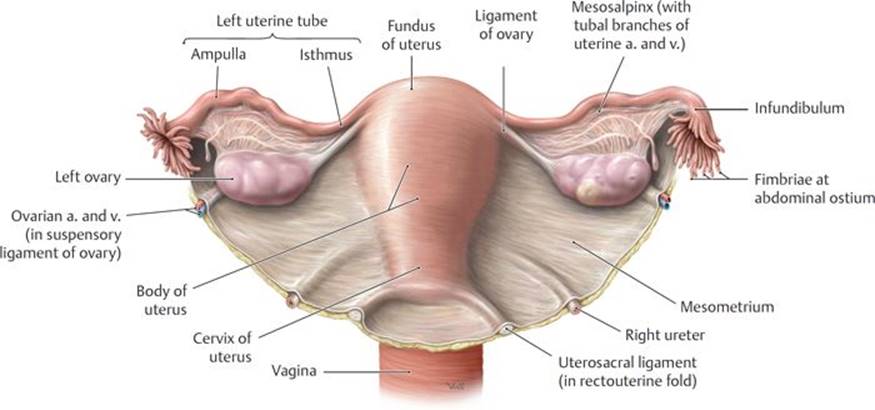
Fig. 11.6 Uterus and uterine tube
A Posterosuperior view.
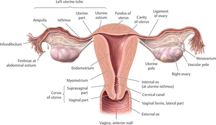
B Coronal section, posterior view with uterus straightened. Removed: Mesometrium.
Bicornuate uterus
The embryonic uterus is formed from the fusion of two paramesonephric ducts. When these ducts fail to fuse properly, a bicornuate uterus results in which the upper part of the uterus is bifurcated. The caudal part of the uterus is usually normal. Although a normal pregnancy is possible with this malformation, there is a higher chance of recurrent pregnancy loss, preterm birth, and malpresentation of the baby (e.g., the baby may be in a breech or transverse position).
– Although the body of the uterus is mobile, its position changes with the fullness of the bladder and rectum. Its normal position is anteflexed and anteverted (Fig. 11.7).
• Flexion describes the angle between the uterine body and isthmus. In an anteflexed uterus, the long axis of the uterine body is tipped anteriorly; a retroflexed uterus is tipped posteriorly.
• Version describes the angle between the cervix and vagina. In an anteverted uterus, the axis of the cervix is bent anteriorly; in a retroverted uterus, the cervix is bent posteriorly.
– Peritoneum covers the body of the uterus, extending as far inferiorly as the cervix on its posterior surface. The uterus is flanked anteriorly by the vesicouterine space and posteriorly by the rectouterine space (pouch) (Fig. 11.8).
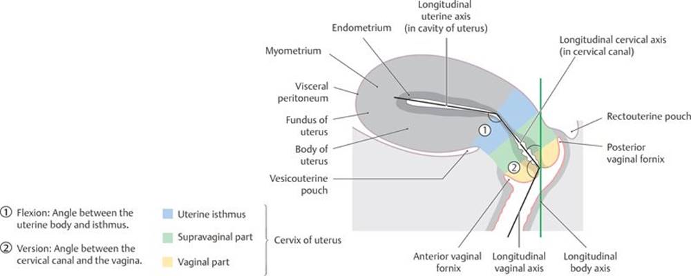
Fig. 11.7 Curvature of the uterus
Midsagittal section, left lateral view. The position of the uterus can be described in terms of flexion (1) and version (2).
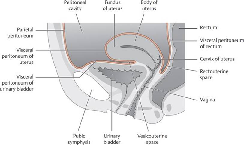
Fig. 11.8 Peritoneum in the female pelvis
– Uterine ligaments that arise from the uterine body include the broad ligament and the round ligaments of the uterus (Fig. 11.9; see also Fig. 11.6A).
• The broad ligament is a double fold of peritoneum that extends laterally from each side of the uterus to the sidewalls of the pelvis. The parts of the broad ligament (Fig. 11.10) are
○ the mesosalpinx, which ensheaths the uterine tube;
○ the mesovarium, a posterior extension, which suspends the ovary; and
○ the mesometrium, which extends from the uterine body below the mesovarium to the sidewall of the pelvis.
• The paired round ligaments of the uterus, which originate near the fundus from each side of the uterus, pass through the deep inguinal rings, traverse the inguinal canal, and insert in the labia majora of the perineum.
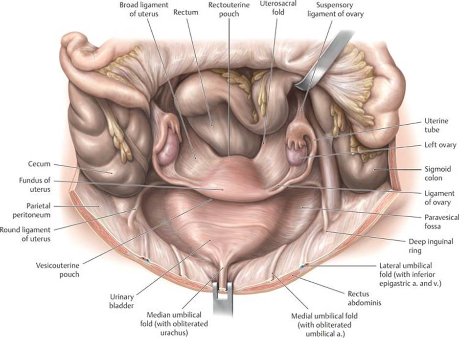
Fig. 11.9 Peritoneal relationships in the female pelvis
Lesser pelvis, anterosuperior view. Retracted: Small intestine loops and colon (portions).
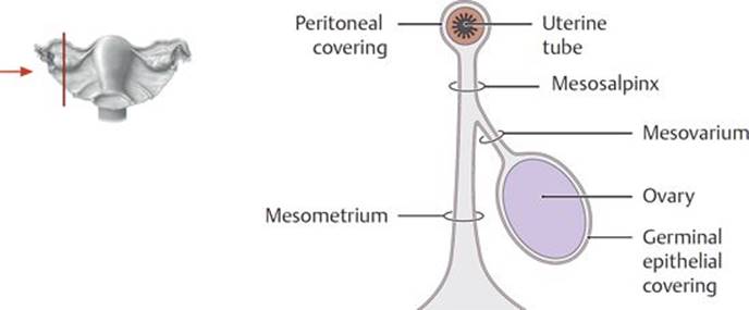
Fig. 11.10 Mesenteries of the broad ligament
Sagittal section. The broad ligament of the uterus is a combination of the mesosalpinx, mesovarium, and mesometrium.
– Uterine ligaments that arise from the cervix include the transverse cervical ligaments and the uterosacral ligaments (Fig. 11.11).
• The paired cardinal (transverse cervical) ligaments are thickenings of endopelvic fascia that connect the uterine cervix to the pelvic sidewall. They are located at the base of the broad ligament and transmit the uterine vessels.
• The paired uterosacral ligaments are thickenings of endopelvic fascia that connect the uterine cervix to the sacrum and help to maintain the anteverted position of the uterus.
– The uterine artery, the primary blood supply to the uterus (see Fig. 10.17), traverses the cardinal ligament and anastomoses superiorly with the ovarian artery and inferiorly with the vaginal artery.
– A uterine venous plexus receives the uterine veins and drains to the internal iliac vein.
– Lymphatic drainage of the uterus is complex but generally follows the uterine veins or the uterine ligaments (see Table 10.4).
• The uterine fundus drains to para-aortic nodes via the ovarian veins.
• The supralateral part of the uterus drains to superficial inguinal nodes via the round ligament.
• The uterine body drains to external iliac nodes via the broad ligament.
• The cervix drains to internal iliac and sacral nodes via the cardinal and uterosacral ligaments.
– The uterovaginal nerve plexus, derived from the inferior hypogastric plexus, innervates the uterus (see Fig. 10.19).
11.2d The Vagina
The vagina is a fibromuscular tube that extends from the cervix of the uterus to the vaginal orifice in the perineum (Figs. 11.12 and 11.13). It serves as the inferior part of the birth canal and the conduit for menstrual fluid, and it accommodates the penis during sexual intercourse.

Fig. 11.11 Ligaments of the female pelvis
Superior view. Removed: Peritoneum, neurovasculature, and superior portion of bladder to demonstrate only the fascial condensations. Deep pelvic ligaments support the uterus within the pelvic cavity and prevent uterine prolapse, the downward displacement of the uterus into the vagina.
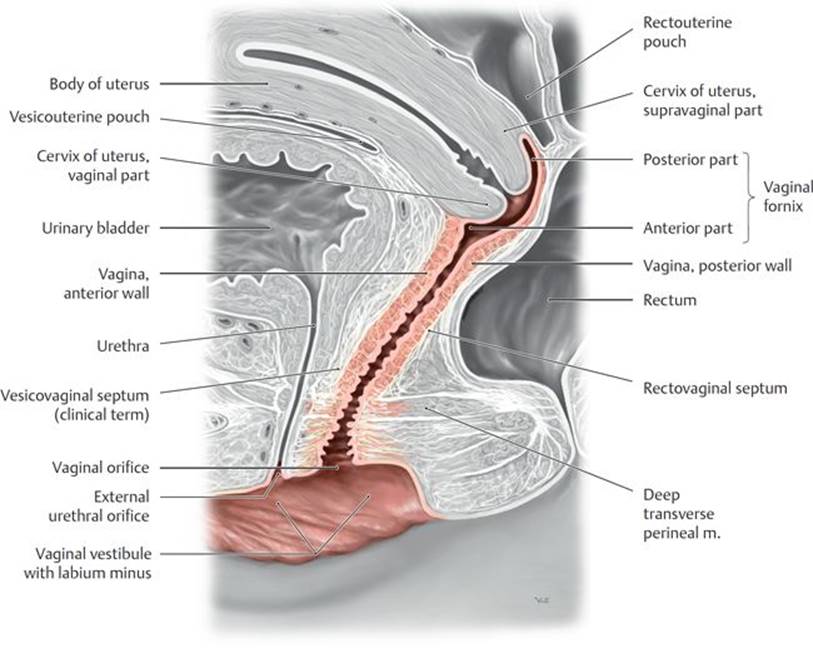
Fig. 11.12 Vagina
Midsagittal section, left lateral view.
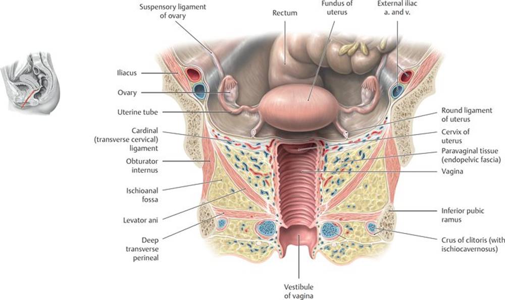
Fig. 11.13 Female pelvis
Coronal section, anterior view.
– The vagina is posterior to the bladder and urethra and anterior to the rectum.
– It is normally flattened with its anterior and posterior walls in contact.
– Connections to the sacrum and side wall of the pelvis via the tendinous arch of the pelvic fascia stabilize the vagina, especially during childbirth.
– The vaginal fornix, which has anterior, lateral, and posterior parts, is a recess that surrounds the lower cervix as it protrudes into the upper vagina (see Figs. 11.5 and 11.6B).
– The posterior fornix is in contact with the rectouterine pouch, thereby providing access to the peritoneal cavity. The anterior fornix is shorter and lies against the posterior wall of the bladder.
– The internal iliac artery supplies the vagina through its uterine, vaginal, and internal pudendal branches (see Figs. 10.14B and 10.17).
– Veins of the vagina contribute to the uterovaginal venous plexus, which drains to the internal iliac vein.
– Lymphatic vessels of the vagina drain to several groups of nodes.
• The superior part of the vagina drains to external or internal iliac nodes.
• The inferior part of the vagina drains to sacral and common iliac nodes.
• The vaginal orifice drains to superficial inguinal nodes.
– The uterovaginal nerve plexus, an extension of the inferior hypogastric plexus, innervates the superior three fourths of the vagina (see Fig. 10.19).
– A deep perineal branch of the pudendal nerve, a branch of the sacral plexus, innervates the lowest vaginal segment (see Fig. 10.15). This somatically innervated segment is the only part of the vagina that is sensitive to touch.
Culdocentesis
Culdocentesis is a procedure in which peritoneal fluid is extracted from the rectouterine pouch by needle aspiration. The needle is advanced through the posterior fornix of the vagina. No fluid or a small amount of clear fluid is normal, but purulent fluid is suggestive of pelvic inflammatory disease (PID). The presence of blood is an indication for emergency surgery.
11.3 Pelvic Urinary Organs
The pelvic urinary organs include the distal ureters, the urinary bladder, and the urethra.
11.3a The Ureters
Each ureter crosses over the brim of the pelvis at the bifurcation of the common iliac artery and descends along the lateral wall near the ischial spine. It runs anteriorly and enters the posterolateral wall of the bladder.
– In males, the ureter passes under the pelvic portion of the ductus deferens and enters the bladder lateral and superior to the free ends of the seminal vesicles (Fig. 11.14; see also Fig. 11.2).
– In females, the ureter passes inferior to the uterine arteries within the cardinal ligament, ~2 cm lateral to the vaginal part of the cervix (Fig. 11.15).
– The most reliable blood supply to the pelvic part of the ureter is the uterine artery in females and the inferior vesical artery in males. Veins of similar names accompany the arteries.
– The pelvic part of the ureter derives its innervation from the inferior hypogastric plexuses.
– Visceral afferent fibers follow sympathetic nerves to spinal cord levels T11–L2; therefore, ureteric pain is usually felt in the ipsilateral inguinal region.
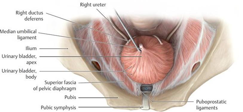
Fig. 11.14 Ureter and bladder in the male pelvis
Superior view.
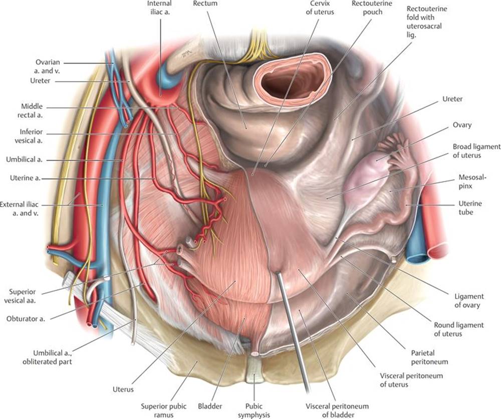
Fig. 11.15 Ureter in the female pelvis
Superior view. Removed from right side: Peritoneum and broad ligament of uterus.
11.3b The Urinary Bladder
The bladder is a muscular reservoir for the temporary storage of urine. Although normally located in the true pelvis, when full it may extend superiorly into the abdomen.
– It lies directly posterior to the pubic symphysis, separated from it by the retropubic space. Posteriorly, it is related to the rectum in the male (see Fig. 11.1) and the upper vagina in the female (see Fig. 11.5).
– It is covered by peritoneum only on its superior surface.
– The bladder is tetrahedral with superior, posterior, and two inferolateral surfaces. It has four parts:
1. The apex points toward the pubic symphysis. The median umbilical ligament extends from the apex to the umbilicus.
2. The fundus forms the bladder base, or posterior wall.
3. The body makes up most of the bladder.
4. The neck is the lowest and least mobile region.
– The neck of the bladder is firmly attached to
• the pubis by anterior extensions of the tendinous arch of the pelvic fascia, the pubovesical ligaments in females and puboprostatic ligaments in males (see Figs. 11.11 and 11.14); and
• the lateral pelvic walls by condensations of endopelvic fascia, the lateral vesicular ligaments (see Fig. 10.12).
– The walls are made up of bundles of smooth muscle fibers. In the male, the muscle fibers near the bladder neck, which form the internal urethral sphincter, contract during ejaculation.
– The internal surface of the base of the bladder is marked by the trigone, a smooth triangular region (Fig. 11.16). The corners of the triangle are formed posterolaterally by the slitlike openings of the right and left ureters and anteriorly by the urethral orifice.
– The bladder is highly distensible and in most individuals may hold up to 600–800 mL (painfully), although micturition (urination) usually occurs at a much smaller volume. Normally, no urine remains in the bladder after voiding.
– The superior vesical arteries, with contributions from the inferior vesical arteries (in males) and vaginal arteries (in females), supply the bladder (see Fig. 10.14A and B).
– The vesical venous plexus surrounds the inferolateral surfaces of the bladder and drains to the internal iliac veins. The plexus communicates with the prostatic plexus in males, with the uterovaginal plexus in females, and with the vertebral venous plexus in both sexes.
– Lymph from the bladder drains to internal and external iliac nodes.

Fig. 11.16 Trigone of the bladder
Coronal section, anterior view.
– The vesical nerve plexus of the bladder is a derivative of the inferior hypogastric plexus (see Fig. 10.19).
• Sympathetic stimulation relaxes the detrusor muscle and contracts the internal sphincter, thus inhibiting micturition.
• Parasympathetic nerves stimulate the detrusor muscle to contract while inhibiting the internal sphincter, thereby facilitating micturition.
• Afferent fibers carrying pain from the inferior bladder follow parasympathetic routes. Pain fibers from the superior bladder follow sympathetic routes.
11.3c The Urethra
The urethra is the muscular conduit for urine in the female and for urine and semen in the male. It extends from the internal urethral orifice at the bladder neck to the external urethral orifice in the perineum.
– The male urethra extends 18 to 22 cm from the urinary bladder to the tip of the glans penis (Fig. 11.17). The male urethra has four parts. Although all are mentioned here, the membranous and spongy parts are located in the perineum and are discussed further in Chapter 12.
1. The preprostatic part at the neck of the bladder contains the internal urethral orifice. Sympathetic nerves of the superior hypogastric plexus control closure of the internal urethral sphincter during ejaculation.
2. The prostatic part is surrounded by the prostate gland and characterized by
○ the urethral crest, a vertical ridge on the posterior wall that contains a central eminence, the seminal colliculus; and
○ ejaculatory ducts that open onto the urethral crest, and prostatic ductules from the prostate that open into recesses on either side of the crest.
3. The membranous part passes through the perineal membrane in the urogenital triangle and is surrounded by the external urethral sphincter.
4. The spongy part passes through the corpus spongiosum, one of the vascular erectile bodies of the penis.
Urethral rupture in males
Fractures of the pelvic girdle may be accompanied by a rupture of the membranous part of the urethra. This allows the extravasation (escape) of urine and blood into the deep perineal space and superiorly through the genital hiatus to the subperitoneal spaces around the prostate and bladder. Rupture of the bulbous part of the spongy urethra may occur from a straddle injury in which there is a forceful blow to the perineum, or from the false passage of a transurethral catheter. In this case urine can leak into the superficial peritoneal space, which is continuous with the scrotal sac, the space around the penis, and the space on the inferior anterior abdominal wall between the abdominal muscles and the membranous layer of the superficial fascia. The attachment of the superficial perineal fascia to the fascia lata (fascia enclosing the thigh muscles) prevents urine from spreading laterally into the thighs. Similarly, it is prevented from spreading into the anal triangle by the attachment of the fascia to the deep perineal fascia and perineal membrane.
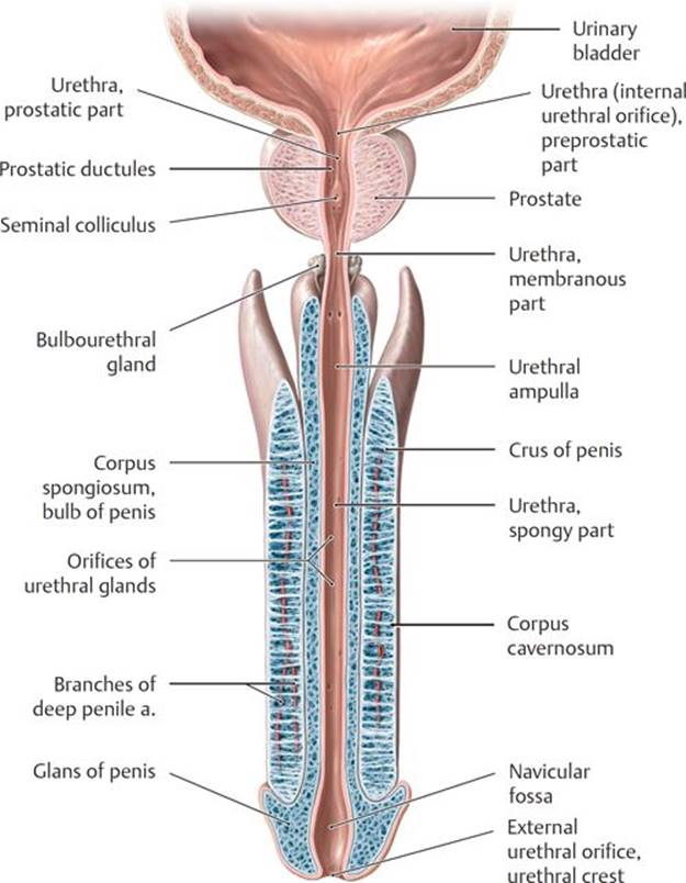
Fig. 11.17 Male urethra
Longitudinal section, anterior view.
– The female urethra extends 4 cm from the internal urethral orifice at the bladder neck to the external urethral orifice in the perineum (Fig. 11.18).
• Within the pelvis it lies anterior to the vagina, forming an elevation within the anterior vaginal wall.
• It passes through the genital hiatus of the pelvic diaphragm, the external urethral sphincter (there is no organized internal urethral sphincter), and the perineal membrane.
• In the perineum, the urethra opens within the vestibule of the vagina directly anterior to the vaginal orifice (see Fig. 12.8).
– Branches of the pudendal artery, as well as the inferior vesical artery in the male and vaginal artery in the female, supply the urethra. In both sexes, a venous plexus that accompanies the arteries drains the urethra.
– The female urethra and proximal parts of the male urethra (preprostatic, prostatic, and membranous) drain to the internal iliac nodes. The spongy urethra of the male (the perineal part) drains to deep inguinal nodes.
– Nerves to the urethra arise from the prostatic nerve plexus in the male and comparable vesical nerve plexus in the female. Sympathetic nerves control the closure of the external urethral sphincter in males.
– Visceral afferent fibers from the pelvic urethra travel with pelvic splanchnic nerves; somatic afferent fibers from the perineal urethra travel with the pudendal nerve.

Fig. 11.18 Female urethra
Coronal section tilted slightly posterior, anterior view.
11.4 The Rectum
The rectum is the continuation of the gastrointestinal tract in the pelvis and functions as a temporary storage site for fecal matter. It is continuous with the sigmoid colon superiorly and the anal canal inferiorly (see Section 12.5a; Figs. 11.19 and 11.20).
– It lies anterior to the lower sacrum and coccyx and rests on the anococcygeal ligament of the pelvic floor.
– Anteriorly, the rectum is related to the bladder, seminal glands, and prostate in males and to the vagina in females. A rectovesical or rectovaginal septum separates the rectum from these anterior structures.
– The rectum has no mesentery. Its superior two thirds are retroperitoneal and form the posterior surface of the rectovesical and rectouterine pouches. The distal third is subperitoneal.
– Unlike the colon, the rectum lacks taeniae coli, haustra, and epiploic appendices.
– The rectum begins at the rectosigmoid junction, the point at which the teniae coli disappear. At this junction, muscle fibers of the bands spread out evenly over the surface of the rectum. This junction usually occurs anterior to the S3 vertebra.
Rectal examination
A rectal examination is performed by inserting a gloved, lubricated finger into the rectum, while the other hand is used to press on the lower abdomen or pelvic region. Palpable structures include the prostate, seminal vesicles, ampulla of the ductus deferens, bladder, uterus, cervix, and ovaries. Pathological anomalies such as hemorrhoids, tumors, enlargements, and changes in consistency of the tissues can be felt. The tonicity of the anal sphincter, mediated by the pudendal nerve (S2–S4), can also be assessed.
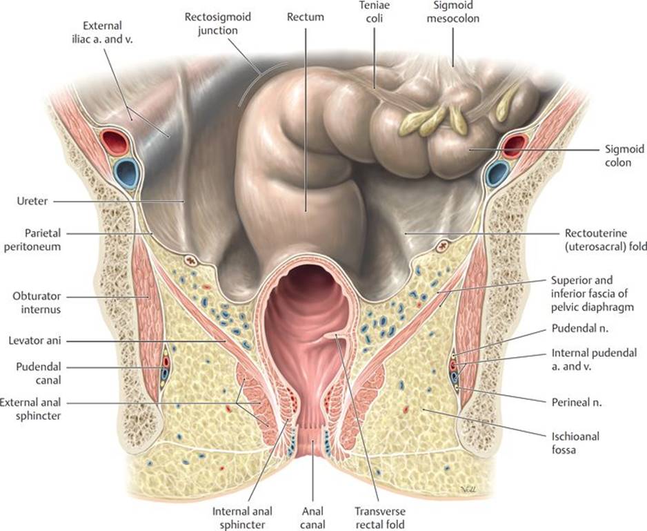
Fig. 11.19 Rectum in situ
Female pelvis, coronal section, anterior view.
– The rectum terminates at the anorectal junction (its junction with the anal canal), where it passes through the pelvic diaphragm adjacent to the tip of the coccyx.
– The internal wall of the rectum has three transverse rectal folds, one on the right and two on the left, which create lateral flexures that are visible externally.
– The ampulla, the most distal segment of the rectum, stores accumulating fecal material until defecation and has an important role in fecal continence. It narrows abruptly as it joins with the anal canal and passes through the pelvic diaphragm.
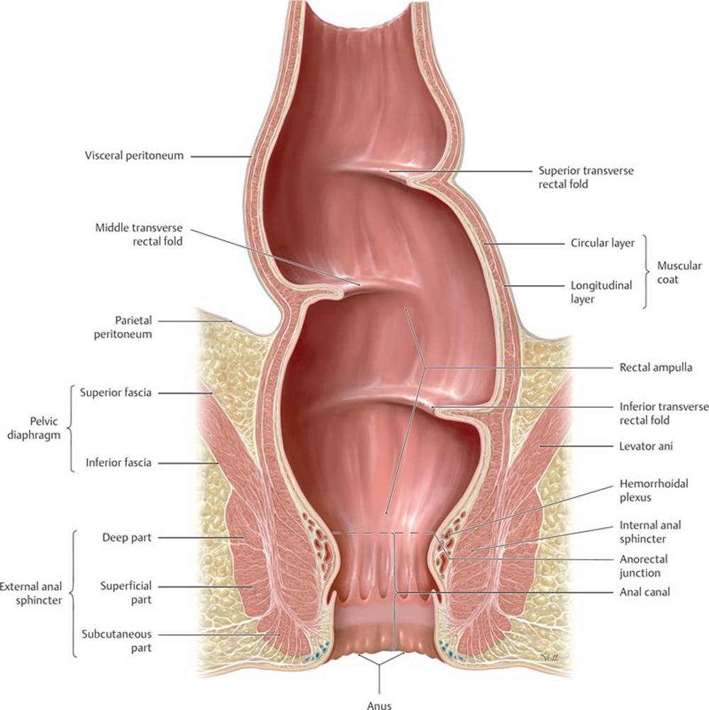
Fig. 11.20 Rectum
Coronal section, anterior view.
– The rectum has a dual blood supply:
• The superior rectal artery, the unpaired terminal branch of the inferior mesenteric artery, supplies the upper rectum (see Fig. 10.18).
• Right and left middle rectal arteries, branches of the internal iliac arteries, supply the lower rectum.
– The rectal veins drain a submucosal rectal venous plexus, which has internal and external (subcutaneous) components.
• The external plexus communicates with other visceral venous plexuses in the pelvis.
• The internal plexus communicates with branches from the rectal arteries (an arteriovenous anastomosis), forming a thickened vascular tissue (hemorrhoidal plexus) that surrounds the anorectal junction. This tissue forms prominent anal cushions in the left lateral, right anterolateral, and right posterolateral positions.
Hemorrhoids
External hemorrhoids are thrombosed veins of the external venous plexus, commonly associated with pregnancy or chronic constipation. They lie below the dentate line and are covered by skin. Because they are somatically innervated, they are more painful than internal hemorrhoids.
Internal hemorrhoids contain dilated veins of the internal rectal plexus. With a breakdown of the muscularis layer, these vascular cushions prolapse into the anal canal and may become ulcerated. Because they lie above the dentate line and are viscerally innervated, these hemorrhoids are painless. They are generally not associated with portal hypertension, but bleeding is characteristically bright red due to the anastomoses between the venous plexus and branches of the rectal arteries.
– Venous blood from the rectum drains into the portal and caval (systemic) venous systems (see Fig. 10.18).
• A superior rectal vein, which drains the upper rectum, is a tributary of the portal system via the inferior mesenteric vein.
• The paired middle and inferior rectal veins that drain the lower rectum (and anal canal) are tributaries of the inferior vena cava via the internal iliac veins.
• Communications between the superior, middle, and inferior rectal veins form clinically important portocaval anastomoses that enlarge in portal hypertension.
– Lymphatic drainage follows venous pathways.
• The upper rectum drains to inferior mesenteric nodes along the course of the superior rectal artery. It eventually drains to lumbar nodes, although some lymph may drain first to sacral nodes.
• The lower rectum drains primarily to sacral nodes or directly to internal iliac nodes.
– Sympathetic innervation to the rectum is carried by lumbar splanchnic nerves to the hypogastric plexuses, as well as by nerves of the inferior mesenteric nerve plexus traveling along the superior rectal artery (see Figs. 10.19 and 10.20).
– Parasympathetic innervation originates in pelvic splanchnic nerves, which also carry visceral afferent fibers.