
The perineum is the space inferior to the pelvic floor, which is divided into a urogenital triangle and an anal triangle. The urogenital triangle contains the male and female external genital structures, and the anal triangle contains the anal canal and anus.
12.1 Perineal Spaces (Figs. 12.1 and 12.2; see also Figs. 10.3 and 10.4)
– The boundaries of the diamond-shaped perineum are
• the pelvic outlet (pubic symphysis, ischiopubic rami, sacrotuberous ligaments, and coccyx), which forms the perimeter;
• the lower parts of the obturator internus muscles and their obturator fasciae, which line the lateral walls;
• the inferior surface of the pelvic diaphragm, which forms the roof; and
• the skin of the perineum, which forms the floor.
– A line connecting the ischial tuberosities separates the perineum into an anterior urogenital triangle and a posterior anal triangle (see Fig. 10.4A and B).
– The perineal membrane, a tough, fibrous sheet, stretches between the ischiopubic rami, extending anteriorly almost to the pubic symphysis and posteriorly to the ischial tuberosities.
• It separates the urogenital triangle into the deep perineal and superficial perineal spaces.
• It forms a platform for the attachment of the cavernous bodies (which become engorged during arousal) of the external genitalia.
– The superficial perineal space is a potential space bounded above by the perineal membrane and below by the superficial perineal fascia (Colles’ fascia), the extension of the membranous layer of superficial fascia (Scarpa’s fascia) on the abdominal wall.
• In both sexes it contains
○ the bulbocavernosus, ischiocavernosus, and superficial transverse perineal muscles and
○ the deep perineal branches of the internal pudendal vessels and pudendal nerve.
• In males it also contains
○ the root of the penis and
○ the proximal portion of the spongy urethra.
• In females it also contains (see Fig. 12.8)
○ the clitoris and associated muscles,
○ the vestibular bulb, and
○ the greater vestibular glands.
– The deep perineal space is bounded inferiorly by the perineal membrane and superiorly by the pelvic diaphragm.
• In both sexes it contains
○ part of the urethra and inferior part of the external urethral sphincter.
○ the anterior recesses of the ischioanal fat pads, and
○ neurovascular structures to the penis or clitoris.
• In males it also contains
○ the membranous part of the urethra,
○ the bulbourethral glands, and
○ the deep transverse perineal muscles.
• In females it also contains
○ compressor urethrae and urethrovaginal sphincter,
○ the proximal part of the urethra, and
○ smooth muscle associated with the perineal body.
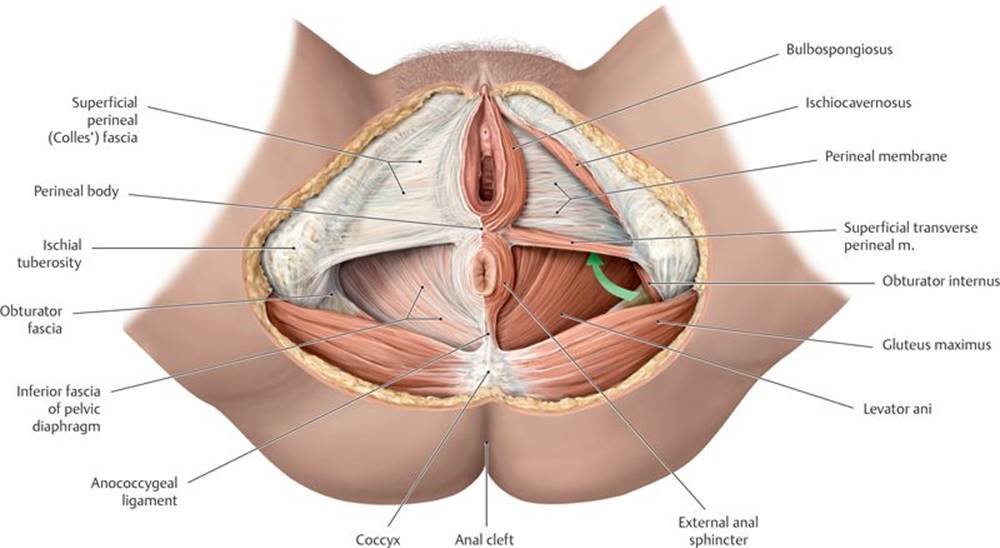
Fig. 12.1  Muscles and fascia of the female perineum
Lithotomy position, caudal (inferior) view. The green arrow is pointing forward to the anterior recess of the ischioanal fossa.
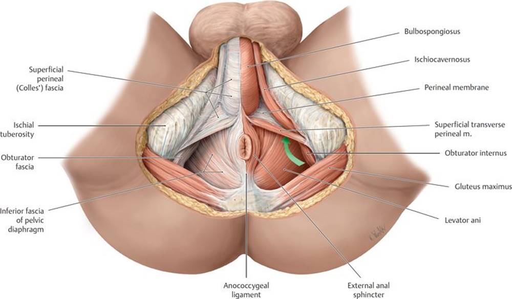
Fig. 12.2 Muscles and fascia of the male perineum
Lithotomy position, caudal (inferior) view. The green arrow is pointing forward to the anterior recess of the ischioanal fossa.
12.2 Muscles of the Perineum
– Muscles of the perineum support the pelvic floor, surround the orifices of the urethra and anus, and assist in the erection of genital structures (Table 12.1, Fig. 12.3A and B; see also Figs. 12.1 and 12.2).
– The perineal body is an irregular subcutaneous mass of fibro-muscular tissue formed by converging fibers of the levator ani, the deep transverse perineal and bulbospongiosus muscles, and the external anal sphincter.
• It lies between the rectum and bulb of the penis in males and between the rectum and vagina in females.
• It supports the pelvic diaphragm and pelvic viscera.
– Perineal branches of the internal pudendal artery supply muscles of the perineum. Venous blood drains to the internal pudendal vein and the internal iliac vein.
– The pudendal nerve (S2–S4) innervates the muscles of the perineum.
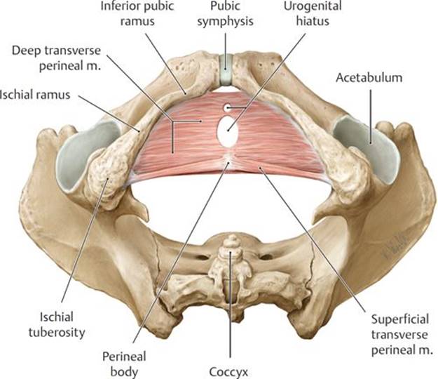
Fig. 12.3 Muscles of the perineum
Inferior view.
A Superficial and deep transverse perineal muscles.
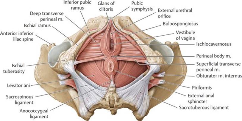
B Sphincter and erector muscles, female.

Episiotomy
During vaginal delivery, pressure on the perineum risks tearing the perineal muscles. A clean incision through the posterior vaginal orifice into the perineal body, known as an episiotomy, is often performed to enlarge the opening and prevent damage to perineal muscles. A median episiotomy extends only into the perineal body, but if further traumatic tearing occurs to extend the cut, it can damage the external anal sphincter (resulting in fecal incontinence) or create an anovaginal fistula. Mediolateral episiotomies often replace the midline incisions. These extend laterally from the vaginal orifice to the superficial perineal muscles, thus avoiding the perineal body and the possible sequelae from extensive tearing.
Prolapse of pelvic organs
The pelvic diaphragm, pelvic ligaments, and perineal body provide important structural support for the pelvic viscera. Stretching or disruption of these tissues often occurs during childbirth and results in prolapse of the uterus into the vagina. A widening of the genital hiatus from an atrophic pelvic floor or a weakened perineal body can allow the bladder (cystocele), rectum (rectocele), or rectovesical pouch (enterocele) to bulge into the vaginal wall.
12.3 Male Urogenital Triangle
The male urogenital triangle contains the scrotum, penis, bulbourethral glands, perineal muscles, and associated neurovasculature (see Figs. 10.3 and 11.3).
12.3a The Scrotum
– The scrotum is a saccular extension of the anterior abdominal wall that encloses the testes and spermatic cord (see Section 7.3).
– The subcutaneous layer of the skin over the scrotum is devoid of fat, but the dartos fascia that underlies the skin is continuous with the deep membranous layer of the superficial fascia (Scarpa’s fascia) of the abdomen and with the superficial perineal fascia (Colles’ fascia) of the perineum.
– An extension of dartos fascia divides the scrotum into right and left compartments and is visible externally as the scrotal raphe.
– Scrotal branches of the internal pudendal and external pudendal arteries, and cremasteric branches of the inferior epigastric artery supply the scrotum. Veins of similar names accompany the arteries (Figs. 12.4A and B and 12.5).
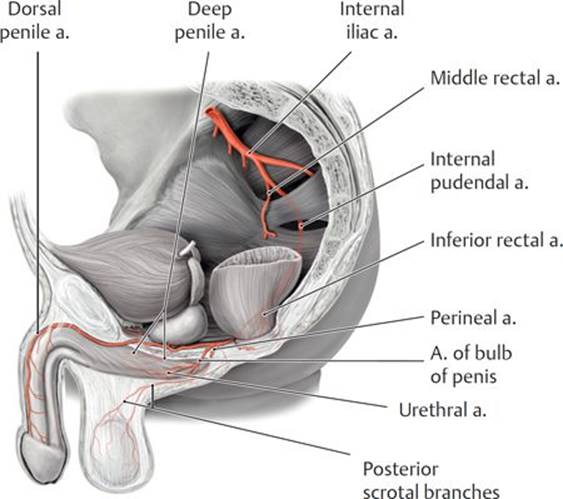
Fig. 12.4 Blood vessels of the male genitalia
Left lateral view.
A Arterial supply.

B Venous drainage.
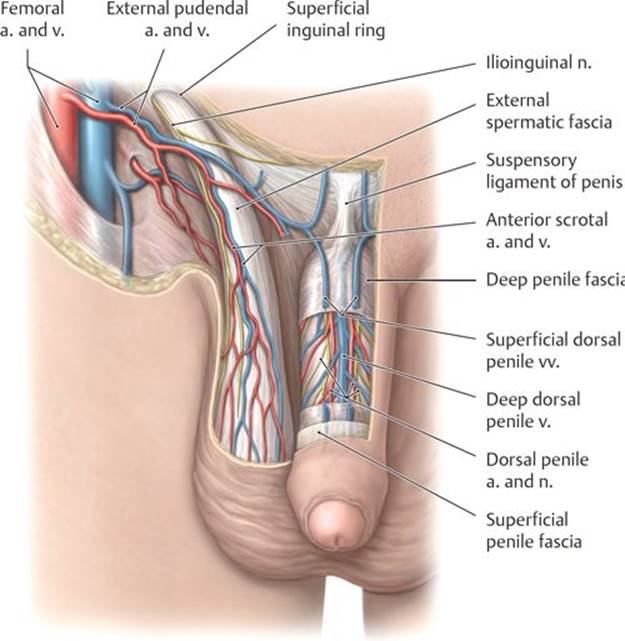
Fig. 12.5 Neurovasculature of the penis and scrotum
Anterior view. Partially removed: Skin and fascia.
– Lymph from the scrotum drains to superficial inguinal nodes. Recall that lymph from the contents within the scrotum (i.e., the testis and epididymis) drains directly to the para-aortic nodes.
– Innervation of the scrotum (see Fig. 10.22B) is supplied by
• the ilioinguinal and genitofemoral (genital branch) nerves, which innervate the anterior scrotum, and
• the pudendal and posterior femoral cutaneous nerves of the thigh, which innervate the posterior scrotum.
12.3b The Penis
The penis is the male copulatory organ, transmitting semen during sexual intercourse. It contains the spongy part of the urethra and therefore also transmits urine from the bladder (Fig. 12.6A and B).
– It has three parts:
1. The root, attached to the perineal membrane and covered by muscles, is composed of
○ paired crura that are attached to the ischiopubic rami and covered by the ischiocavernosus muscles, and
○ the bulb of the penis, which is attached to the perineal membrane and covered by the bulbospongiosus muscle. The spongy urethra enters on its dorsal surface.
2. The body, pendulous and not covered by muscle, is made up of three cylindrical bodies of erectile tissue. A tunica albuginea, a dense fibrous coat, surrounds each erectile body, and distally a deep penile fascia (Buck’s fascia) binds the three bodies together. The three erectile bodies include
○ two corpora cavernosa, continuations of the crura, which lie side by side on the dorsum of the penis, and
○ one corpus spongiosum, a continuation of the bulb of the penis, which lies ventral to the two corpora cavernosa and is traversed by the penile urethra.
3. The glans penis (also known as the glans), an expansion of the distal end of the corpus spongiosum, is characterized by
○ the corona, which overhangs the distal ends of the corpora cavernosa, and
○ the external urethral orifice at its tip.
– The external pudendal artery supplies the skin and subcutaneous tissue of the penis. Venous drainage of this tissue passes through the superficial dorsal veins, which drain to the external pudendal veins (see Fig. 12.5).
– The internal pudendal artery supplies the deep penile structures. Its branches (Fig. 12.7; see also Fig. 12.6B) include
• the artery of the bulb of the penis, which supplies the bulb of the penis, the urethra within the bulb, and the bulbourethral glands;
• the dorsal penile artery, which runs between the deep penile fascia and tunica albuginea to supply the penile fascia and skin and the glans; and
• the deep penile artery, which runs within the corpus cavernosum and gives off helicine arteries that supply the erectile tissue and are responsible for engorgement of the corpora during erection.
– The erectile bodies are drained by a venous plexus that empties into the single deep dorsal vein, which passes under the pubic symphysis to join the prostatic venous plexus in the pelvis.
– Lymphatic drainage areas of the penis include
• the erectile bodies of the penis, which drain to internal iliac nodes;
• the glans penis, which drains to deep inguinal nodes; and
• the urethra, which drains to internal iliac and deep inguinal nodes.
– The glans of the penis is richly innervated by sensory fibers via the dorsal penile nerve, a branch of the pudendal nerve. Sympathetic fibers from the hypogastric nerve plexus are also carried along this route.

Fig. 12.6 Penis
A Inferior (ventral) view.

B Cross section through the body of the penis.

Fig. 12.7. Neurovasculature on the dorsum of the penis
Superior (dorsal) view. Removed: Skin.
– Parasympathetic fibers carried by the cavernous nerves derived from the prostatic plexus innervate the helicine arteries within the erectile tissue and are responsible for penile erection.
12.3c Bulbourethral Glands
The bulbourethral glands are paired mucus-secreting glands (see Figs. 11.2 and 11.3).
– They lie on either side of the urethra below the prostate surrounded by the urethral sphincter.
– Their ducts open into the proximal part of the spongy urethra.
– They are active during sexual arousal.
12.3d Erection, Emission, and Ejaculation
The sexual responses of erection, emission, and ejaculation involve sympathetic, parasympathetic, and somatic (via the pudendal nerve) pathways.
– During erection
• constriction of the helicine arteries, normally maintained by sympathetic innervation, is inhibited by parasympathetic stimulation. As the arteries relax, the cavernous spaces dilate and become engorged, and
• contractions of the bulbocavernosus and ischiocavernosus muscles, innervated by the pudendal nerve, impede venous outflow and maintain the erection.
– During emission
• parasympathetic stimulation mediates the secretion of seminal fluid from the seminal glands, bulbourethral glands, and prostate gland, and
• sympathetic stimulation mediates emission, the movement of seminal fluid through the ducts, by initiating peristalsis of the ductus deferens and seminal glands. This propels the seminal fluid into the prostatic urethra, where additional fluid is added as the prostate contracts.
– During ejaculation
• sympathetic stimulation constricts the internal urethral sphincter, which prevents the seminal fluid from entering the bladder (retrograde ejaculation);
• parasympathetic stimulation contracts the urethral muscles; and
• the pudendal nerve contracts the bulbospongiosus muscle.
Retrograde ejaculation
Relaxation of the detrusor muscle and contraction of the internal urethral sphincter in males are controlled by sympathetic fibers from the superior hypogastric plexus. Closure of the sphincter occurs during ejaculation, preventing the retrograde flow of semen into the bladder. Disruption of the sympathetic nerves can result in retrograde ejaculation. This can occur during the repair of an abdominal aortic aneurysm, for example, if the aneurysm involves the bifurcation of the aorta.
12.4 Female Urogenital Triangle
As in the male perineum, the female perineum contains erectile bodies, secretory glands, and their associated neurovasculature. In addition, it contains paired folds of skin that surround the urethral and vaginal orifices. These external genitalia are known collectively as the vulva (Figs. 12.8, 12.9, and 12.10).
– The mons pubis, a superficial mound of fatty subcutaneous tissue that is continuous with the superficial fatty layer of the abdominal wall, lies anterior to the pubic symphysis and is continuous with the labia majora.
– Labia majora, bilateral folds of fatty subcutaneous tissue, flank the pudendal cleft, the opening between the labia. The labia join anteriorly at the anterior commissure and posteriorly at the posterior commissure. Pigmented skin and coarse pubic hair cover the labia’s outer surface; the inner surface is smooth and hairless.
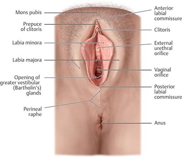
Fig. 12.8 Female external genitalia
Lithotomy position with labia minora separated.
– Labia minora, bilateral folds of hairless skin within the pudendal cleft, flank the vestibule of the vagina.
– The clitoris, a highly sensitive erectile organ, is located at the anterior junction of the paired labia minora.
• Paired erectile bodies, the corpora cavernosa, make up the crura, which join to form the body of the clitoris. The prepuce (hood) covers the body.
• The glans at the tip of the clitoris is its most sensitive part.
– The vestibule of the vagina is a space surrounded by the two labia minora. It contains the urethral and vaginal orifices and the openings of the ducts of the greater vestibular and lesser vestibular glands.
– Vestibular bulbs are paired masses of erectile tissue that lie deep to the labia minora and are covered by the bulbospongiosus muscles.
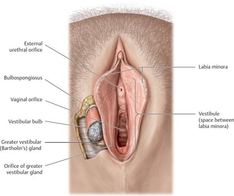
Fig. 12.9 Vestibule and vestibular glands
Lithotomy position with labia separated.

Fig. 12.10 Erectile muscles and tissue of the female genitalia
Lithotomy position. Removed: Labia, skin, and perineal membrane; Removed from left side: Ischiocavernosus and bulbospongiosus muscles; greater vestibular (Bartholin’s) gland.
– Greater vestibular (Bartholin’s) glands, small glands that lie under the posterior end of the vestibular bulbs, help to lubricate the vestibule during sexual arousal.
– Small lesser vestibular glands lie on each side of the vestibule and secrete mucus to moisten the labia and vestibule.
– The external pudendal artery supplies the skin over the mons pubis and labia majora. Similar to those in the male, these superficial structures drain to the external pudendal vein.
– The internal pudendal artery supplies most of the external genitalia through branches that are similar to those in the male perineum (Fig. 12.11A).
• The perineal artery supplies perineal muscles and labia minora.
• The artery of the vestibular bulb supplies the bulb of the vestibule and greater vestibular gland.
• The dorsal clitoral artery supplies the glans of the clitoris.
• The deep clitoral artery supplies the corpus cavernosum and is responsible for their engorgement during arousal.
– Tributaries of the pudendal vein drain most perineal structures and accompany the arteries. A single deep dorsal clitoral vein drains the venous plexuses of the erectile tissue and passes under the pubic symphysis to join the venous plexus in the pelvis (Fig. 12.11B).
– Most lymph from the female perineum drains to superficial inguinal lymph nodes. Exceptions include
• the clitoris, bulbs of the vestibule, and anterior labia, which drain to deep inguinal or internal iliac nodes; and
• the urethra, which drains to sacral or internal iliac nodes.
– The pudendal nerve is the primary nerve of the perineum (see Fig. 10.21B). It supplies
• the perineal nerve to the vaginal orifice and superficial perineal muscles;
• the dorsal clitoral nerve to deep perineal muscles and sensation from the clitoris, especially the glans; and
• the posterior labial nerves to the posterior vulva.
– As in the innervation of the male scrotum, the anterior vulva receives sensory innervation (see Fig. 10.21B) from
• the ilioinguinal nerve and genital branch of the genitofemoral nerve, which supply branches to the mons pubis and anterior labia; and
• the posterior femoral cutaneous nerve, which supplies the posterolateral vulva.
– Sympathetic fibers to the perineum travel with the hypogastric nerve plexus; parasympathetic fibers travel with cavernous nerves of the uterovaginal plexus. Both innervate the erectile tissue of the clitoris and bulbs of the vestibule.
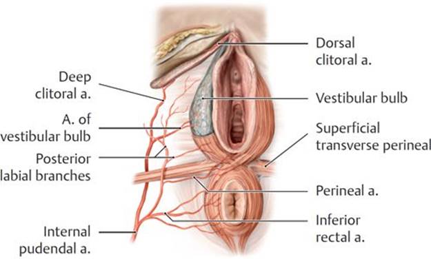
Fig. 12.11 Blood vessels of the female external genitalia
Inferior view.
A Arterial supply.
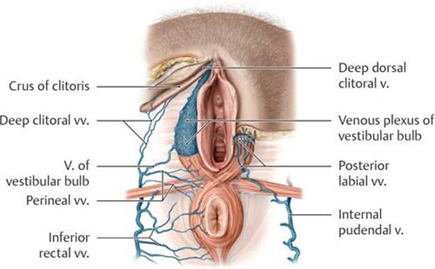
B Venous drainage.
12.5 Anal Triangle
The anal triangle contains the anal canal and ischioanal fossae.
12.5a The Anal Canal
The anal canal, the terminal part of the gastrointestinal tract, controls fecal continence and the defecation response. It extends from the anorectal junction at the pelvic diaphragm to the anus (Fig. 12.12).
– The puborectalis muscle forms a sling around the anorectal junction, pulling it anteriorly and creating the perineal flexure (Fig. 12.13). From this angle the anal canal descends inferiorly and posteriorly between the anococcygeal ligament (levator plate) and the perineal body (see Fig. 12.1A and B).

Fig. 12.12 Anal canal
Coronal section, anterior view.

Fig. 12.13 Closure of the rectum
Left lateral view. The puborectalis acts as a muscular sling that kinks the anorectal junction. It functions in the maintenance of fecal continence.
– Two sphincters surround the anal canal:
1. The internal anal sphincter is a thickening of the circular muscular layer that surrounds the upper part of the anal canal.
○ It is an involuntary sphincter.
○ It remains contracted via sympathetic innervation except in response to distension of the rectal ampulla. Parasympathetic innervation relaxes the sphincter.
2. The external anal sphincter is a broad band of muscle that extends anteriorly to merge with the perineal body, posteriorly to attach to the coccyx (through the anococcygeal ligament), and superiorly to merge with the puborectalis muscle of the pelvic floor (see also Figs. 11.1, 12.1, and 12.2).
○ It is a voluntary sphincter.
○ Although the anal sphincter is described as having deep, superficial, and subcutaneous parts, they are functionally, and often anatomically, indistinct.
○ The inferior rectal nerve, a branch of the pudendal nerve, innervates this sphincter.
– The inner surface of the anal canal is characterized by
• anal columns, vertical ridges formed by underlying branches of the superior mesenteric vessels;
• anal valves that connect the inferior edges of the anal columns; and
• anal sinuses, recesses at the base of the anal columns that secrete mucus to facilitate defecation.
– The dentate (pectinate) line is an irregular ridge at the base of the anal columns.
• It divides the anal canal into a superior part derived from the embryonic hindgut and an inferior part derived from the ectoderm.
• It divides the anal canal in its blood supply, lymphatic drainage, and innervation.
– Below the dentate line, a smooth lining devoid of glands and hair, the anal pecten, extends down to the anocutaneous line, or intersphincteric groove.
– Below the anocutaneous line, the anal canal is lined by hair-bearing skin that is continuous with perianal skin that surrounds the anus.
Anal fissures
Anal fissures are tears in the mucosa around the anus (usually in the posterior midline) that are caused by the passing of hard or large stools. Because they are below the pectinate line and innervated by the inferior rectal nerves, these lesions are painful. Most anal fissures heal spontaneously within a few weeks if care is taken to prevent constipation. Perianal abscesses that develop from the fissures can spread into the adjacent ischioanal fossae.
– Above the dentate line the neurovasculature of the anal canal is similar to that of the distal gastrointestinal tract (see Figs. 10.18 and 10.20).
• It receives its blood supply from the superior rectal artery, a branch of the inferior mesenteric artery.
• The rectal venous plexus drains through the superior rectal vein into the portal venous system.
• Lymph drains to the internal iliac nodes.
• Visceral innervation is transmitted via rectal plexuses to the inferior hypogastric plexus.
○ Sympathetic stimulation maintains the tone of the sphincter.
○ Parasympathetic innervation relaxes the sphincter and stimulates peristalsis of the rectum.
○ Visceral afferent fibers travel with pelvic splanchnic (parasympathetic) nerves and convey only sensation of stretching (no sensitivity to pain).
– Below the dentate line the neurovasculature of the anal canal is similar to that of the perineum (see Fig. 10.15).
• It receives its blood supply from the right and left inferior rectal arteries, branches of the internal iliac arteries.
• The rectal venous plexus drains into inferior rectal veins, which in turn drain to internal iliac veins of the venae caval system.
• Lymph drains to superficial inguinal nodes.
• Somatic innervation is transmitted via the inferior rectal nerve, a branch of the pudendal nerve.
○ Somatic efferent fibers stimulate contraction of the external anal sphincter.
○ Somatic afferent fibers transmit pain, touch, and temperature.
12.5b Ischioanal Fossae and Pudendal Canal
– The ischioanal fossae are paired wedge-shaped spaces on either side of the anal canal bounded by the pelvic diaphragm superiorly and the skin of the anal region inferiorly (see Fig. 11.19).
• Fat and loose connective tissue, strengthened by strong, fibrous bands, fill the fossae. These tissues support the anal canal but can be displaced readily when the anal canal distends with feces.
• The inferior rectal vessels and nerve, branches of the internal pudendal vessels and pudendal nerve, traverse the fossae.
• The ischioanal fossae extend anteriorly into the urogenital triangle superior to the perineal membrane.
– The pudendal canal is a passageway formed by the splitting of the fascia of the obturator internus muscle on the lateral wall of the ischioanal fossa.
• The internal pudendal artery and vein and the pudendal nerve enter the canal after exiting the lesser sciatic foramen and giving off inferior rectal branches.