
The cranial meninges continuous with the meninges of the spinal cord, as well as the 12 cranial nerves that arise from the brain, are essential parts of gross anatomy of the head and neck region and are discussed in detail in this unit. The study of the brain, however, is generally confined to the neuroanatomy curriculum, and only a brief overview is provided here.
18.1 The Meninges
The cranial meninges, coverings that protect the brain, consist of the external fibrous dura mater, the thin intermediate arachnoid mater, and the delicate inner pia mater (Fig. 18.1).
18.1a Dura mater
– The dura mater, or dura, a tough outer membrane surrounding the brain, is composed of a periosteal layer and a meningeal layer. The two layers are inseparable except where they enclose the venous sinuses that drain the brain (e.g., the superior sagittal sinus shown in Fig. 18.3).
• The outer periosteal layer, formed by the periosteum of the skull, adheres tightly to the inner surface of the skull, particularly at the sutures. This layer ends at the foramen magnum and is not continuous with the dura around the spinal cord.
• The inner meningeal layer, a strong membranous sheet that adheres to the inner surface of the periosteal layer, provides sheaths for the cranial nerves as they pass through the skull foramina. It is closely applied, although not attached, to the underlying arachnoid mater (see Fig. 18.1). It continues into the vertebral canal as dura of the spinal cord.
– The middle meningeal arteries, branches of the maxillary arteries, supply most of the dura, with contributions from the ophthalmic, occipital, and vertebral arteries. Veins accompany the arteries and drain into the pterygoid venous plexus.
– Branches of the trigeminal nerve (CN V) transmit sensation from the dura of the anterior and middle cranial fossa. Spinal nerves C1, C2, and C3 and small branches of the vagus nerve (CN X) innervate the dura of the posterior cranial fossa.

Fig. 18.1  Layers of the meninges
Opened cranium, superior view. Left side: Dura mater (outer layer) cut to reveal arachnoid mater (middle layer). Right side: Dura mater and arachnoid mater removed to reveal pia mater (inner layer) lining the surface of the brain. Note: Arachnoid granulations, sites for loss of cerebral spinal fluid into the venous blood, are protrusions of the arachnoid layer of the meninges into the venous sinus system.
18.1b Dural Partitions
Infoldings of the meningeal layer of the dura form incomplete membranous partitions that separate and support parts of the brain (Fig. 18.2).
– The falx cerebri, a vertical sickle-shaped partition separating the right and left cerebral hemispheres, is attached anteriorly to the crista galli and the inner crest of the frontal bone and is continuous posteriorly with the tentorium cerebelli. The inferior, free edge of the falx cerebri is unattached.
– The tentorium cerebelli, a horizontal continuation of the falx cerebri, separates the occipital lobes of the cerebrum from the cerebellar hemispheres in the posterior cranial fossae.
• It is attached to the posterior clinoid processes and the petrous part of the temporal bones anteriorly and to the parietal and occipital bones posterolaterally.
• A U-shaped tentorial notch separates the attachments to the petrous ridge on each side and connects the middle and posterior cranial fossae.
Tentorial herniation
Increased pressure within the middle cranial fossa created by a space-occupying lesion such as a tumor can squeeze the brain tissue and force part of the temporal lobe to herniate through the tentorial notch. Pressure on the adjacent brainstem can be fatal in this situation. The oculomotor nerve (CN III) can also be stretched or damaged, leading to fixed pupil dilation (loss of parasympathetic function) and a “down and out” gaze due to paralysis of most of the extraocular muscles.
– The falx cerebelli, a vertical partition separating the cerebellar hemispheres, is continuous superiorly with the tentorium cerebelli and is attached posteriorly to the occipital crest.
– The diaphragma sellae, a small dural fold attached to the anterior and posterior clinoid processes, forms a roof over the sella turcica, which encloses the hypophysis (pituitary gland).
18.1c Dural Venous Sinuses
Dural venous sinuses are valveless venous spaces that form as a result of the separation of the periosteal and meningeal layers of the dura. Most of the large veins of the brain, skull, orbit, and inner ear drain through the dural sinuses and into the internal jugular veins in the neck (Figs. 18.3 and 18.4; Table 18.1).
– The confluence of sinuses at the posterior edge of the tentorium cerebelli is a junction of the superior sagittal, straight, occipital, and transverse sinuses.
– The superior sagittal sinus runs in the attached superior border of the falx cerebri and ends in the confluence of sinuses.
– The inferior sagittal sinus runs in the free inferior edge of the falx cerebri and ends in the straight sinus.
– The straight sinus runs in the space formed by the union of the falx cerebri and tentorium cerebelli. It receives the inferior sagittal sinus and great cerebral vein and drains into the confluence of sinuses.
– The paired transverse sinuses run along the attached posterolateral margins of the tentorium cerebelli. Posteriorly, they join at the confluence of sinuses, and anteriorly they drain into the sigmoid sinuses, forming grooves in the occipital and parietal bones along their course.
– The paired sigmoid sinuses run in deep grooves of the occipital and temporal bones and drain into the internal jugular veins at the jugular foramen.
– The occipital sinus runs in the free edge of the falx cerebelli and ends in the confluence of sinuses.
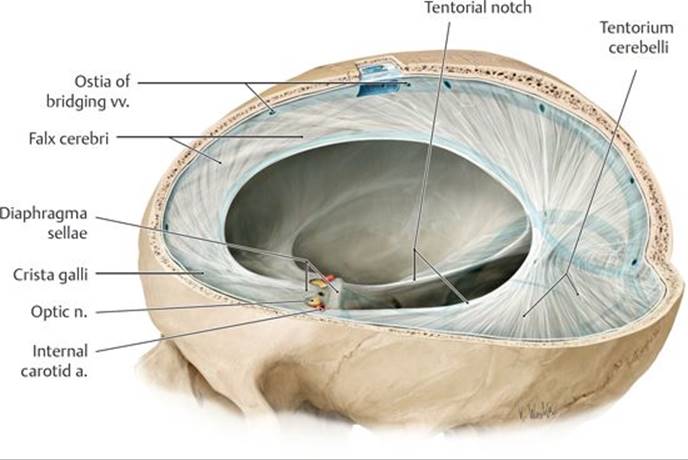
Fig. 18.2 Dural septa (folds)
Left anterior oblique view.
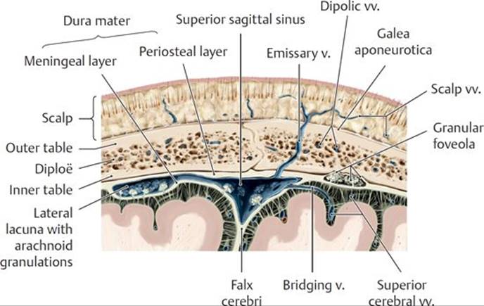
Fig. 18.3 Structure of a dural sinus
Superior sagittal sinus, coronal section, anterior view.
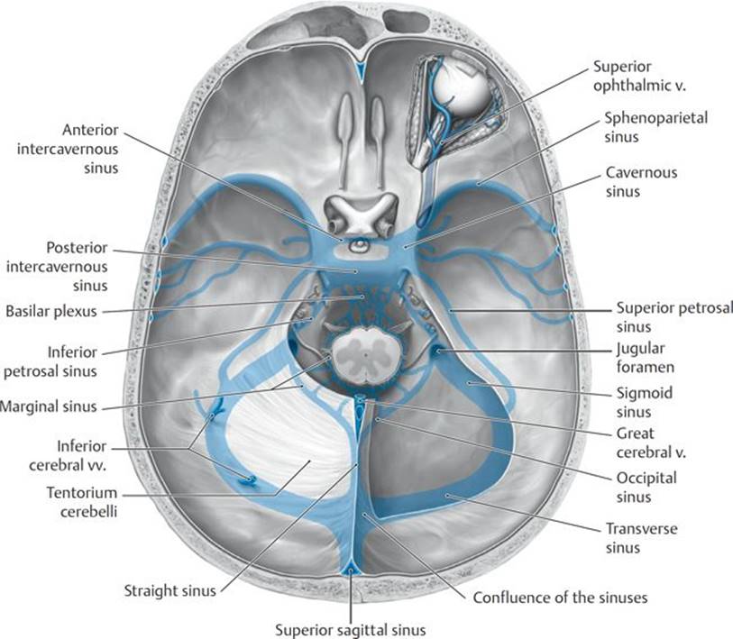
Fig. 18.4 Dural sinuses in the cranial cavity
Opened cranial cavity with dural sinus system ghosted in blue, superior view. Removed: Tentorium cerebelli.
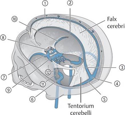
TABLE 18.1  Principal Dural Sinuses
|
Upper Group |
Lower Group |
|
|
|
|
|
|
|
|
|
|
|
|
|
|
|
|
|
|
The occipital sinus is also included in the upper group.
– The paired cavernous sinuses, located on either side of the sella turcica, have characteristics that distinguish them from other dural sinuses (Figs. 18.5 and 18.6).
• Each cavernous sinus contains a large plexus of thin-walled veins.
• Several important structures are associated with each cavernous sinus:
○ Internal carotid artery, which is surrounded by the sympathetic internal carotid nerve plexus
○ Oculomotor nerve (CN III)
○ Trochlear nerve (CN IV)
○ Ophthalmic and maxillary divisions (CN V1, V2) of the trigeminal nerve
○ Abducent nerve (CN VI)
• The cavernous sinuses receive the superior and inferior ophthalmic veins, the sphenoparietal sinuses, the superficial middle cerebral veins, and the central veins of the retina.
• The cavernous sinuses drain into the superior and inferior petrosal sinuses posteriorly and the pterygoid venous plexus inferiorly.
• Anterior and posterior intercavernous sinuses (see Fig. 18.4) connect the right and left cavernous sinuses.
Cavernous sinus thrombophlebitis
Cavernous sinus thrombophlebitis can occur secondary to thrombophlebitis of the facial vein. Although blood from the angle of the eye, lips, nose, and face usually drains inferiorly, it can also drain through the veins of the orbit to the cavernous sinus. Infections from the face, particularly from the danger triangle of the face (which extends from the bridge of the nose to the angles of the mouth) can spread infected thrombi to the cavernous sinus. This can affect the nerves that traverse the sinus (CN III, CN IV, CN V1 and V2, and CN VI) and result in acute meningitis.
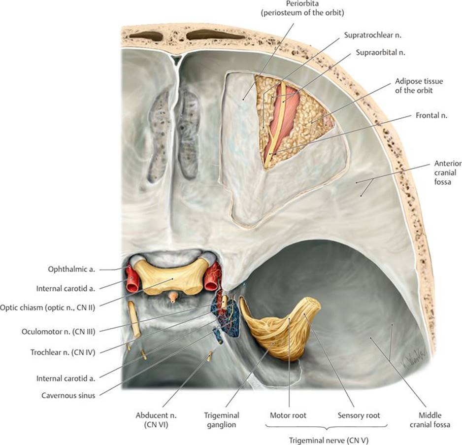
Fig. 18.5 Cavernous sinus and cranial nerves
Left anterior and middle cranial fossae, superior view. Removed: Lateral dural wall and roof of the cavernous sinus. The trigeminal ganglion is cut and retracted laterally following removal of its dural covering.

Fig. 18.6 Cavernous sinus
Middle cranial fossa, coronal section, anterior view.
– Paired superior petrosal sinuses, which drain the cavernous sinuses, travel within the attached margins of the tentorium cerebelli along the top of the petrous part of the temporal bones and empty into the sigmoid sinuses.
– Paired inferior petrosal sinuses drain the cavernous sinuses, passing through a groove between the petrous part of the temporal bones and the basilar part of the occipital bone and emptying into the sigmoid sinuses at the origin of the internal jugular veins. The inferior petrosal sinuses communicate, through a basilar plexus, with the vertebral venous plexus.
18.1d Arachnoid Mater and Pia Mater (Fig. 18.7; see Figs. 18.1 and 18.3)
– Arachnoid mater, or arachnoid, is a thin, avascular, fibrous layer underlying the meningeal layer of the dura.
• Cerebrospinal fluid presses the arachnoid against the dura, but the two layers are not attached. Weblike arachnoid trabeculae attach the arachnoid to the underlying pia mater.
• Delicate fingers of the arachnoid layer, the arachnoid villi, pierce the dura to allow the reabsorption of cerebrospinal fluid into the venous circulation and are especially numerous in the superior sagittal sinus. They form aggregations called arachnoid granulations that protrude into the largest dural venous sinuses and can push the dura ahead of them into the parietal bone, forming “pits.”
• Congregations of arachnoid granulations also occur in lateral lacunae, lateral expansions of the superior sagittal sinus.
– Pia mater, or pia, is a thin, highly vascular layer that adheres to the surface of the brain and closely follows its contours.

Fig. 18.7 Meningeal spaces
Meninges, coronal section, anterior view.
18.1e Meningeal Spaces
– The epidural space between the cranium and dura is not a natural space because the dura adheres to the skull. Meningeal vessels that supply the skull and dura travel in this space (see Fig. 18.7).
– The subdural space between the dura and arachnoid is a potential space, open only in pathological conditions such as a subdural hematoma. Superficial cerebral veins (“bridging veins”) cross this space, connecting the venous circulation of the brain with the dural venous sinuses (see Fig. 18.7).
– The subarachnoid space, between the arachnoid and pia layers, contains cerebrospinal fluid, arteries, and veins (see Fig. 18.7).
• Subarachnoid cisterns are spaces that form where the subarachnoid space enlarges around large infoldings of the brain. The largest of these include the cerebellomedullary, pontomedullary, interpeduncular, chiasmatic, quadrigeminal, and ambient cisterns (see Section 18.2b; see Fig. 18.10).
Extracerebral hemorrhage
Bleeding from vessels between the bony skull and the brain (extracerebral hemorrhage) increases intracranial pressure and can damage brain tissue. Three types of cerebral hemorrhages are distinguished based on their relationship to the meningeal layers.
Epidural hemorrhages commonly originate from a torn middle meningeal artery following a skull fracture at the pterion and result in bleeding into the epidural space. The hemorrhagic spread is usually limited by suture lines because the dura is attached to the skull at these points. As a result, the local accumulation of blood causes compression of the brain in that area.
Subdural hematomas result from tearing of the bridging veins as they traverse the gap between the dural sinus and cerebral cortex. The elderly are more susceptible to this type of hemorrhage because with brain shrinkage these veins bridge a larger gap and are more vulnerable to injury from head trauma. This condition may mimic a slowly evolving stroke with a fluctuating level of consciousness and localizing neurologic signs.
Most subarachnoid hemorrhages occur due to the rupture of aneurysms associated with vessels of the circle of Willis and most frequently with vessels of the anterior cerebral circulation. These hemorrhages into the subarachnoid space begin with a sudden, severe headache, neck stiffness, and drowsiness but can progress to severe consequences such as hemiplegia and coma.
18.2 The Brain
The brain, enclosed within the bony skull, is the largest part of the central nervous system. It communicates with the peripheral nervous system through the spinal cord and spinal nerves and through the 12 pairs of cranial nerves.
18.2a Regions of the Brain
The major regions of the brain are the cerebrum, diencephalon, brainstem (mesencephalon, pons, medulla oblongata), and cerebellum (Fig. 18.8A, B, and C).
– The cerebrum is the largest part of the brain and the center for integration within the central nervous system.
• The falx cerebri lies in a longitudinal fissure between the right and left cerebral hemispheres.
• Each cerebral hemisphere is further divided into frontal, parietal, occipital, and temporal lobes that occupy the anterior and middle cranial fossae.
• Posteriorly, the cerebrum rests on the tentorium cerebelli.
• The surface layer of the cerebrum forms gyri (folds) separated by sulci (grooves).
– The diencephalon forms the central core of the brain and consists of the thalamus, hypophysis, and hypothalamus.
– The mesencephalon, the most anterior part of the brainstem, passes through the tentorial notch between the middle and posterior cranial fossae.
• It is associated with the oculomotor (CN III) and trochlear (CN IV) nerves.
– The pons, the middle part of the brainstem, lies in the anterior part of the posterior cranial fossa below the mesencephalon.
• Several ascending and descending fiber tracts connect the pons to the cerebellum.
• The pons is associated with the trigeminal (CN V), abducent (CN VI), and facial (CN VII) nerves.
– The medulla oblongata, the most posterior part of the brainstem, connects the brain and spinal cord.
• It contains nuclei for the vestibulocochlear (CN VIII), glossopharyngeal (CN IX), vagus (CN X), and hypoglossal (XII) nerves.
– The cerebellum, which occupies most of the posterior cranial fossa, lies inferior to the cerebrum and is separated from it by the tentorium cerebelli.
• It consists of paired hemispheres and a small middle section, the vermis.
18.2b Ventricular System and Cerebrospinal Fluid
The brain and spinal cord are suspended in cerebrospinal fluid (CSF). The buoyant environment created by the CSF reduces the pressure of the brain on the nerves and vessels on its inferior surface.
– CSF is produced in the choroid plexuses, vascular networks within four ventricles (spaces) of the brain. The first two of these ventricles are large and paired; the third and fourth are smaller and lie in the midline (Fig. 18.9).
• The 1st and 2nd (lateral) ventricles, paired cavities that occupy a large portion of each cerebral hemisphere, communicate with the 3rd ventricle through the interventricular foramina.
• The 3rd ventricle, a slitlike space between the two halves of the diencephalon, communicates posteriorly with the 4th ventricle through a narrow passage, the cerebral aqueduct, which passes through the mesencephalon.
• The 4th ventricle, a pyramidally shaped space that extends from the pons to the medulla oblongata, is continuous with the spinal canal inferiorly and with the subarachnoid space through the median and lateral apertures in its roof.

Fig. 18.8 Adult brain
CN, cranial nerve.
A Left lateral view.
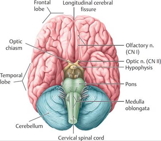
B Basal view.
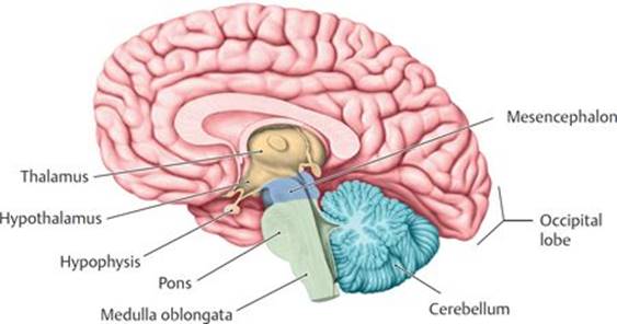
C Midsagittal section showing the right hemisphere.

Fig. 18.9 Ventricular system in situ
Ventricular system with neighboring structures, left lateral view.

Fig. 18.10 Circulation of cerebrospinal fluid (CSF)
– CSF circulates through the ventricles and passes into the subarachnoid space and subarachnoid cisterns through the median and lateral apertures of the 4th ventricle. It flows superiorly through the fissures and sulci of the cerebrum and is reabsorbed into the venous circulation through the arachnoid granulations that protrude into the superior sagittal sinus (Fig. 18.10).
Hydrocephalus
Hydrocephalus, an excessive accumulation of cerebrospinal fluid (CSF) in the ventricles of the brain, can occur as a result of partial obstruction of the flow of CSF within the ventricular system, interference of CSF reabsorption into the venous circulation, or, in rare cases, overproduction of CSF. Excess CSF in the ventricles causes them to dilate and exert pressure on the surrounding cortex, causing the bones of the calvaria to separate, thus creating the characteristic increase in head size. Treatment involves the placement of a shunt between the ventricles and the abdomen, which allows CSF to drain to the peritoneal cavity, where it can be easily absorbed.
18.2c Arteries of the Brain
As a result of its high metabolic demand, the brain receives one sixth of the cardiac output and one fifth of the oxygen consumed by the body at rest. This blood supply, derived from the internal carotid and vertebral arteries, is divided into anterior and posterior cerebral circulations (Fig. 18.11), which unite on the ventral surface of the brain to form a cerebral arterial circle (of Willis) (Fig. 18.12).
– The internal carotid artery supplies the anterior cerebral circulation.
• Its petrous part has a tortuous course as it enters the skull and follows the carotid canal horizontally and medially within the temporal bone. Small branches pass into the middle ear and pterygoid canal.
• The cavernous part crosses over the foramen lacerum and runs anteriorly within the cavernous sinus. Small branches supply the meninges, hypophysis, and cranial nerves within the cavernous sinus.
• The cerebral part in the middle cranial fossa gives off the ophthalmic artery (see Fig. 20.8) and immediately makes a U-turn to run posteriorly, where it divides into the anterior cerebral and middle cerebral arteries.
– The vertebral and basilar arteries supply the posterior cerebral circulation.
• The vertebral artery enters the skull through the foramen magnum and supplies branches to the spinal cord and cerebellum before merging with the opposite vertebral artery to form a single basilar artery.
○ Intracranial branches of the vertebral artery include the posterior inferior cerebellar artery and the anterior and posterior spinal arteries.

Fig. 18.11 Internal carotid artery
Left lateral view.

Fig. 18.12 Arteries of the brain
Inferior (basal) view.
• The basilar artery ascends on the ventral surface of the brainstem, distributing branches to the brainstem, cerebellum, and cerebrum. It terminates as the right and left posterior cerebral arteries.
○ Major branches of the basilar artery are the anterior inferior cerebellar artery and the superior cerebellar artery.
– The cerebral arterial circle (of Willis), an important arterial anastomosis on the ventral surface of the brain, supplies the brain and connects the circulations of the internal carotid and vertebral arteries.
• A small anterior communicating artery connects the two anterior cerebral arteries, linking the right and left anterior cerebral circulations.
• A pair of posterior communicating arteries connects the internal carotid and posterior cerebral arteries on each side, completing the communication between the anterior and posterior cerebral circulations.
• The branches of the circle are
○ the anterior communicating arteries,
○ the anterior cerebral arteries,
○ the internal carotid arteries,
○ the posterior communicating arteries, and
○ the posterior cerebral arteries.
– The cerebral arteries that arise from the cerebral arterial circle provide the blood supply to the cerebral hemispheres (Table 18.2).
TABLE 18.2 Distribution of the Cerebral Arteries
|
Artery |
Origin |
Distribution |
|
Anterior cerebral |
Internal carotid artery |
Frontal pole and medial and superior surfaces of the cerebral hemispheres |
|
Middle cerebral |
Internal carotid artery |
Most of the lateral surface of the cerebral hemispheres |
|
Posterior cerebral |
Basilar artery |
Occipital pole and inferior part of temporal lobe |
Stroke
A stroke is the manifestation of a neurologic deficiency resulting from a cerebral vascular impairment. Ischemic strokes are usually caused by an embolus obstructing one of the major cerebral arteries. Although the vessels of the circle of Willis can provide collateral circulation to circumvent the obstruction, anastomoses between the vessels are often incomplete or of insufficient size to provide adequate flow. Hemorrhagic strokes are usually due to rupture of an aneurysm, most often a saccular, or berry, aneurysm that bleeds into the subarachnoid space. Symptoms occur shortly after the cerebral event and relate to the area of brain affected. They may include difficulty speaking, understanding language, or walking; vision problems; contralateral paralysis or numbness; and headache.
18.2d Veins of the Brain
Veins that drain the brain are thin-walled and valveless and usually drain into one of the dural venous sinuses (Fig. 18.13A and B).
– Superficial (external) veins that drain the cerebral hemispheres include
• the superior cerebral veins, which drain the supralateral and medial aspects. These “bridging veins” traverse the subdural space and drain into the superior sagittal sinus;
• the middle cerebral veins, which drain the lateral hemispheres and empty into the cavernous sinus and from there into the petrosal and transverse sinuses; and
• the inferior cerebral veins, which drain inferior aspects of the brain and join either the superior cerebral or the basilar veins.
– Basilar veins drain the small anterior cerebral veins and deep middle cerebral veins.
– Internal cerebral veins drain the 3rd and 4th ventricles and deep parts of the cerebrum. These unite to form the great cerebral vein.
– The great cerebral vein receives the basilar veins and merges with the inferior sagittal sinus to form the straight sinus.
– Superior and inferior cerebellar veins drain the cerebellum into adjacent dural sinuses or superficially into the great cerebral vein.

Fig. 18.13 Cerebral veins
A Lateral view of the left hemisphere.

B Medial view of the right hemisphere.
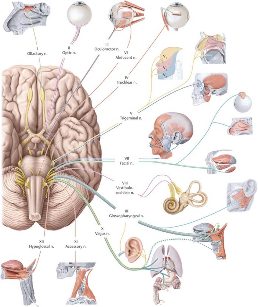
Fig. 18.14 Cranial nerves
Inferior (basal) view. The 12 pairs of cranial nerves (CN) are numbered according to their emergence from the brainstem.
18.3 Cranial Nerves
The 12 cranial nerves arise from the brainstem (Fig. 18.14; Table 18.3). Like spinal nerves, cranial nerves can stimulate muscles or transmit sensation from a peripheral structure to the central nervous system. Some cranial nerves also carry fibers from the cranial portion of the parasympathetic nervous system. Seven types of nerve fibers are found (alone or in combination) in cranial nerves.
TABLE 18.3 Classification of Cranial Nerve Fibers
|
Fiber Type |
Function |
|
General somatic motor |
Innervate voluntary muscle |
|
General visceral motor |
Constitute the cranial component of the parasympathetic system, innervate involuntary muscles and glands |
|
Special visceral motor (branchial motor) |
Innervate muscles that developed from the primitive pharynx (pharyngeal arches) |
|
General somatic sensory |
Carry sensations such as touch, temperature, pain, and pressure |
|
Special somatic sensory |
Carry impulses from the eye for sight and from the ear for hearing and balance |
|
General visceral sensory |
Transmit information from viscera such as carotid bodies, the heart, esophagus, trachea, and gastrointestinal tract |
|
Special visceral sensory |
Transmit information regarding smell and taste |
CN I, the olfactory nerve, carries special sensory fibers that transmit sensation of smell from the superior aspect of the lateral and septal walls of the nasal cavity (Fig. 18.15).
– Olfactory neurons pass through the cribriform plate of the ethmoid bone and synapse with secondary neurons in the olfactory bulbs.
• The axons of these secondary neurons form the olfactory tracts.
• The olfactory bulbs and tracts are extensions of the cerebral cortex.
CN II, the optic nerve, is a collection of special sensory nerve fibers that originate on the retina of the eye and converge at the optic disc at the back of the eyeball (Fig. 18.16; see also Section 20.1e).
– The nerve exits the orbit through the optic canal and joins the contralateral optic nerve to form the optic chiasm.
– The optic chiasm is a redistribution center where nerve fibers from the medial half of each optic nerve cross to the opposite side.
– Two optic tracts diverge from the chiasm. Each tract contains nerve fibers from the medial half of one eye and the lateral half of the other eye.

Fig. 18.15 Olfactory nerve (CN I)
Olfactory fibers, bulb, and tract. Portion of left nasal septum and lateral wall of right nasal cavity, left lateral view.

Fig. 18.16 Optic nerve (CN II)
Optic nerve in the left orbit, left lateral view.
CN III, the oculomotor nerve; CN IV, the trochlear nerve; and CN VI, the abducent nerve innervate structures of the orbit (Fig. 18.17; see also Section 20.1e). They pass through the cavernous sinus before entering the orbit through the superior orbital fissure.
– The oculomotor nerve has somatic and visceral components.
• General somatic motor fibers innervate four of the extraocular muscles (superior rectus, medial rectus, inferior rectus, and inferior oblique), which move the eyeball, and the levator palpebrae superioris muscle, which elevates the eyelid.
• General visceral motor fibers carry preganglionic parasympathetic fibers that synapse in the ciliary ganglion and innervate the pupillary sphincter muscle (which constricts the pupil) and ciliary body(which changes the curvature of the lens of the eye) (see Fig. 20.5).
– The trochlear nerve carries general somatic motor fibers and innervates the superior oblique muscle, which turns the eye upward and outward.
– The abducent nerve carries general somatic motor fibers and innervates the lateral rectus muscle, which abducts the eye.
CN V, the trigeminal nerve, is the primary sensory nerve of the face (Fig. 18.18). Its small motor component innervates the muscles of mastication (chewing).
– The general somatic sensory neurons, which form the sensory root, synapse in the trigeminal ganglion located in a dural space on the petrous part of the temporal bone.
– A small motor root in the mandibular division (CN V3) contains branchial motor fibers.
– Branches of the trigeminal nerve are associated with the parasympathetic ganglia of the head and distribute postganglionic parasympathetic fibers to their target organs.
– The trigeminal nerve has three divisions:
1. The ophthalmic division (CN V1) (see Section 20.1e)
○ contains only somatic sensory fibers;
○ passes through the cavernous sinus and superior orbital fissure into the orbit;
○ is associated with the ciliary ganglion (see Section 18.4);
○ distributes visceral motor fibers from the facial nerve (CN II) to the lacrimal gland via the lacrimal nerve;
○ innervates the orbit, the cornea, and the skin on the top of the nose, forehead, and scalp;
○ functions as the sensory component of the corneal reflex via the nasociliary branch; and
○ has lacrimal, frontal, and nasociliary branches.

Fig. 18.17 Oculomotor (CN III), trochlear (IV), and abducent (VI) nerves
Course of the nerves innervating the extraocular muscles, right orbit, lateral view.
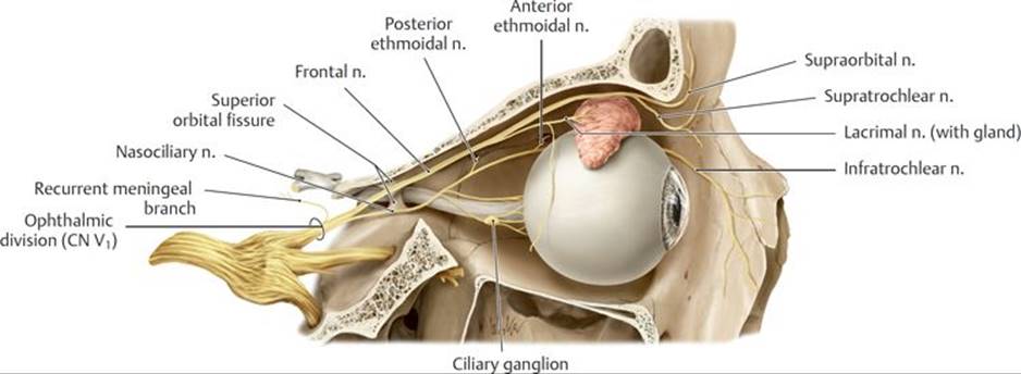
Fig. 18.18 Trigeminal nerve (CN V)
Course of the trigeminal nerve divisions.
A Ophthalmic division (CN V1), partially opened right orbit.

B Maxillary division (CN V2), partially opened right maxillary sinus with the zygomatic arch removed.

C Mandibular division (CN V3), partially opened mandible with the zygomatic arch removed. Note: The mylohyoid nerve (not shown) branches from the inferior alveolar nerve just before the mandibular foramen.
2. The maxillary division (CN V2) (see Section 19.6)
○ contains only somatic sensory fibers;
○ travels through the cavernous sinus and the foramen rotundum to enter the pterygopalatine fossa;
○ is associated with the pterygopalatine ganglion (see Section 18.4);
○ innervates the skin of the midface (from the lower eyelid to the upper lip) and structures associated with the maxilla, such as the maxillary sinus, palate, nasal cavity, and maxillary teeth; and
○ has infraorbital, zygomatic, greater and lesser palatine, superior alveolar, and nasopalatine branches (see also Fig. 19.18).
3. The mandibular division (CN V3) (see Section 19.4 and 19.5)
○ contains somatic sensory and branchial motor fibers;
○ passes through the foramen ovale into the infratemporal fossa;
○ is associated with the otic and submandibular ganglia (see Section 18.4);
○ distributes visceral motor fibers of the facial nerve (CN VII) to the submandibular and sublingual glands via the lingual nerve;
○ distributes visceral motor fibers of the glossopharyngeal nerve (CN IX) to the parotid gland via the auriculotemporal nerve;
○ has a sensory component that innervates skin over the lower jaw and lateral face and structures associated with the mandible, such as the lower teeth, temporomandibular joint, floor of the mouth, and anterior tongue;
○ has a motor component that innervates the digastric (anterior belly), mylohyoid, tensor veli palatini, and tensor tympani muscles and the muscles of mastication (see Sections 19.2, 19.8a, and 19.8b); and
○ has meningeal, buccal, auriculotemporal, lingual, inferior alveolar, and muscular branches (to muscles noted above).
Trigeminal neuralgia
Trigeminal neuralgia, a pathology of the sensory root of the trigeminal nerve (CN V), most commonly affects the maxillary division (CN V2) and least frequently affects the ophthalmic division (CN V1). The disorder is characterized by unilateral electric shock–like pain in the area supplied by the nerve. It usually lasts several seconds to several minutes. As the condition progresses, the pain may last longer, and there may be a shorter period between attacks. The pain may be initiated by touching a trigger point in the face by eating, talking, brushing teeth, or shaving. It is believed that trigeminal neuralgia is caused by the loss of myelin on the sensory root due to the pressure from an abnormal blood vessel. Surgery to destroy the nerve root or ganglion may be effective but may lead to permanent facial numbness.
CN VII, the facial nerve, is the primary motor nerve of the face but also has sensory and visceral components (Figs. 18.19 and 18.20). It contains a motor root that innervates the muscles of facial expression, and an intermediate nerve that carries special sensory (for taste) and visceral motor fibers (parasympathetic) and somatic sensory fibers. Both the motor root and the intermediate nerve pass through the internal acoustic meatus into the facial canal of the temporal bone, where three branches arise: the greater petrosal nerve, stapedial nerve, and chorda tympani.
– The motor root
• exits the skull through the stylomastoid foramen and
• contains branchial motor fibers, which
○ innervate the stylohyoid, stapedius, and digastric (posterior belly) muscles (see Table 19.7, Figs. 19.21 and 20.12, and Section 19.2) and
○ form the parotid plexus within the parotid gland, which innervates the muscles of facial expression (see Section 19.1). Branches of the parotid plexus include the posterior auricular, temporal, zygomatic, buccal, marginal mandibular, and cervical branches.
– The intermediate nerve contains the following:
• The greater petrosal nerve (parasympathetic), which passes through the middle cranial fossa and combines with the deep petrosal nerve (sympathetic) to form the nerve of the pterygoid canal (see Section 19.6). The visceral motor (parasympathetic) fibers synapse in the pterygopalatine ganglion and are distributed to glands of the nasal mucosa and palate and to the lacrimal gland.
• The chorda tympani, which passes through the middle ear cavity, exits through the stylomastoid foramen and travels with the lingual nerve of CN V3. It carries
○ visceral motor fibers that synapse in the submandibular ganglion and supply the submandibular and sublingual salivary glands, and
○ special sensory fibers for taste from the anterior part of the tongue and palate.
• General somatic sensory fibers, which transmit sensations from the external ear to the geniculate ganglion, the sensory ganglion of the facial nerve, located in the temporal bone.

Fig. 18.19 Course of the facial nerve
Visceral motor (parasympathetic) and special visceral sensory (taste) fibers shown in black, right lateral view.

Fig. 18.20 Facial nerve (CN VII)
Branches of the facial nerve, right lateral view.
A Facial nerve in the temporal bone.

B Parotid plexus.

Fig. 18.21 Vestibulocochlear nerve (CN VIII)
A Vestibulocochlear nerve in the temporal bone, medial wall of the tympanic cavity, oblique sagittal section.

B Vestibular and cochlear (spiral) ganglia.
Bell’s palsy
Bell’s palsy is paralysis of the facial muscles due to a lesion of the facial nerve (CN VII). Symptoms usually begin suddenly and affect one side of the face only. They include drooping of the corner of the mouth, eyebrow, and lower eyelid and the inability to smile, whistle, blow out cheeks, wrinkle the forehead, blink, or close the eyes forcefully. Taste is impaired on the anterior two thirds of the tongue (due to involvement of the chorda tympani), decreased tear production leads to dry eyes (due to involvement of the greater petrosal nerve), sensitivity to sounds is increased (due to paralysis of the stapedius), and the lower jaw and tongue deviate to the opposite side (due to paralysis of the posterior belly of the digastric muscle).
CN VIII, the vestibulocochlear nerve, is the sensory nerve of hearing and balance. The nerve enters the temporal bone with the facial nerve through the internal acoustic meatus.
– The two branches of the vestibulocochlear nerve carry special sensory fibers (Fig. 18.21A and B; see Section 20.2c):
• The cochlear root supplies the cochlea and its spiral organ, the organ of hearing.
• The vestibular root, which contains the vestibular ganglia, supplies the utricle, saccule, and semicircular ducts, the organs of balance.
CN IX, the glossopharyngeal nerve, leaves the skull through the jugular foramen and contains special sensory (taste), visceral sensory, somatic motor, and visceral motor components (Figs. 18.22, 18.23Aand B; Table 18.4).
– Somatic motor fibers innervate the stylopharyngeus muscle.
– Visceral motor fibers arise with the tympanic nerve, a branch of the glossopharyngeal nerve. Carrying sensory and visceral motor fibers, it runs through the tympanic cavity of the middle ear (see Section 20.2b), where it contributes to the tympanic plexus. It gives rise to the lesser petrosal nerve.
○ The lesser petrosal nerve passes through the middle cranial fossa and foramen ovale carrying visceral motor fibers that synapse in the otic ganglion. Postganglionic fibers travel with the auriculotemporal nerve (CN V3) to innervate the parotid gland.
○ Sensory fibers of the tympanic plexus supply the tympanic cavity and pharyngotympanic (auditory) tube.
– Special sensory fibers transmit taste from the posterior third of the tongue.
– Visceral sensory fibers transmit information from the tonsils, soft palate, and posterior third of the tongue and, via the branch to the carotid sinus, from receptors in the carotid body and carotid sinus at the bifurcation of the common carotid artery.
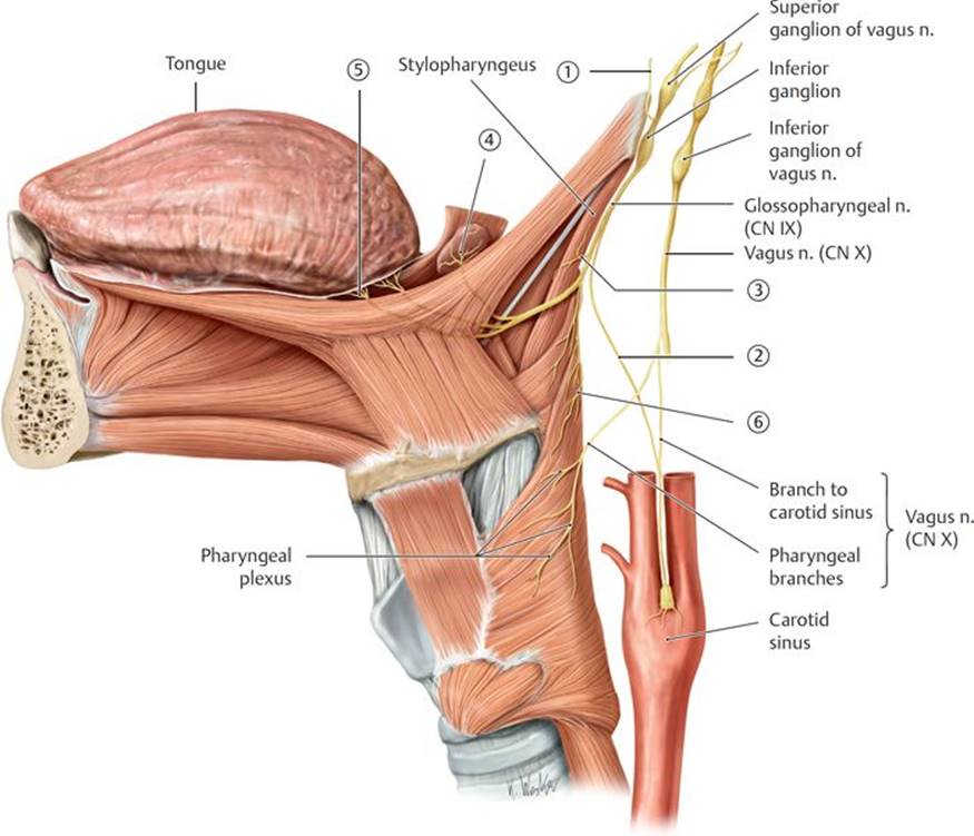
Fig. 18.22 Glossopharyngeal nerve (CN IX)
Course of the glossopharyngeal nerve, left lateral view.
TABLE 18.4 Glossopharyngeal Nerve Branches
|
|
Tympanic n. |
|
|
Branch to carotid sinus |
|
|
Branch to stylopharyngeus muscle |
|
|
Tonsillar branches |
|
|
Lingual branches |
|
|
Pharyngeal branches |

Fig. 18.23 Branches of the glossopharyngeal nerve
A Glossopharyngeal nerve in the tympanic cavity, left anterolateral view.

B Visceral motor fibers of the glossopharyngeal nerve, right lateral view.
CN X, the vagus nerve, has the most extensive distribution of the cranial nerves (Fig. 18.24; Table 18.5).
– Branchial motor fibers innervate the muscles of the soft palate (except tensor veli palatini), pharynx (except stylopharyngeus), and larynx, and the palatoglossus muscle of the tongue.
– Visceral motor fibers innervate smooth muscle and glands of the pharynx, larynx, thoracic organs, and abdominal foregut and midgut.
– General somatic sensory fibers transmit sensation from the dura in the posterior cranial fossa, the skin of the external ear, and the external auditory canal.
– Visceral sensory fibers transmit sensation from mucosa of the lower pharynx, the larynx, lungs and airway, the heart, the abdominal foregut and midgut, and the chemoreceptors of the aortic body and baroreceptors of the aortic arch.
– Special sensory fibers carry taste from the epiglottis.
– The vagus nerve has cervical, thoracic, and abdominal segments.
• In the neck
○ each vagus nerve leaves the skull through the jugular foramen and descends within the carotid sheath of the neck, and
○ its branches are pharyngeal branches, the superior laryngeal nerve, cervical cardiac (parasympathetic) branches, and the right recurrent laryngeal nerve (which arises from the right vagus and recurs around the right subclavian artery).
• In the thorax
○ the right and left vagus nerves enter the thorax posterior to the sternoclavicular joints and merge on the surface of the esophagus as the esophageal plexus (see Section 3.2d), and
○ their branches are the left recurrent laryngeal nerve (which arises from the left vagus and recurs around the arch of the aorta) and the thoracic cardiac and pulmonary branches (parasympathetic).
• In the abdomen
○ right and left vagal trunks arise from the esophageal plexus and pass through the esophageal hiatus of the diaphragm as the anterior and posterior vagal trunks; and
○ their parasympathetic branches are distributed to organs of the foregut, midgut, and retroperitoneum.

Fig. 18.24 Vagus nerve (CN X)
Branches of the vagus nerve in the neck, anterior view.
TABLE 18.5 Vagus Nerve Branches in the Neck
|
|
Pharyngeal branches |
|
|
Superior laryngeal n. |
|
|
Right recurrent laryngeal n. |
|
|
Left recurrent laryngeal n. |
|
|
Cervical cardiac branches |

Fig. 18.25 Accessory nerve (CN XI)
Brainstem with the cerebellum removed, posterior view.
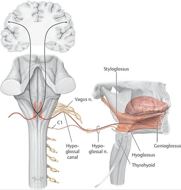
Fig. 18.26 Hypoglossal nerve (CN XII)
Brainstem with the cerebellum removed, posterior view. Note: C1, which innervates the thyrohyoid and geniohyoid, runs briefly with the hypoglossal nerve.
CN XI, the accessory nerve, contains general somatic motor fibers, which originate in a nucleus of the upper segments of the spinal cord (Fig. 18.25).
– The nerve emerges with the upper five or six cervical spinal nerves and ascends within the vertebral canal. It enters the skull through the foramen magnum and exits through the jugular foramen with the vagus and glossopharyngeal nerves.
– It innervates the sternocleidomastoid muscle in the neck and then crosses the lateral region of the neck to innervate the trapezius muscle.
CN XII, the hypoglossal nerve, contains only general somatic motor fibers (Fig. 18.26).
– The hypoglossal nerve leaves the skull through the hypoglossal canal and runs forward, medial to the angle of the mandible, to enter the oral cavity.
– It innervates all of the muscles of the tongue except the palatoglossus.
Injury to the hypoglossal nerve
Injury to the hypoglossal nerve causes ipsilateral paralysis of half of the tongue. When the tongue is protruded, the tip deviates toward the paralyzed side because the action of the genioglossus muscle on the unaffected side is unopposed. Symptoms mainly manifest as slurring of speech. Over time, the tongue becomes weak and atrophies.
18.4 Autonomic Nerves of the Head
– Sympathetic nerves of the head arise as postganglionic fibers from the superior cervical ganglia (see Section 21.3c).
• The internal carotid plexus of sympathetic fibers surrounds the internal carotid artery and its branches within the skull. A similar external carotid plexus follows the branches of the external carotid artery on the face.
• Sympathetic fibers often travel with the parasympathetic nerves, but they do not synapse in the parasympathetic ganglia.
– The cranial portion of the parasympathetic (visceral motor) system is associated with the oculomotor (CN III), facial (CN VII), glossopharyngeal (CN IX), and vagus (CN X) nerves.
• Preganglionic parasympathetic fibers traveling with the oculomotor (CN III), facial (CN VII), and glossopharyngeal (CN IX) nerves synapse in the four parasympathetic ganglia of the head: the ciliary, pterygopalatine, submandibular, and otic ganglia (Table 18.6; see Figs. 18.17, 18.19, and 18.23B).
• Parasympathetic nerves traveling with the vagus nerve (CN X) extend into the thorax and abdomen and synapse in ganglia of nerve plexuses in those regions.
• Parasympathetic ganglia of the head are usually attached to, or in close association with, a branch of the trigeminal nerve (CN V). The postganglionic parasympathetic fibers travel to their target organ by “piggybacking” on these trigeminal branches.

 Superior sagittal sinus
 Cavernous sinus
 Inferior sagittal sinus
 Anterior intercavernous sinus
 Straight sinus
 Posterior intercavernous sinus
 Confluence of the sinuses
 Sphenoparietal sinus
 Transverse sinus
 Superior petrosal sinus
 Sigmoid sinus
 Inferior petrosal sinus


