
The anatomy of the head can be divided into smaller regions that lie anterior and lateral to the neurocranium and form the superficial and deep structures of the face. They include the scalp; the parotid region; the temporal, infratemporal, and pterygopalatine fossae; and the nasal and oral cavities.
19.1 The Scalp and Face
The scalp covers the neurocranium and extends from the superior nuchal lines of the occipital bone, which mark the superior limit of the neck, to the supraorbital ridge of the frontal bone. The face extends from the forehead to the chin and to the ears on either side.
– The scalp is composed of five layers:
• Skin
• Connective tissue containing the vessels of the scalp
• Aponeurosis of the occipitofrontalis, temporoparietalis, and superior auricular muscles
• Loose areolar tissue
• Periosteum of the skull
The first letter of each layer spells “SCALP.”
– Muscles of facial expression lie in the loose connective tissue layer of the face and scalp. Their origin on bones of the face and insertion into the overlying skin allows them to create facial movements (Fig. 19.1A and B; Tables 19.1 and 19.2).
– Most arteries that supply the face and scalp are branches of the external carotid artery (see Fig. 17.15) and include the following:
• The superior and inferior labial arteries, the lateral nasal artery, and the angular branches of the facial artery, which supply the face between the eye and lower lip
• The mental branch of the inferior alveolar artery, which supplies the chin
• The superficial temporal, posterior auricular, and occipital arteries, which supply the lateral and posterior parts of the scalp
• The supratrochlear and supraorbital branches of the ophthalmic artery (a branch of the internal carotid artery), which supply the anterior scalp. These arteries anastomose with the angular artery on the face and form a connection between internal carotid and external carotid circulations (see Fig. 20.8).
– Superficial veins of the head drain the face and scalp. Most of these veins follow the arteries of similar name and territory but drain to the facial and retromandibular veins, which terminate in the internal and external jugular veins.
– Veins of the scalp have deep connections to (see Fig. 18.3)
• diploic veins, which run within the diplöe layer of the skull, and
• emissary veins, which drain through the skull from the dural venous sinuses.
Scalp infections
Infections of the scalp spread easily over the calvaria through the loose connective tissue layer. Spread into the posterior neck is inhibited by the attachment of the occipitofrontalis muscle to the occipital and temporal bones. Laterally, spread is inhibited beyond the zygomatic arches by the attachment of the epicranial aponeurosis to the zygomatic arches via the temporal fascia. Anteriorly, however, infections can spread to the eyelids and nose under the frontalis muscle. Additionally, emissary veins may carry infections intracranially to the dural sinuses and can result in meningitis.

Fig. 19.1  Muscles of facial expression
A Anterior view. Muscle origins (O) and insertion (I) indicated on left side of face.
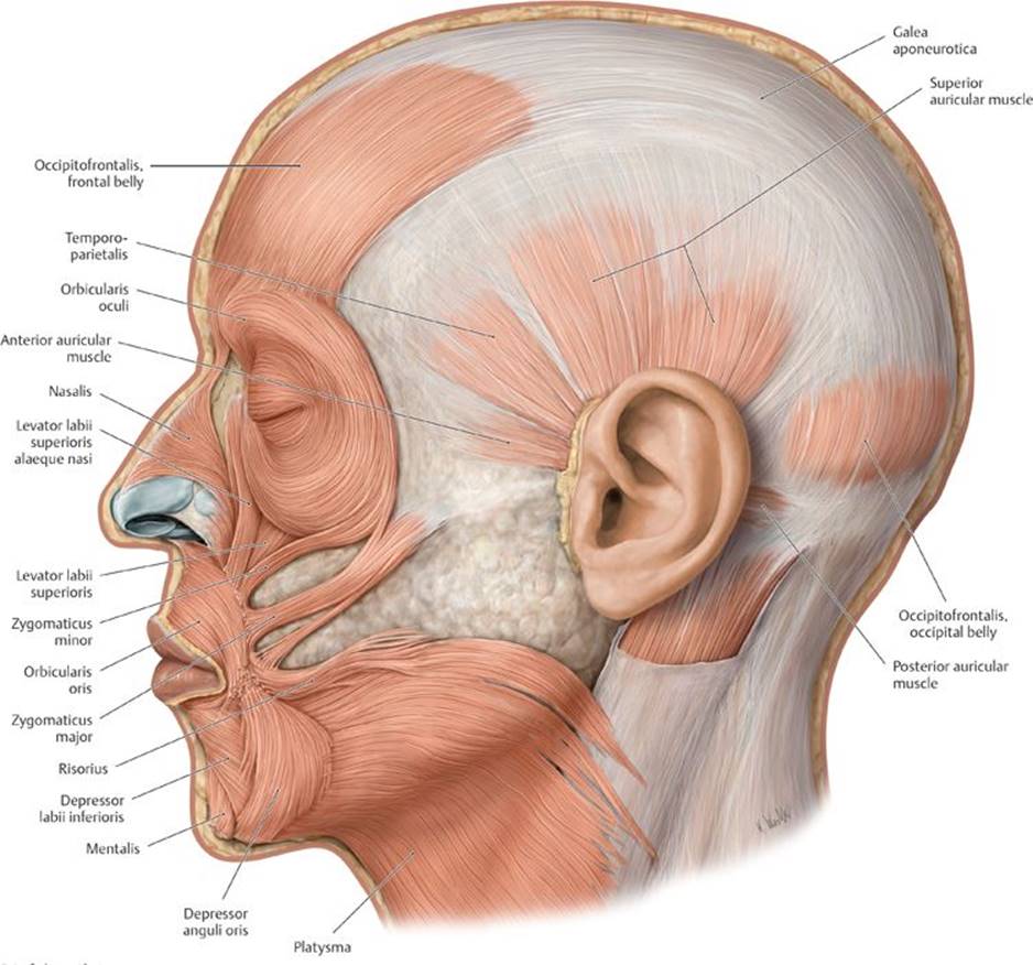
B Left lateral view.


– Primary sensory nerves of the face and scalp (Fig. 19.2A and B) include
• the supraorbital and supratrochlear nerves (CN V1);
• the infraorbital nerve and zygomaticotemporal and zygomaticofacial nerves (V2);
• the auriculotemporal, buccal, and mental (a branch of the inferior alveolar nerve) nerves (V3);
• the great auricular and lesser occipital nerve, which are anterior rami of C2 and C3 via the cervical plexus; and
• the greater occipital and third occipital nerves, which are posterior rami of C2 and C3, respectively.
– Motor nerves of the face and scalp (see Figs. 19.2B and 18.18C) include
• the temporal, zygomatic, buccal, and marginal mandibular branches of the facial nerve (CN VII), which innervate muscles of the face;
• the temporal and posterior auricular branches of the facial nerve, which innervate muscles of the scalp; and
• the muscular branches of the mandibular division of the trigeminal nerve (CN V3), which innervate the muscles of mastication.

Fig. 19.2 Cutaneous innervation of the face
A Cutaneous innervation of the head and neck, left lateral view. The occiput and nuchal regions are supplied by the dorsal rami (blue) of the spinal nerves (the greater occipital nerve is the dorsal ramus of C2).

B Divisions of the trigeminal nerve, left lateral view.
19.2 The Temporomandibular Joint and Muscles of Mastication
– The temporomandibular joint (TMJ), which joins the mandible to the skull, is a synovial articulation between the head of the mandible and the mandibular fossa and articular tubercle of the temporal bone (Figs. 19.3 and 19.4Aand B).
• An articular disk divides the joint cavity into a separate upper part for gliding movement and a lower part for hinge movement.
• The lateral ligament strengthens the fibrous capsule that surrounds the joint.
• Sphenomandibular and stylomandibular ligaments support the joint during chewing.

Fig. 19.3 Temporomandibular joint
The head of the mandible articulates with the mandibular fossa in the temporomandibular joint. Temporomandibular joint, sagittal section, left lateral view.

Fig. 19.4 Ligaments of the temporomandibular joint
A Lateral view of the left temporomandibular joint.

B Medial view of the right temporomandibular joint.
– Muscles of mastication, which include the masseter, temporalis, lateral pterygoid, and medial pterygoid muscles, act at the temporomandibular joint to move the mandible during chewing (Figs. 19.5, 19.6, and 19.7; Table 19.3). They reside in the parotid region and in the temporal and infratemporal fossae.
Dislocation of the temporomandibular joint
In some individuals yawning (or other activities in which the mouth opens very widely) may cause the condyle of the mandible to slide in front of the anterior tubercle, where it locks the mandible in the protruded position. The ligaments supporting the joint become stretched, which causes severe spasm (trismus) of the masseter, medial pterygoid, and temporalis muscles.
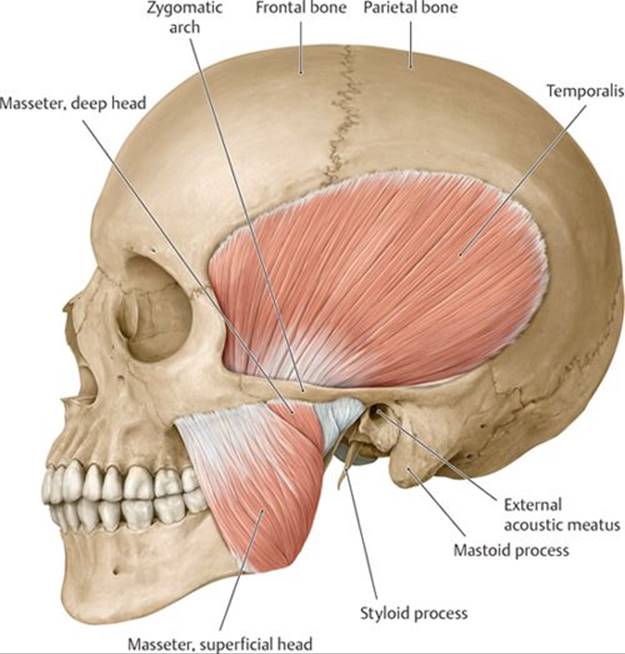
Fig. 19.5 Masseter and temporalis muscles
Left lateral view.

Fig. 19.6 Lateral pterygoid muscle
Left lateral view. Removed: Coronoid process of mandible.

Fig. 19.7 Medial pterygoid muscles
Oblique posterior view. Revealed: Muscular sling formed by the masseter and medial pterygoid muscles that embed the mandible.

19.3 Parotid Region
The parotid region lies superficial to the ramus of the mandible and includes the parotid gland and its surrounding structures (Figs. 19.8 and 19.9).
– The parotid gland, the largest of the three salivary glands, lies anterior to the ear on the lateral side of the face.
• A superficial part of the gland lies on the masseter muscle.
• A deep part of the gland curves around the posterior edge of the ramus of the mandible.
• A tough parotid fascia derived from the deep cervical fascia encloses the gland.
• Secretomotor (parasympathetic) fibers to the parotid gland arise with the glossopharyngeal nerve (CN IX) and synapse in the otic ganglion. Postganglionic fibers hitchhike on the auriculotemporal nerve of CN V3.
– The parotid duct crosses the masseter muscle superficially to pierce the buccinator muscle and enter the mouth, where it opens opposite the second upper molar tooth.

Fig. 19.8 Parotid gland
Left lateral view. Note: The parotid duct penetrates the buccinator muscle to open opposite the second upper molar.
– The parotid plexus, formed by the facial nerve (CN VII), is embedded within the parotid gland and gives rise to five branches that supply muscles of the face: the temporal, zygomatic, buccal, marginal mandibular, and cervical branches (see Fig. 18.20B).
– Structures traversing, or embedded within, the parotid gland include
• the parotid plexus of the facial nerve (CN VII);
• the retromandibular vein, formed by the superficial temporal and maxillary veins;
• the external carotid artery and the origin of its terminal branches, the superficial temporal and maxillary arteries; and
• the parotid lymph nodes, which drain the parotid gland, the external ear, the forehead, and the temporal region.
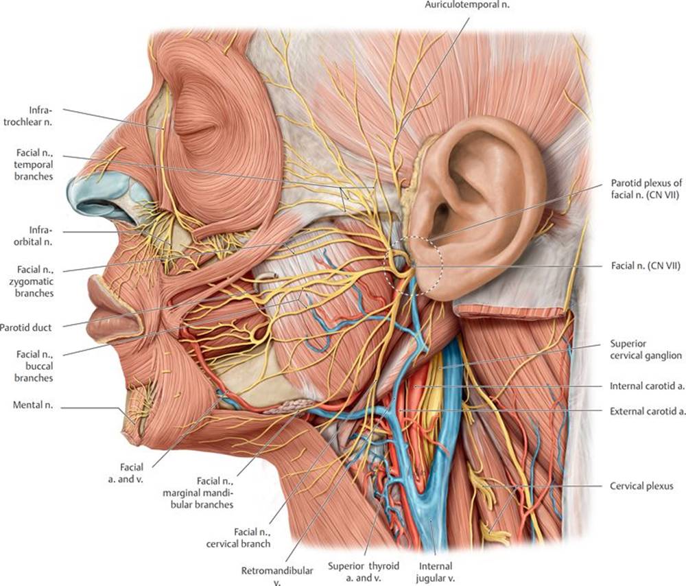
Fig. 19.9 Parotid region
Left lateral view. Removed: Parotid gland, sternocleidomastoid, and veins of the head. Revealed: Parotid bed and carotid triangle.
19.4 Temporal Fossa
The temporal fossa lies superior and medial to the parotid region and covers the lateral aspect of the head (Fig. 19.10, see also Fig. 17.3).
– The boundaries of the temporal fossa are
• anteriorly, the frontal process of the zygomatic bone and zygomatic process of the frontal bone;
• laterally, the zygomatic arch;
• medially, the frontal bone, parietal bone, greater wing of the sphenoid bone, and squamous part of the temporal bone; and
• inferiorly, the infratemporal fossa.
– The temporal fossa contains
• the temporalis muscle and temporal fascia,
• the superficial temporal artery and vein,
• the deep temporal branches of the maxillary artery, and
• the deep temporal nerves and auriculotemporal nerve of the mandibular division of the trigeminal nerve (CN V3).

Fig. 19.10 Temporal and infratemporal fossae
Left lateral view. Removed: Ramus of mandible. Note: The mylohyoid nerve branches from the inferior alveolar nerve just before the mandibular foramen.
19.5 Infratemporal Fossa
The infratemporal fossa lies deep to the ramus of the mandible and is continuous superiorly with the temporal fossa (Fig. 19.11; see also Figs. 17.3, 17.6, and 19.10).
– The bony boundaries of the infratemporal region are
• anteriorly, the posterior wall of the maxilla;
• posteriorly, the mandibular fossa of the temporal bone;
• medially, the lateral pterygoid plate of the sphenoid bone;
• laterally the ramus of the mandible; and
• superiorly, the temporal bone and greater wing of the sphenoid bone.
– The infratemporal fossa communicates with the orbit anteriorly, the pterygopalatine fossa medially, and the middle cranial fossa superiorly.
– The contents of the infratemporal fossa include
• the temporomandibular joint,
• the medial and lateral pterygoid muscles and the inferior part of the temporalis muscle,
• the maxillary artery and its branches (Table 19.4),
• the pterygoid venous plexus,
• the mandibular division of the trigeminal nerve (CN V3) and its branches,
• the otic ganglion, and
• the chorda tympani of the facial nerve (CN VII).

Fig. 19.11 Infratemporal fossa: Deep dissection
Left lateral view. Removed: Lateral pterygoid muscle (both heads). Revealed: Deep infratemporal fossa and mandibular nerve as it enters the mandibular canal via the foramen ovale in the roof of the fossa.
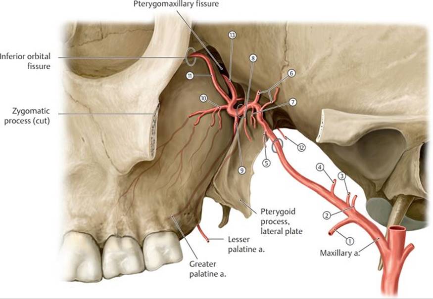
Branches of the maxillary artery, left lateral view.
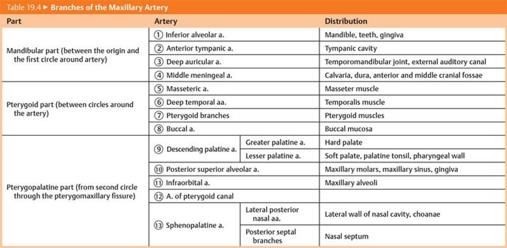
– The mandibular nerve (CN V3), the nerve of the infratemporal fossa and the only division of the trigeminal nerve that carries general sensory and somatic motor fibers, distributes postganglionic parasympathetic (visceral motor) fibers that arise from the otic and submandibular ganglia (Fig. 19.12, Table 19.5; see Fig. 18.18C).
19.6 Pterygopalatine Fossa
The pterygopalatine fossa, a narrow space located medial to the infratemporal fossa, is an important distribution center for branches of the maxillary division of the trigeminal nerve (CN V2) and the accompanying branches of the maxillary artery.
– The bony boundaries of the pterygopalatine fossa are (see Fig. 20.1B)
• superiorly, the apex of the orbit;
• anteriorly, the maxillary sinus;
• posteriorly, the lateral pterygoid plate of the sphenoid bone;
• laterally, the pterygomaxillary fissure; and
• medially, the vertical plate of the palatine bone.
– Contents of the pterygopalatine fossa include
• the pterygopalatine part of the maxillary artery, its branches, and its accompanying veins;
• the nerve of the pterygoid canal;
• the pterygopalatine ganglion; and
• the maxillary division of the trigeminal nerve (CN V2) and its branches.
– The pterygopalatine fossa communicates anteriorly with the orbit, medially with the nasal cavity and palate, and posteriorly with the middle cranial fossa and base of the skull (Table 19.6).
– The maxillary artery passes from the infratemporal fossa to the pterygopalatine fossa through the pterygomaxillary fissure (see Table 19.4). Its branches accompany the branches of the maxillary nerve (CN V2) to supply the nose, palate, and pharynx.

Fig. 19.12 Mandibular nerve (CN V3)) in the infratemporal fossa
Medial view.
– The nerve of the pterygoid canal, which enters the pterygopalatine fossa from the middle cranial fossa, is an autonomic nerve that carries
• preganglionic parasympathetic fibers from the greater petrosal nerve, a branch of the facial nerve (CN VII); and
• postganglionic sympathetic fibers from the deep petrosal nerve, which arises from the internal carotid plexus.
– The pterygopalatine ganglion receives general sensory fibers from the maxillary nerve (CN V2) and parasympathetic and sympathetic fibers from the nerve of the pterygoid canal. Only the parasympathetic fibers synapse in the ganglion; general sensory and sympathetic fibers pass through the ganglion but do not synapse there (Table 19.7).
TABLE 19.5  Nerves of the Infratemporal Fossa
|
Nerve |
Nerve Fibers |
Distribution |
|
Muscular branches (CN V3) |
Branchial motor |
Muscles of mastication; mylohyoid; tensor tympani; tensor veli palatini; anterior belly of digastric |
|
Auriculotemporal (CN V3) |
General sensory |
Auricle, temporal region, and temporomandibular joint |
|
Visceral motor from glossopharyngeal n. (CN IX) |
Parotid gland |
|
|
Inferior alveolar (CN V3) |
General sensory |
Mandibular teeth; mental branch supplies skin of lower lip and chin |
|
Lingual (CN V3) |
General sensory |
Anterior two thirds of tongue, floor of mouth |
|
Buccal (CN V3) |
General sensory |
Skin and mucous membrane of cheek |
|
Meningeal (CN V3) |
General sensory |
Dura of middle cranial fossa |
|
Chorda tympani (CN VII) |
Special sensory taste |
Anterior two thirds of tongue |
|
Visceral motor |
Submandibular and sublingual glands via submandibular ganglion and lingual n. (CN V3). |
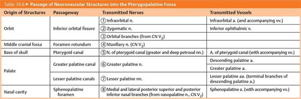

TABLE 19.7 Nerves of the Pterygopalatine Fossa
|
Nerve |
Motor Innervation |
Sensory Distribution |
|
|
Skin of the midface, maxillary sinus, teeth, and gingiva |
|
|
|
Visceral motor (postganglionic) to lacrimal gland |
Skin of lateral cheek, temple |
|
|
Orbit, ethmoid and sphenoid sinuses |
|
|
|
||
|
|
Visceral motor (preganglionic) to glands of the nasal mucosa, and palate; lacrimal gland |
|
|
|
Hard palate |
|
|
|
Soft palate, palatine tonsil |
|
|
|
Nasal septum, upper lateral nasal wall, ethmoid sinuses |
|
|
|
Maxillary sinus, cheeks, gingiva, maxillary molars |
– The maxillary division of the trigeminal nerve (CN V2)
• passes from the middle cranial fossa to the pterygopalatine fossa through the foramen rotundum;
• suspends the pterygopalatine ganglion by two small ganglionic nerves, which transmit general sensory fibers of the maxillary nerve;
• distributes postganglionic parasympathetic (secretomotor) and sympathetic (vasoconstrictive) fibers to the lacrimal, nasal, palatial, and pharyngeal glands; and
• distributes general sensory fibers to the midface, maxillary sinus, maxillary teeth, nasal cavity, palate, and superior pharynx.
19.7 Nasal Cavity
The nasal cavity is located in the middle of the face between the orbits and maxillary sinuses and above the oral cavity.
19.7a Structure of the Nasal Cavity
The nose has an external portion and paired internal nasal cavities that are separated by a nasal septum (Figs. 19.13A and B and 19.14A and B).
– The external nose consists of
• anteriorly, the alar and lateral nasal cartilages, which form the nasal ala and crura surrounding the nostrils and the apex, or tip of the nose; and
• posteriorly, the frontal, maxillary, and nasal bones, which form the root, or bridge, of the nose.
– The nasal cavities are pyramidally shaped spaces that communicate anteriorly with the outside through the nares (nostrils) and posteriorly with the nasopharynx through the choanae.
– The lateral walls of the nasal cavities are formed by the superior and middle nasal conchae of the ethmoid bone; the inferior nasal conchae; and the maxillary, palatine, lacrimal, and nasal bones.
• The superior, middle, and inferior nasal conchae are scroll-like bony processes that project into the nasal cavity.
• The superior, middle, and inferior nasal meatuses are recesses below the respective nasal conchae.
– The nasal septum forms the medial wall of each nasal cavity and consists of the vomer, the perpendicular plate of the ethmoid bone, and the septal cartilage.
– The hard palate, consisting of the maxilla and palatine bones, forms the floor of the nasal cavities and separates them from the oral cavity (see Section 19.8b and Figs. 19.19 and 19.20).

Fig. 19.13 Skeleton of the nose
The skeleton of the nose is composed of an upper bony portion and a lower cartilaginous portion. The proximal portions of the nostrils (alae) are composed of connective tissue with small embedded pieces of cartilage.
A Left lateral view.

B Inferior view.

Fig. 19.14 Bones of the nasal cavity
The left and right nasal cavities are flanked by lateral walls and separated by the nasal septum. Air enters the nasal cavity through the anterior nasal aperture and travels through three passages: the
A Nasal septum in left nasal cavity, para-sagittal section, viewed from left side.

B Lateral wall of the right nasal cavity, sagittal section, medial view. Removed: Nasal septum. Note: The superior and middle conchae are parts of the ethmoid bone, whereas the inferior nasal conchae is a superior, middle, and inferior meatuses (arrows). These passages are separated by the superior, middle, and inferior conchae.

Fig. 19.15 Location of the paranasal sinuses
The paranasal sinuses (frontal, ethmoid, maxillary, and sphenoid) are air-filled cavities that reduce the weight of the skull.
A Anterior view.
B Left lateral view.
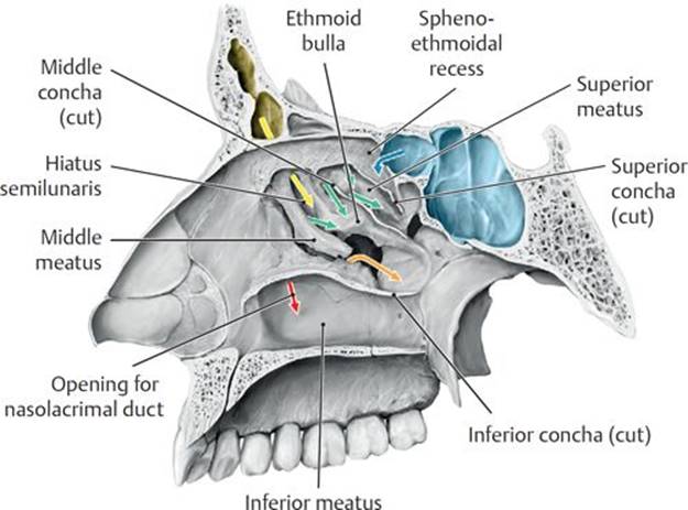
Fig. 19.16 Paranasal sinuses
Openings of the paranasal sinuses and nasolacrimal duct. Right nasal cavity, sagittal section, medial view. Mucosal secretions from the sinuses and nasolacrimal duct drain the nose.
TABLE 19.8 Opening of Nasal Structures into the Nose
|
Nasal Passage |
Sinuses/Duct |
|
Sphenoethmoid recess |
Sphenoid sinus (blue) |
|
Superior meatus |
Posterior ethmoid sinus (green) |
|
Middle meatus |
Anterior and middle ethmoid sinus (green) |
|
Frontal sinus (yellow) |
|
|
Maxillary sinus (orange) |
|
|
Inferior meatus |
Nasolacrimal duct (red) |
– Paranasal sinuses are air-filled cavities within bones of the skull that communicate with the nasal cavities (Figs. 19.15A and B and Fig. 19.16; Table 19.8).
• Paired frontal sinuses, which are usually asymmetrical, lie above the root of the nose and drain into the middle meatus through a frontonasal duct into the hiatus semilunaris.
• Sphenoid sinuses, which form within the body of the sphenoid bone, lie between the right and left cavernous sinuses and drain into the sphenoethmoidal recess in the posterosuperior part of the nasal cavities above the superior concha.
• Ethmoid sinuses comprise the medial wall of the orbit and are formed by numerous thin-walled ethmoid air cells. They lie between the orbits above the nasal cavities and drain into the superior and middle meatuses.
• Paired maxillary sinuses, the largest of the paranasal sinuses, lie on either side of the nasal cavities, inferior to the orbits, and drain into the middle meatuses.
Infection of the maxillary sinuses
Infections arising in the nasal cavity may spread to any of the paranasal sinuses, but the maxillary sinuses are the most commonly affected. Mucus builds up within the maxillary sinuses and is unable to drain because their ostia are located high on the superomedial walls. The ostia are also commonly obstructed by inflammation of the mucous membranes of the sinuses (maxillary sinusitis). Maxillary sinusitis usually follows the common cold or influenza virus but may also arise from the spread of infections from posterior maxillary teeth.
– A nasolacrimal duct drains tears from the medial corner of each eye and empties into the inferior meatus on each side.
19.7b Neurovasculature of the Nasal Cavity
– The external carotid artery via its maxillary and facial branches, and the internal carotid artery via its ophthalmic branch, supply the nasal cavity. The area of overlap between the external and internal circulations is referred to as Kiesselbach’s area (Fig. 19.17A and B).
• Branches of the maxillary artery include
○ the lateral posterior nasal and posterior septal arteries, branches of the sphenopalatine artery; and
○ the greater palatine artery, a branch of the descending palatine artery.
• Branches of the facial artery include the lateral nasal artery and a septal artery, a branch of the superior labial artery.
• Branches of the ophthalmic artery include anterior and posterior ethmoidal arteries.
– Veins that drain the nasal cavity form a submucosal venous plexus that drains into the ophthalmic, facial, and sphenopalatine veins.
Epistaxis
Bleeding, or epistaxis, of the highly vascular nasal mucosa is a common consequence of nasal trauma, and even minor trauma can disrupt the veins in the vestibule. Most bleeding originates from the arteries of Kiesselbach’s area in the anterior third of the nasal cavity. These include the sphenopalatine, greater palatine, anterior ethmoidal, and superior labial arteries.
– The olfactory nerve (CN I) and the ophthalmic (CN V1) and maxillary (CN V2) divisions of the trigeminal nerve innervate the nose (Fig. 19.18A and B).
• Olfactory nerves, which are responsible for smell, arise from the olfactory epithelium in the roof of the nasal cavity. They pass through the cribriform plate and end in the olfactory bulbs.
• Infratrochlear and anterior ethmoid branches of CN V1 and the infraorbital branch of CN V2 innervate the external nose.
• The anterior and posterior ethmoidal branches of CN V1 innervate the external nose and the mucosa of the anterosuperior nasal cavity through internal, external, medial, and lateral nasal branches.
• The posterior nasal branches of the nasopalatine nerve on the septum and nasal branches of the greater palatine nerve on the lateral walls (both branches of CN V2) innervate the mucosa of the posteroinferior nasal cavity.

Fig. 19.17 Arteries of the nasal cavity
A Arteries of the nasal septum, left lateral view.

B Arteries of the right lateral nasal wall, medial view.

Fig. 19.18 Nerves of the nasal cavity
A Nerves of the nasal septum, left lateral view.

B Nerves of the right lateral nasal wall, medial view.
19.8 Oral Region
The oral cavity, located below the nasal cavity and anterior to the pharynx, is bounded superiorly by the palate, inferiorly by the tongue and muscular floor, anteriorly by the lips, posteriorly by the uvula, and laterally by the cheeks (Fig. 19.19).
19.8a Lips, Cheeks, Gingiva, Teeth, and Oral Cavity
– The lips frame the mouth and surround the oral fissure, the opening into the oral cavity.
• The lips contain the sphincter-like obicularis oris muscle and superior and inferior labial muscles. Externally, they are covered by skin; internally, they are covered by the mucous membrane of the oral cavity.
• The philtrum, an external midline depression on the upper lip, extends superiorly to the nasal septum.
• Labial frenula are midline folds of mucous membrane that attach the inner surfaces of the upper and lower lips to the gums.
Cleft lip
Cleft lip is a congenital defect that occurs early in embryonic life when the maxillary prominence and the median nasal prominence fail to fuse. It is seen in ~1:1000 live births and is more common in males than females. The cleft may be unilateral or bilateral and is described as complete when it extends into the nose and incomplete when it appears as a notch in the lip. Corrective surgery is usually performed when the infant is around 10 weeks old.

Fig. 19.19 Oral cavity and pharynx
Midsagittal section, left lateral view.
– The cheeks, continuous with the lips, form the walls of the mouth and the buccal region of the face.
• The buccinator muscle, innervated by the buccal branch of the facial nerve (CN VII), forms the moveable wall of the cheek.
• Buccal fat pads, encapsulated cushions of fat that lie superficial to the buccinator muscles, are proportionately large in infants and reduced in adults.
• The zygomatic bone and zygomatic arch form the “cheek bone.”
– Teeth are anchored in alveoli (sockets) of the maxillary and mandibular dental arches (see Figs. 17.6 and 17.10).
• Children have 20 deciduous teeth, which are replaced at predictable intervals between ages 7 and 25 years.
• Adult have 32 teeth, consisting of incisors, canines, premolars, and molars.
– The gingivae, or gums, made of fibrous tissue covered by the mucous membrane of the oral cavity, are firmly attached to the maxilla and mandible.
– The oral cavity, or mouth, is divided into two regions: the oral vestibule and the oral cavity proper (Fig. 19.20).
• The oral vestibule is the narrow space between the lips and cheeks and the dental arches of the maxilla and mandible.
• The oral cavity proper is the space bounded anteriorly and laterally by the upper and lower dental arches. Superiorly, the palate forms the roof, and inferiorly, the tongue rests on a muscular floor.
– The oral cavity connects posteriorly to the pharynx through a narrow space, the faucial isthmus.
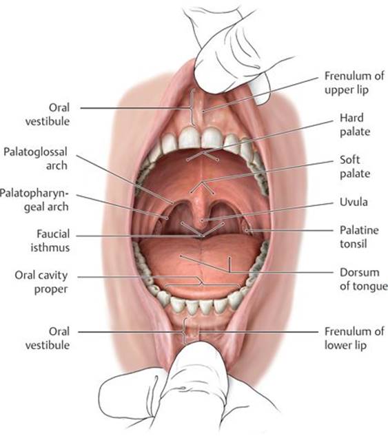
Fig. 19.20 Divisions of the oral cavity
Anterior view.
– Muscles that form the floor of the mouth, the suprahyoid muscles, are attached to the hyoid bone in the neck (Fig. 19.21A and B; Table 19.9). They have a complex innervation with contributions from the trigeminal and facial nerves and the C1 spinal nerve via the hypoglossal nerve.
– The lingual, facial, and maxillary branches of the external carotid artery supply the lips, cheeks, floor of the mouth, and upper and lower teeth.

Fig. 19.21 Muscles of the oral floor: Suprahyoid muscles
A Left lateral view.

B Superior view of the mandible and hyoid bone.

– The trigeminal nerve (CN V) transmits sensation from the mouth.
• The superior alveolar branch of the maxillary division (CN V2) innervates the upper teeth.
• Inferior alveolar, lingual, and buccal branches of the mandibular division (CN V3) innervate the cheek, lower teeth, and floor of the mouth.
– Visceral motor fibers carried by the chorda tympani (CN VII) synapse in the submandibular ganglion in the floor of the mouth. Postganglionic fibers travel via the lingual nerve to innervate the submandibular and sublingual glands.
19.8b The Palate
The palate forms the roof of the oral cavity and the floor of the nasal cavity and separates the oral cavity from the pharynx posteriorly.
– Nasal mucosa covers the superior surface, and oral mucosa, densely packed with mucus-secreting palatine glands, covers the inferior surface.
– The palate has anterior and posterior regions.
• The hard palate, formed by the palatine processes of the maxillary bones and the horizontal processes of the palatine bones, makes up the anterior two thirds of the palate (see Fig. 17.6).
• The soft palate, the posterior third of the palate, has an anterior aponeurotic part that is attached to the hard palate and a posterior muscular part with an unattached posterior margin that ends in a conical projection, the uvula(see Fig. 19.19).
Cleft palate
Cleft palate is a congenital defect that occurs early in embryonic life when the lateral palatine processes fail to fuse with each other, with the nasal septum, and/or with the median palatine processes. It is seen in ~1:2500 live births and is more common in females than males. The cleft (a fissure or opening) is described as complete when it involves the soft and hard palate and incomplete when it appears as a “hole” in the roof of the mouth (usually in the soft palate). In each case the uvula is usually also split. The cleft connects the oral cavity directly to the nasal cavity. Initial treatment involves the use of a prosthetic device called a palatal obturator to seal the cleft until corrective surgery is performed when the infant is between 6 and 12 months old.
– During swallowing, muscles of the soft palate can tense and elevate it against the posterior pharyngeal wall to prevent food from passing into the nasal cavity. They can also draw the palate downward against the tongue to prevent food from entering the pharynx (Fig. 19.22; Table 19.10). Muscles of the soft palate include the tensor veli palatini, levator veli palatini, musculus uvulae, palatoglossus, and palatopharyngeus.
– The paired palatoglossal and palatopharyngeal arches, formed by the palatoglossus and palatopharyngeus muscles, anchor the soft palate to the tongue and pharynx (see Fig. 19.20).
– Greater palatine, lesser palatine, and sphenopalatine branches of the maxillary artery supply the palate (Fig. 19.23, see also Fig. 19.17B).
– The greater palatine, lesser palatine, and nasopalatine branches of the maxillary division (CN V2) carry sensory innervation from the palate.
19.8c The Tongue
The tongue is a muscular organ that is involved in speech, taste, and manipulation of food during the initial phase of swallowing. Two thirds of the tongue lies in the oral cavity; the remainder forms the anterior wall of the oropharynx, the part of the pharynx posterior to the oral cavity.
– The tongue has three parts (Fig. 19.24):
• the root, the attached posterior portion;
• the body, the largest portion, between the root and apex; and
• the apex, the anterior tip.
– The sulcus terminalis, a groove on the dorsum of the tongue, divides the anterior two thirds of the tongue from the posterior third. A small pit at the center of the groove, the foramen cecum, marks the embryonic origin of the thyroid gland.
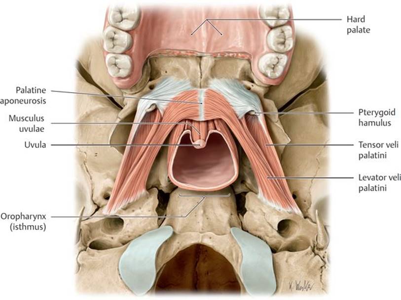
Fig. 19.22 Muscles of the soft palate
Inferior view. The soft palate forms the posterior boundary of the oral cavity, separating it from the oropharynx.


Fig. 19.23 Neurovasculature of the hard palate
Inferior view. The hard palate receives sensory innervation primarily from terminal branches of the maxillary division of the trigeminal nerve (CN V2). The arteries of the hard palate arise from the maxillary artery.

Fig. 19.24 Structure of the tongue
Superior view. The V-shaped sulcus terminalis divides the tongue into an anterior (oral, presulcal) and a posterior (pharyngeal, postsulcal) part.
– Numerous lingual papillae, many containing taste buds, give the mucosa of the anterior two thirds of the tongue its rough texture.
• Vallate papillae, the largest of the lingual papillae, are arranged in a row directly anterior to the sulcus terminalis.
• Foliate papillae are found on the small lateral folds of the lingual mucosa and are not well developed.
• Filiform papillae contain afferent nerve endings that are sensitive to touch but not to taste. These are highly keratinized and facilitate rasplike licking activity.
• Fungiform papillae are most numerous at the apex and margins of the tongue.
– The lingual tonsil is a collection of lymphoid nodules distributed over the mucosa of the posterior part of the tongue. Lingual tonsils have crypts that are flushed by glands below them.
– The frenulum of the tongue, a midline mucosal fold, attaches the inferior surface of the tongue to the floor of the oral cavity and restricts its movement.
– Extrinsic muscles of the tongue, which originate outside the tongue and are responsible primarily for tongue movements, include the genioglossus, hyoglossus, palatoglossus, and styloglossus muscles (Fig. 19.25).
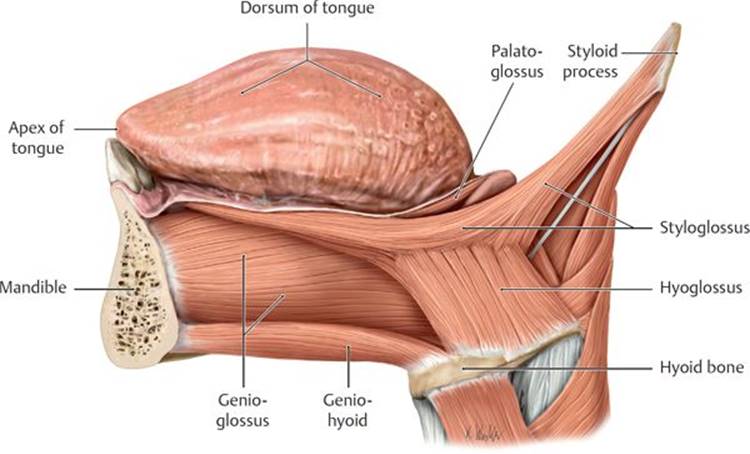
Fig. 19.25 Extrinsic muscles of the tongue
Left lateral view.
– Intrinsic muscles of the tongue have no bony attachments and are responsible for changing the shape of the tongue. They include the superior and inferior longitudinal muscles and the transverse and vertical muscles.
– All extrinsic and intrinsic muscles of the tongue, except the palatoglossus, are innervated by the hypoglossal nerve (CN XII). The palatoglossus is innervated by the vagus nerve (CN X).
– The lingual arteries, branches of the external carotid arteries, supply the tongue. The lingual veins accompany the arteries and drain into the internal jugular vein.
– Lymph from the tongue ultimately drains to deep cervical and jugular nodes in the neck, although the drainage from the various parts of the tongue follows four different pathways (Fig. 19.26A and B; Table 19.11). These drainage routes can be clinically important in the metastasis of lingual tumors.
– The five cranial nerves (trigeminal, facial, glossopharyngeal, vagus, and hypoglossal) that innervate the tongue carry motor, general sensory, and special sensory fibers (Table 19.12; Fig. 19.27).

Fig. 19.26 Lymphatic drainage of the tongue and oral floor
Lymph flows to the submental and submandibular lymph nodes of the tongue and oral floor, which ultimately drain into the jugular lymph nodes along the internal jugular vein. Because the lymph nodes receive drainage from both the ipsilateral and contralateral sides (B), tumor cells may become widely disseminated in this region (e.g., metastatic squamous cell carcinoma, especially on the lateral border of the tongue, frequently metastasizes to the opposite side).
A Left lateral view.

B Anterior view.
19.8d Salivary Glands
Three pairs of salivary glands, the parotid, sublingual, and submandibular, produce and secrete saliva into the mouth (Fig. 19.28; see Fig. 19.8).
– The sublingual gland, the smallest of the three glands, lies deep to the mucosa of the floor of the mouth, forming the sublingual folds, onto which it secretes saliva through numerous small ducts.
– The submandibular gland has a superficial part located in the neck and a deep part located in the floor of the mouth, which connect around the posterior border of the mylohyoid muscle. The submandibular (Wharton’s) ductopens via the sublingual papilla at the base of the lingual frenulum.
– The parotid gland, the largest of the three glands, is located in the parotid region on the lateral side of the head, anterior to the ear. The parotid duct opens into the vestibule of the mouth at the parotid papilla located adjacent to the second maxillary molar (see Section 19.3).
TABLE 19.11  Lymphatic Drainage of the Tongue
|
Area of the Tongue |
Drainage Pattern |
Primary Nodes |
|
Root |
Bilaterally |
Superior deep cervical |
|
Medial part of the body |
Bilaterally |
Inferior deep cervical |
|
Lateral parts of the body |
Ipsilaterally |
Submandibular |
|
Apex and frenulum |
Midline—bilaterally Sides—ipsilaterally |
Submental |
TABLE 19.12 Innervation of the Tongue
|
Nerve |
Nerve Fibers |
Distribution |
|
Lingual nerve (CN V3) |
General sensory |
Anterior two thirds of the tongue |
|
Chorda tympani (CN VII) |
Special sensory |
Anterior two thirds of the tongue |
|
Glossopharyngeal nerve (CN IX) |
General sensory and special sensory |
Posterior third of the tongue |
|
Vagus nerve (CN X) |
General sensory and special sensory |
Root of the tongue |
|
Hypoglossal nerve (CN XII) |
Somatic motor |
Muscles of the tongue, except the palatoglossus, which is innervated by the vagus nerve (CN X) |
– The facial, lingual, maxillary, and superficial temporal arteries supply the salivary glands. Veins of similar names accompany the arteries and drain into the retromandibular vein.
– The sublingual and submandibular glands receive parasympathetic secretomotor fibers via the chorda tympani (a branch of CN VII) and submandibular ganglion.
– The parotid gland receives secretomotor (parasympathetic) fibers of the glossopharyngeal nerve (CN IX) that synapse in the otic ganglion.
– Sympathetic innervation to the glands arises from the superior cervical ganglion and follows the branches of the external carotid artery.

Fig. 19.27 Somatosensory and taste innervation of the tongue
Anterior view.

Fig. 19.28 Salivary glands
Submandibular and sublingual glands, superior view with tongue removed.
 Infraorbital n. (CN V2)
 Zygomatic n. (CN V2)–communicating branch from zygomaticotemporal n.
 Orbital branches (CN V2)
 Maxillary (CN V2)
 N. of the pterygoid canal (CN VII)
 Greater palatine n. (CN V2)
 Lesser palatine n. (CN V2)
 Medial and lateral posterior superior and inferior nasal nn. (CN V2)
 Superior alveolar n. (CN V2)