
The neck extends from the base of the skull to the clavicles and manubrium. It contains vital neurovascular structures that supply the head, the thorax, and the upper limb. The neck also contains viscera of the gastrointestinal, respiratory, and endocrine systems.
21.1 Deep Fascia of the Neck
Layers of deep cervical fascia surround and compartmentalize the structures of the neck (Table 21.1).
– The investing (superficial) layer of the deep fascia of the neck, a thin layer that lies deep to the skin, surrounds the entire neck but splits to enclose the sternocleidomastoid and trapezius muscles and contains cutaneous nerves, superficial vessels, and superficial lymphatics of the neck.
– The pretracheal fascia in the anterior neck has a muscular lamina (layer) that encloses the infrahyoid muscles and a visceral lamina that encloses the viscera of the anterior neck.
– The prevertebral fascia surrounds the vertebral column and the deep neck muscles and is continuous with the nuchal fascia posteriorly.
– The carotid sheath, a condensation of the pretracheal, prevertebral, and investing fasciae, forms a narrow cylindrical tube that encloses the neurovascular bundle of the neck: the internal jugular vein, common carotid artery, and vagus nerve.
– The retropharyngeal space, a potential space between the visceral layer of the pretracheal fascia and the prevertebral fascia, extends from the base of the skull superiorly to the superior mediastinum inferiorly.
TABLE 21.1  Deep Cervical Fascia
|
The deep cervical fascia is divided into four layers that enclose the structures of the neck. |
||
|
Layer |
Type of Fascia |
Description |
|
|
Muscular |
Envelopes entire neck; splits to enclose sternocleidomastoid and trapezius muscles |
|
Pretracheal layer |
|
Encloses infrahyoid muscles |
|
|
Surrounds thyroid gland, larynx, trachea, pharynx, and esophagus |
|
|
|
Muscular |
Surrounds cervical vertebral column and associated muscles |
|
|
Neurovascular |
Encloses common carotid artery, internal jugular vein, and vagus nerve |
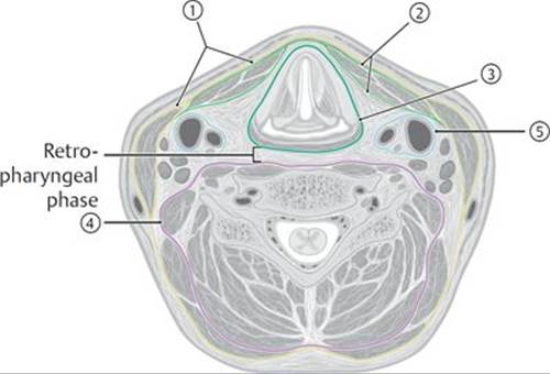
A Transverse section at level of C5 vertebra.

B Midsagittal section, left lateral view.
21.2 Muscles of the Neck
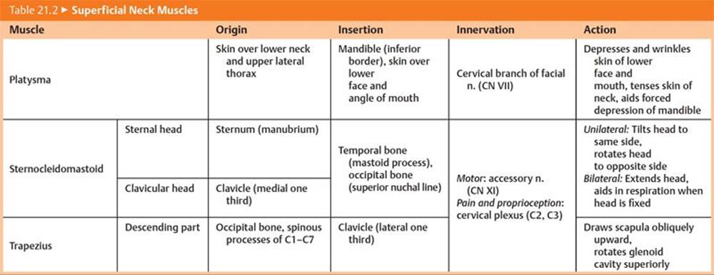
– Three muscles form the most superficial muscular layer of the neck (Fig. 21.1; Table 21.2):
• The platysma, a muscle of facial expression, and therefore a subcutaneous muscle, that extends onto the anterolateral surfaces of the neck
• The sternocleidomastoid muscle, a visible landmark that divides the neck into anterior and lateral cervical regions
• The trapezius, a muscle of the shoulder girdle that extends onto the neck and forms the posterior border of the lateral cervical region
Congenital torticollis
Congenital torticollis (wryneck) is a condition in which one of the sternocleidomastoid muscles (SCM) is abnormally short, causing the head to tilt to one side with the chin pointing upward to the opposite side. This shortening is thought to be the result of trauma at birth (tears or stretching of the SCM) causing bleeding and swelling within the muscle and subsequent scar tissue formation.

Fig. 21.1  Superficial musculature of the neck
Left lateral view.
– Muscles attached to the hyoid bone lie between the superficial and deep neck muscles.
• Suprahyoid muscles, the digastric, stylohyoid, mylohyoid, geniohyoid, and hyoglossus, form the floor of the mouth and elevate the hyoid and larynx during swallowing and phonation (see Table 19.9).
• Infrahyoid muscles in the neck, omohyoid, sternohyoid, sternothyroid, and thyrohyoid, depress the hyoid and larynx during swallowing and phonation (Fig. 21.2; Table 21.3).
– The deep muscles of the neck lie deep to the prevertebral fascia and include the prevertebral and scalene muscles (Fig. 21.3; Table 21.4).

Fig. 21.2 Suprahyoid and infrahyoid muscles
Anterior view. The sternohyoid has been cut on the right side. See Table 19.9 for the suprahyoid muscles.
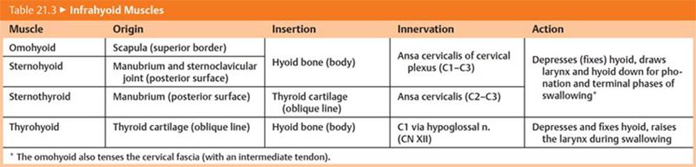
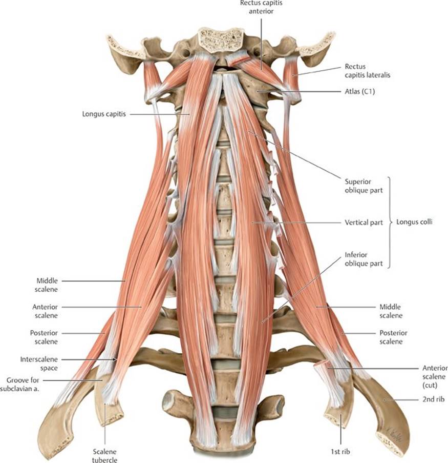
Fig. 21.3 Deep muscles of the neck
Prevertebral and scalene muscles, anterior view. Removed: Longus capitis and anterior scalene.

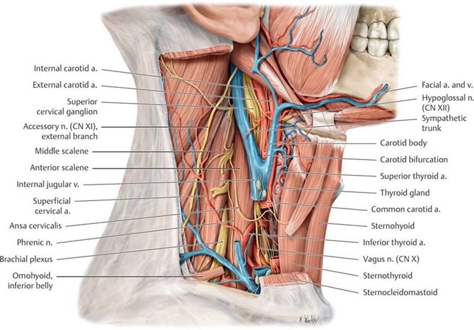
Fig. 21.4 Deep lateral cervical region
Right lateral view with sternocleidomastoid windowed.


21.3 Nerves of the Neck
Nerves of the neck include cervical and thoracic spinal nerves, nerves from the cervical sympathetic trunk, and cranial nerves (Fig. 21.4).
21.3a Cervical Nerves
The C1–C4 spinal nerves supply the regions of the neck (Table 21.5).
– Posterior rami of cervical spinal nerves C1 through C3 form three nerves that provide motor and cutaneous innervation to the posterior neck and scalp: the suboccipital (C1), greater occipital (C2), and 3rd occipital nerves (C3) (Fig. 21.5).
– Anterior rami of C1–C4 cervical spinal nerves form the cervical plexus, which has sensory and motor components.
• The sensory nerves of the plexus, the lesser occipital (C1), great auricular (C2–C3), transverse cervical (C2–C3), and supraclavicular nerves (C3–C4), innervate the skin of the anterior and lateral neck and lateral scalp. They emerge from behind the midpoint of the posterior border of the sternocleidomastoid muscle, a location known as the nerve point of the neck (or Erb’s point) (Fig. 21.6).
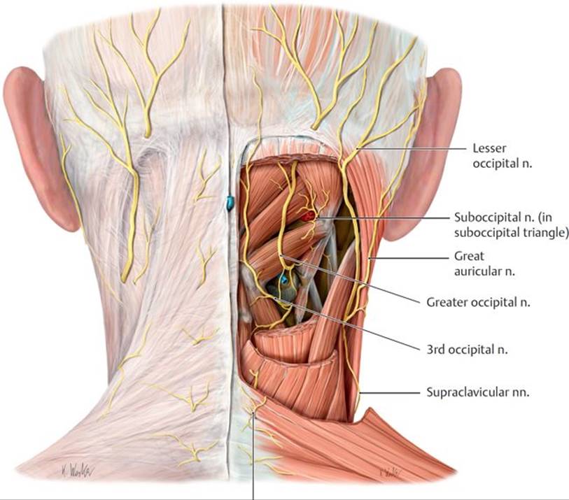
Fig. 21.5 Innervation of the nuchal region
Posterior view.
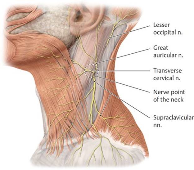
Fig. 21.6 Sensory innervation of the anterolateral neck
Sensory branches of the cervical plexus, left lateral view.
• The ansa cervicalis (C1–C3), the motor part of the plexus, which has a superior and inferior root, innervates all of the infrahyoid muscles except the thyrohyoid and usually lies anterior to the internal jugular vein (Fig. 21.7).
– The phrenic nerve, which arises from anterior rami of C3–C5 spinal nerves, descends on the surface of the anterior scalene muscle and enters the thorax, where it supplies the diaphragm with sensory and motor innervation.
21.3b Brachial Plexus
The brachial plexus, which innervates the upper limb, forms from the anterior rami of C5–T1. It emerges through the interscalene groove (the space between the anterior and middle scalene muscles) in the lateral region of the neck before continuing into the axilla (see Section 13.4d).
21.3c The Cervical Sympathetic Trunk
The cervical sympathetic trunk, a continuation of the thoracic sympathetic trunk, extends into the neck to the level of the C1 vertebra, where it lies anterolateral to the vertebral column and posterior to the carotid sheath.
– The cervical sympathetic trunk receives no white rami communicantes from cervical spinal nerves. The preganglionic fibers that synapse in the cervical ganglia originate in thoracic spinal nerves and ascend in the sympathetic trunk to the cervical region.
– Postganglionic fibers from the cervical sympathetic ganglia are distributed along three routes:
• Via gray rami communicantes to join cervical spinal nerves
• Via cervical cardiac (cardiopulmonary) nerves to the cardiac plexus in the thorax
• Via sympathetic nerve plexuses surrounding vessels (periarterial plexuses), especially along the external carotid, internal carotid, and vertebral arteries, to supply structures of the head and neck
– The cervical sympathetic trunk has superior, middle, and inferior ganglia.
• The superior cervical ganglion lies anterior to C1 vertebra and posterior to the internal carotid artery. Its branches include
○ the superior cervical sympathetic cardiac nerve,
○ branches to the pharyngeal nerve plexus,
○ an internal carotid nerve that forms the internal carotid plexus, and
○ an external carotid nerve that forms the external carotid plexus.
○ gray rami communicantes to anterior rami of C1–C4 spinal nerves.

Fig. 21.7 Motor innervation of the anterolateral neck
Left lateral view.
• The middle cervical ganglion lies at the level of the C6 vertebra and gives off the middle cervical sympathetic cardiac nerve, which joins the cardiac plexus in the thorax, and gray rami communicantes to anterior rami of C5 and C6 spinal nerves.
• The inferior cervical ganglion usually combines with the most superior thoracic ganglion (T1) to form the stellate ganglion, which lies anterior to the transverse process of the C7 vertebra. It gives off the inferior cervical sympathetic cardiac nerve, which descends into the thorax, and gray rami communicantes to anterior rami of C7 and C8 spinal nerves.
21.3d Cranial Nerves in the Neck
Four of the cranial nerves are found in the neck.
– The glossopharyngeal nerve (CN IX) sends branches to the tongue and pharynx in the head and descends into the neck to innervate the carotid body and carotid sinus (see Fig. 18.22). It innervates one muscle, the stylopharyngeus.
– The vagus nerve (CN X) descends within the carotid sheath in the neck before entering the thorax. Its branches to cervical structures arise from both thoracic and cervical parts of the nerve (see Fig. 18.24).
• A superior laryngeal nerve arises from the cervical part of each vagus nerve and innervates the upper larynx through its internal and external branches.
• The right recurrent laryngeal nerve arises from the inferior cervical part of the right vagus nerve and recurs around the subclavian artery in the neck.
• The left recurrent laryngeal nerve arises from the thoracic part of the left vagus nerve. It recurs around the arch of the aorta and ascends in the neck between the trachea and esophagus.
• Cervical cardiac nerves carry visceral motor (presynaptic parasympathetic) fibers and visceral sensory fibers to the cardiac plexus.
– The spinal accessory nerve (CN XI), which is derived from roots of the upper cervical spinal cord segments, enters the skull through the foramen magnum. After exiting the skull through the jugular foramen, it innervates the sternocleidomastoid muscle, then crosses the lateral region of the neck to innervate the trapezius muscle (see Fig. 18.25).
• The cranial root joins the vagus nerve.
• The spinal root innervates the sternocleidomastoid muscle, then crosses the lateral region of the neck to innervate the trapezius muscle.
– The hypoglossal nerve (CN XII), which exits the skull through the hypoglossal canal and courses anteriorly to the submandibular region, enters the oral cavity to innervate muscles of the tongue.
• Along its course, fibers from C1 join the hypoglossal nerve briefly, eventually diverging from it to innervate the geniohyoid and thyrohyoid muscles and to form the superior root of the ansa cervicalis (see Fig. 21.7).
21.4 The Pharynx
The pharynx, a fibromuscular tube that forms part of the upper airway and the upper digestive tract, transmits air from the nasal cavity to the trachea and food from the oral cavity to the esophagus.
21.4a Regions of the Pharynx
The pharynx, which extends from the base of the skull to the inferior aspect of the larynx (cricoid cartilage), is divided into three regions (Figs. 21.8 and 21.9; see also Fig. 19.19): the nasopharynx, the oropharynx, and the laryngopharynx.
– The nasopharynx, the uppermost part of the pharynx, lies posterior to the nasal cavity and above the soft palate. Anteriorly, the nasopharynx communicates with the nasal cavity through the paired choanae.
• The orifices (openings) of the pharyngotympanic tubes are located on the lateral pharyngeal wall. Above the orifice, the cartilaginous part of the tube protrudes into the pharynx to form a bulge, the torus tubarius.
• A salpingopharyngeal fold, formed by the salpingopharyngeus muscle deep to the mucosa, extends inferiorly from the torus tubarius.
– The oropharynx, which lies posterior to the oral cavity, extends superiorly to the soft palate and inferiorly to the top of the larynx. The posterior tongue forms the anterior boundary.
• Two arches, the anterior palatoglossal arch (fold) and the posterior palatopharyngeal arch (fold), formed by the palatoglossus and palatopharyngeal muscles, respectively, separate the oropharynx from the oral cavity (see Fig. 19.20). Both muscles are innervated by the vagus nerve (CN X).
• The palatine tonsil lies between these arches in the tonsillar fossa.
• A median fold of mucosa divides the space between the base of the tongue and the epiglottis. The spaces on either side of the fold are called valleculae.

Fig. 21.8 Regions of the pharynx
Midsagittal section, left lateral view. The blue arrow indicates flow of air and the red arrow, passage of food.
– The laryngopharynx, which lies posterior to the larynx, extends from the epiglottis to the inferior border of the cricoid cartilage, where it narrows and is continuous with the esophagus.
• The laryngopharynx communicates with the larynx through the laryngeal inlet.
• Aryepiglottic folds separate the laryngeal inlet from mucus-lined fossae on the lateral walls of the pharynx called piriform recesses.
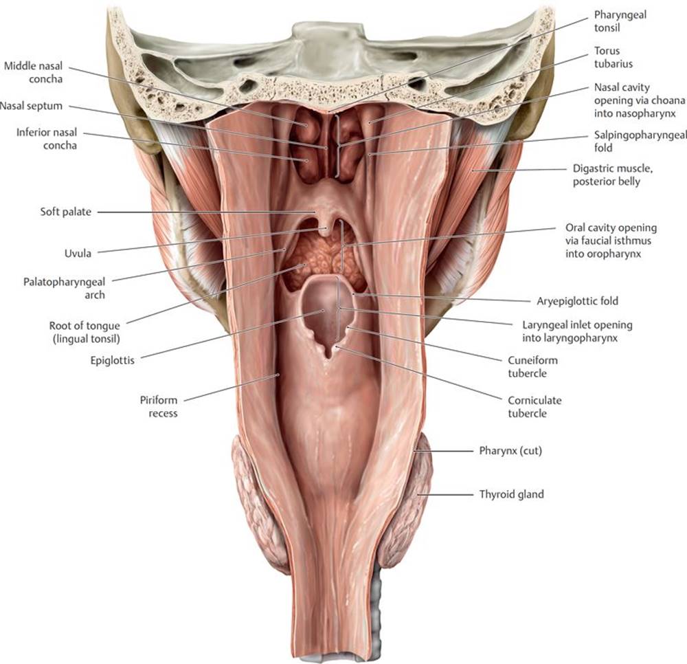
Fig. 21.9 Pharynx
Posterior view of opened pharynx.
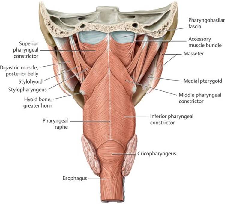
Fig. 21.10 Pharyngeal muscles
Posterior view.
A Muscles in the posterior pharynx.

B Muscles in the opened pharynx.
21.4b Muscles of the Pharynx
An outer circular layer and inner longitudinal layer of skeletal muscles form the walls of the pharynx (Fig. 21.10A and B; Table 21.6). Pharyngeal muscles function during swallowing.
– Circular superior, middle, and inferior pharyngeal constrictor muscles propel a bolus of food downward through the oropharynx and laryngopharynx.
– Longitudinal muscles, the salpingopharyngeus and palatopharyngeus innervated by the vagus nerve (CN X) and pharyngeal plexus, and the stylopharyngeus innervated by the glossopharyngeal nerve (CN IX), elevate the soft palate to prevent food from entering the nasopharynx.
21.4c Tonsils
– Tonsils, masses of lymphoid tissue found in the mucosal lining of the pharynx, form an incomplete circular ring called the pharyngeal lymphatic ring (Waldeyer’s ring) around the superior part of the pharynx (Fig. 21.11). They include
• pharyngeal tonsils (also known as adenoids) in the mucous membrane of the roof and posterior wall of the pharynx,
• tubal tonsils near the orifices of the pharyngotympanic tube,
• palatine tonsils in the fossae between the palatoglossal and palatopharyngeal arches, and
• lingual tonsils on the posterior one third of the dorsum of the tongue.
– Tonsillar lymphatic vessels drain to the jugulodigastric node near the angle of the mandible before draining to the deep cervical lymph nodes.
Tonsillectomy
During a tonsillectomy, the palatine tonsils are removed from the tonsillar bed along with their accompanying fascia. The glossopharyngeal nerve, which lies on the lateral wall of the pharynx, is vulnerable to injury during the procedure and may result in loss of sensation and taste from the posterior one third of the tongue. Bleeding may occur from the large external palatine vein or from the tonsillar branches of the facial, ascending pharyngeal, maxillary, and lingual arteries. The internal carotid artery may also be vulnerable because it runs just lateral to the tonsil.
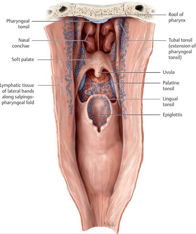
Fig. 21.11 Tonsils: Waldeyer’s ring
Posterior view of the opened pharynx.
TABLE 21.6 Muscles of the Pharynx
|
Muscles |
Innervation |
Action |
|
Longitudinal muscles |
||
|
Salpingopharyngeus |
Pharyngeal plexus–CN X |
Elevates pharynx |
|
Palatopharyngeus |
Elevates pharynx and larynx |
|
|
Stylopharyngeus |
CN IX |
Elevates pharynx |
|
Circular muscles |
||
|
Superior constrictor |
Pharyngeal plexus–CN X |
Constricts pharynx |
|
Middle constrictor |
||
|
Inferior constrictor |
Pharyngeal plexus, superior laryngeal (external br.), recurrent laryngeal nn.–CN X |
|
21.4d Neurovasculature of the Pharynx
– Direct and indirect branches of the external carotid artery supply the pharynx. These include the facial, lingual, ascending palatine, descending palatine, and ascending pharyngeal arteries (see Fig. 17.14).
– Venous drainage of the pharynx passes through the pharyngeal venous plexus to the internal jugular vein (see Fig. 17.20).
– Sensory innervation of the pharynx varies by region.
• The maxillary nerve (CN V2) innervates the nasopharynx.
• The glossopharyngeal nerve (CN IX) innervates the oropharynx.
• The vagus nerve (CN X) via its internal laryngeal branch innervates the laryngopharynx.
Gag reflex
The gag reflex is a reflexive contraction of the pharyngeal muscles that protects the airway by preventing accidental ingestion and aspiration. It is elicited by touching the soft palate or back of the tongue. The glossopharyngeal nerve (CN IX) is the afferent limb, and the vagus nerve (CN X) serves as the efferent limb of the reflex.
21.5 Esophagus
The esophagus is a muscular tube that connects the pharynx in the neck to the stomach in the abdomen (see Section 5.6 and Fig. 21.10A and B).
– The cervical esophagus begins at the C6 vertebral level, which corresponds to the inferior border of the cricoid cartilage. The cervical esophagus is posterior to the trachea, anterior to the cervical vertebrae, and continuous with the laryngopharynx superiorly.
– At the pharyngoesophageal junction, the cricopharyngeus, the lowest part of the inferior pharyngeal constrictor, forms the superior esophageal sphincter.
– The inferior thyroid arteries, branches of the subclavian arteries via the thyrocervical trunks, supply the cervical part of the esophagus. Veins of similar name accompany the arteries.
– Lymphatic vessels from the cervical esophagus drain to paratracheal and deep cervical nodes.
– The recurrent laryngeal nerves, branches of the vagus nerve (CN X), and vasomotor fibers from the cervical sympathetic trunk innervate the esophagus in the neck.
21.6 Larynx and Trachea
The larynx, part of the upper airway, is responsible for sound production. It communicates superiorly with the pharynx and inferiorly with the trachea and lies anterior to the C3–C6 vertebrae. The trachea, the upper part of the tracheobronchial tree, descends into the thorax, where it is continuous with the bronchi of the lungs.
21.6a Laryngeal Skeleton
Three single cartilages and two sets of paired cartilages form the laryngeal skeleton (Figs. 21.12, 21.13, and 21.15B and D). All except the epiglottis (elastic cartilage) are formed of hyaline cartilage.
– The thyroid cartilage, the largest of the nine cartilages, has two laminae that join at the midline to form a laryngeal prominence (Adam’s apple). The superior horn of the thyroid cartilage attaches to the hyoid bone, and the inferior horn articulates with the cricoid cartilage at the cricothyroid joint.
– The cricoid cartilage, the only part of the laryngeal skeleton to form a complete ring around the airway, articulates superiorly with the thyroid cartilage and is attached inferiorly to the first tracheal ring. The anterior part of the cricoid cartilage, the arch, is short, and the lamina, its posterior part, is tall.
– The epiglottis, a leaf-shaped cartilage that forms the anterior wall of the laryngeal inlet at the root of the tongue, is attached inferiorly to the thyroid cartilage and anteriorly to the hyoid bone.
– The paired pyramidal arytenoid cartilages, which articulate with the superior border of the cricoid lamina, have an apex that articulates with the tiny corniculate cartilages and a vocal process that attaches to the thyroid cartilage through the vocal ligaments.
– The small paired corniculate and cuneiform cartilages appear as tubercles within the aryepiglottic fold. Although the corniculate cartilages articulate with the arytenoids, the cuneiform cartilages do not articulate with the other cartilages.
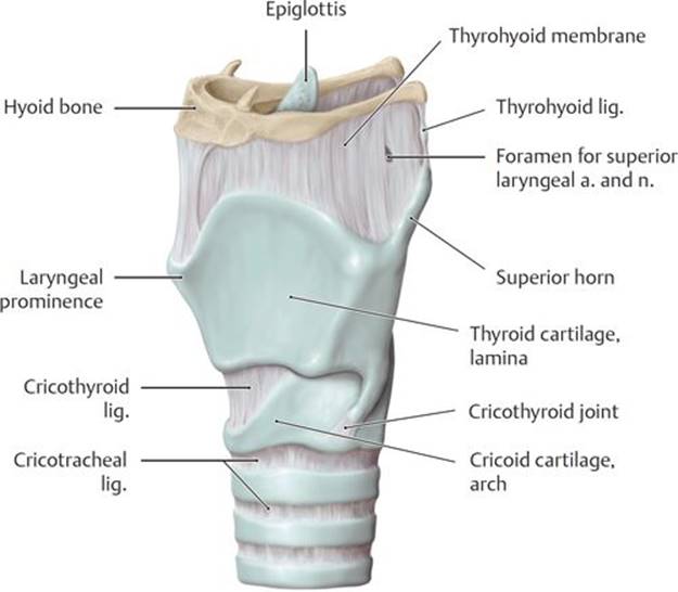
Fig. 21.12 Structure of the larynx
A Left anterior oblique view.
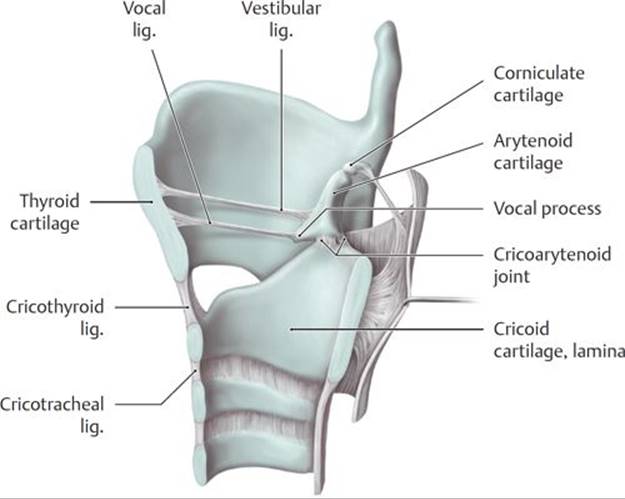
B Sagittal section viewed from the left medial aspect. Removed: Hyoid bone and thyrohyoid ligament. The arytenoid cartilage alters the position of the vocal folds during phonation.

C Posterior view. Arrows indicate the direction of movement in the various joints.

D Superior view of thyrohyoid, cricoid, and corniculate cartilages.
21.6b Membranes and Ligaments of the Larynx
Membranes of the larynx connect the laryngeal cartilages to each other and to the hyoid bone and trachea (Figs. 21.13 and 21.14; see also Fig. 21.12A and B).
– The thyrohyoid membrane attaches the thyroid cartilage to the hyoid bone superiorly.
– The cricotracheal ligament attaches the cricoid cartilage to the first tracheal ring inferiorly.
– The quadrangular membrane extends posteriorly from the lateral border of the epiglottis to the arytenoid cartilage on each side.
• The superior free margin of this membrane forms the aryepiglottic ligament, which, when covered by mucosa, is known as the aryepiglottic fold.
• The inferior free margin is the vestibular ligament, which, when covered by mucosa, is known as the vestibular fold or false vocal cord.
– The cricothyroid membrane connects the cricoid and thyroid cartilages and extends superiorly deep to the thyroid cartilage as the conus elasticus (see Figs. 21.12D and 21.15C).
– The free superior border of the conus elasticus forms the vocal ligament, which extends from the midpoint of the thyroid cartilage to the vocal processes of the arytenoid cartilage. The vocal ligament and the vocalis muscle form the vocal fold or vocal cord.
Tracheostomy and cricothyroidotomy
When the upper airway is obstructed, airway access can be reestablished using two different approaches. Tracheostomy is a surgical procedure in which a tracheostomy tube is inserted through an incision in the proximal trachea. This procedure is generally used for long-term management of the airway. Cricothyroidotomy is a related procedure in which an incision is made in the cricothyroid membrane. This procedure, usually performed in emergency situations, is less technically difficult than tracheostomy and has fewer complications.
21.6c Laryngeal Cavity
The laryngeal cavity begins at the laryngeal inlet and extends to the inferior border of the cricoid cartilage (see Figs. 21.13 and 21.14).
– The vestibular and vocal folds define spaces within the laryngeal cavity.
• The supraglottic space (laryngeal vestibule) or supraglottic space, lies above the vestibular folds.
• The rima vestibuli is the aperture between the two vestibular folds.
• The laryngeal ventricles are recesses of the laryngeal cavity between the vestibular and vocal folds.
• Laryngeal saccules are the blind ends of the laryngeal ventricles.
• The rima glottidis is the aperture between the two vocal folds.
• The subglottic space (infraglottic cavity) is the inferior part of the laryngeal cavity, which lies below the vocal folds and extends to the inferior border of the cricoid cartilage.
– Sound is produced as air passes through the laryngeal cavity between the vocal folds. Variations in sound arise from changes in the position, tension, and length of these folds.
– The vestibular folds protect the airway but have no role in sound production.
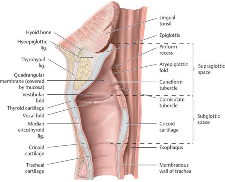
Fig. 21.13 Cavity of the larynx
Midsagittal section viewed from the left side.

Fig. 21.14 Vestibular and vocal folds
Coronal section, superior view.

Fig. 21.15 Laryngeal muscles
The laryngeal muscles move the laryngeal cartilages relative to one another, affecting the tension and/or position of the vocal folds.
A Extrinsic laryngeal muscles, left lateral oblique view. Removed: Epiglottis.

B Intrinsic laryngeal muscles, left lateral view. Removed: Thyroid cartilage (left lamina). Revealed: Epiglottis and external thyroarytenoid muscle.
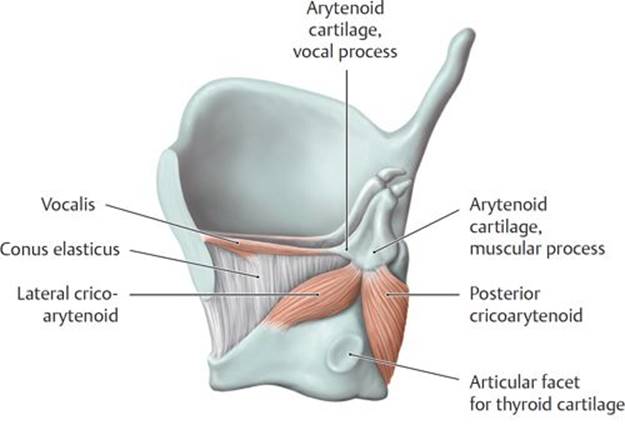
C Left lateral view. Removed: Thyroid cartilage (left lamina) and epiglottis.

D Posterior view.

A Laryngeal muscles, superior view.
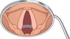
B Open rima glottidis.

C Closed rima glottidis.
TABLE 21.7 Actions of the Laryngeal Muscles
|
Muscle |
Action |
Effect on Rima Glottidis |
|
|
Tightens the vocal folds |
None |
|
|
||
|
|
Adducts the vocal folds |
Closes |
|
|
||
|
|
Abducts the vocal folds |
Opens |
|
|
Adducts the vocal folds |
Closes |
|
* The cricothyroid m. is the only extrinsic laryngeal muscle. |
||
21.6d Muscles of the Larynx
The larynx has extrinsic and intrinsic muscle groups.
– Extrinsic muscles are attached to the hyoid bone and move the larynx and hyoid bone together. These include the suprahyoid muscles (see Table 19.9), which elevate the larynx, and the infrahyoid muscles (see Table 21.3), which depress the larynx.
– Intrinsic muscles move the laryngeal cartilages, which changes the length and tension of the vocal ligaments and size of the rima glottidis (Table 21.7; Fig. 21.15A, B, C, and D).
• The posterior cricoarytenoid muscle is the only muscle that abducts the vocal folds and opens the rima glottidis.
• The cricothyroid muscle, innervated by the external branch of the superior laryngeal nerve, is the only intrinsic muscle that is not innervated by the inferior laryngeal nerve (a continuation of the recurrent laryngeal nerve).
21.6e Neurovasculature of the Larynx (Figs. 21.16 and 21.17A and B)
– The superior and inferior laryngeal arteries are branches of the superior and inferior thyroid arteries, respectively. Veins of the larynx follow the laryngeal arteries and join the thyroid veins.
– The superior and inferior laryngeal branches of the vagus nerve (CN X) provide all of the motor and sensory innervation of the larynx.
• The superior laryngeal nerve splits into an internal sensory branch, which supplies the mucosa of the vestibule and superior surface of the vocal folds, and an external motor branch, which innervates the cricothyroid muscle.
• The inferior laryngeal nerve, a continuation of the recurrent laryngeal nerve, supplies the mucosa of the infraglottic larynx and innervates all intrinsic muscles of the larynx except the cricothyroid.
Recurrent laryngeal nerve paralysis with thyroidectomy
The recurrent laryngeal nerves in the neck are vulnerable to damage during thyroidectomy because they run immediately posterior to the thyroid gland. Unilateral damage results in hoarseness; bilateral damage causes respiratory distress and aphonia (inability to speak). Aspiration pneumonia may occur as a complication.
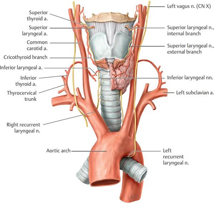
Fig. 21.16 Arteries and nerves of the larynx, thyroid, and parathyroids
Anterior view.
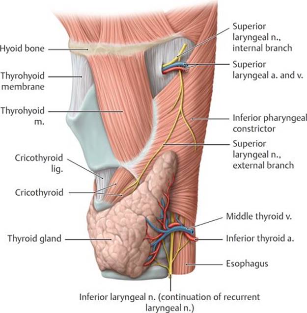
Fig. 21.17 Neurovasculature of the larynx
Left lateral view.
A Superficial layer.
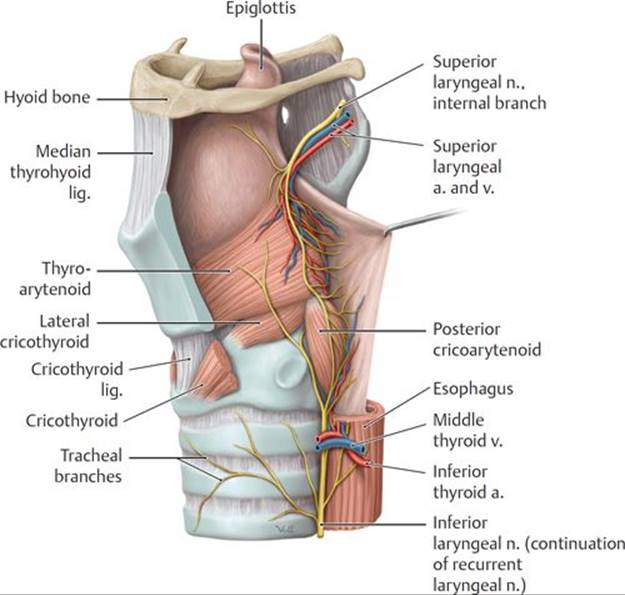
B Deep layer. Removed: Cricothyroid muscle and left lamina of thyroid cartilage. Retracted: Pharyngeal mucosa.
21.6f The Trachea
The trachea, the extension of the airway inferior to the larynx, extends from the inferior border of the cricoid cartilage to the level of the T4–T5 intervertebral disks in the thorax, where it bifurcates into the two primary bronchi of the lungs (see Section 5.8).
21.7 Thyroid and Parathyroid Glands
The thyroid and parathyroid glands are endocrine glands that reside in the anterior cervical region.
21.7a The Thyroid Gland
The thyroid gland, the largest endocrine gland in the body, secretes thyroid hormone, which regulates the rate of metabolism, and calcitonin, which regulates the metabolism of calcium (Fig. 21.18A).
– The thyroid lies deep to the sternohyoid and sternothyroid muscles (infrahyoid muscles) and anterolateral to the larynx and trachea between C5 and T1 vertebral levels.
– The thyroid has right and left (lateral) lobes connected by a narrow isthmus anterior to the second and third tracheal rings.
– A pyramidal lobe, present in ~50% of the population, is a remnant of the embryonic thyroglossal duct that extends from the isthmus toward the hyoid bone.
– A fibrous capsule encloses the thyroid gland. The pretracheal fascia of the neck lies outside the thyroid capsule.
Thyroglossal duct cyst
A thyroglossal duct cyst is a fluid-filled cavity in the midline of the neck, just inferior to the hyoid bone. It results from the proliferation of epithelium left behind from the thyroglossal duct during the descent of the thyroid gland from its embryonic origin on the tongue to its postnatal position in the neck. The cyst can enlarge and press on the trachea and esophagus, in which case it can be surgically removed.
21.7b The Parathyroid Glands
Parathyroid glands, small, ovoid endocrine glands that lie on the posterior surface of the thyroid gland, secrete parathormone, which regulates the metabolism of phosphorus and calcium (Fig. 21.18B).
– There are normally four glands, two superior and two inferior, although the number can range from two to six glands.
– The superior parathyroid glands are constant in position near the lower border of the cricoid cartilage. The position of the inferior parathyroid glands may vary, ranging from the lower pole of the thyroid gland to the superior mediastinum.
21.7c Neurovasculature of the Thyroid and Parathyroid Glands
– The superior thyroid artery, a branch of the external carotid artery, and the inferior thyroid artery from the thyrocervical trunk supply the thyroid gland (see Fig. 21.16). The inferior thyroid artery is usually the main supply to the parathyroid glands.
– Superior and middle thyroid veins drain into the internal jugular veins; inferior thyroid veins drain into the brachiocephalic veins in the mediastinum (Fig. 21.19). Venous drainage from the parathyroid glands joins the thyroid veins.
– Lymphatic vessels of the thyroid may drain directly into the superior and inferior deep cervical nodes or indirectly, passing first through prelaryngeal, pretracheal, and paratracheal nodes.
– The parathyroid glands drain with the thyroid into the inferior deep cervical and paratracheal lymph nodes.
– Cardiac and superior and inferior thyroid sympathetic plexuses arise from the superior, middle, and inferior cervical sympathetic ganglia to supply vasomotor innervation to the thyroid and parathyroid glands.
– The thyroid and parathyroid glands are under hormonal control and therefore lack secretomotor innervation.
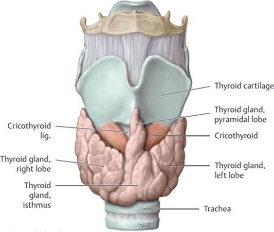
Fig. 21.18 Thyroid and parathyroid glands
A Thyroid gland. Anterior view.

B Thyroid and parathyroid glands, posterior view.

Fig. 21.19 Veins of the larynx, thyroid, and parathyroid glands
Left lateral view. Note: The inferior thyroid vein generally drains into the left brachiocephalic vein.
21.8 Topography of the Neck
The regions of the neck, defined by muscular and skeletal boundaries, are largely descriptive rather than functional, but they are useful for understanding the topographic relationships in the neck, which often play a significant role in medical practice (Table 21.8; Figs. 21.20A and B and 21.21A, B, and C).
– The anterior cervical region (anterior triangle) extends from the midline of the neck to the anterior border of the sternocleidomastoid muscle.
• The region is further divided into submandibular, sub-mental, muscular, and carotid triangles.
• The anterior region contains most of the cervical viscera, which includes the lower pharynx, esophagus, larynx, trachea, thyroid, and parathyroid glands.
– The sternocleidomastoid region is a narrow area defined by the anterior and posterior borders of the sternocleidomastoid muscle.
• Inferiorly, the sternal and clavicular heads of the muscle define the small lesser supraclavicular fossa.
• This region contains parts of the major vascular structures of the neck.
– The lateral cervical region (posterior triangle) extends from the posterior border of the sternocleidomastoid muscle to the anterior border of the trapezius muscle.
• The posterior belly of the omohyoid muscle divides this region into omoclavicular and occipital triangles.
• The scalene muscles, and cervical and brachial plexuses are located in this region.
– The posterior cervical region extends from the anterior border of the trapezius muscle to the posterior midline of the neck.
• It contains the trapezius and suboccipital muscles, the vertebral artery, and posterior branches of the cervical plexus.
– The root of neck, a transition area for structures passing between the thorax and neck, is enclosed by the superior thoracic aperture, which is formed by the manubrium, the first ribs and their costal cartilages, and the first thoracic vertebra.
• It contains the trachea, esophagus, common carotid and subclavian arteries, brachiocephalic veins, vagus and phrenic nerves, the sympathetic trunk, the thoracic duct, and the apex of each lung.

A Right anterior oblique view.

B Left posterior oblique view.
TABLE 21.8 Regions of the Neck
|
Region |
Divisions |
Contents |
|
|
Submandibular (digastric) triangle |
Submandibular gland and lymph nodes, hypoglossal n. (CN XII), facial a. and v. |
|
Submental triangle |
Submental lymph nodes |
|
|
Muscular triangle |
Sternothyroid and sternohyoid muscles, thyroid and parathyroid glands |
|
|
Carotid triangle |
Carotid bifurcation, carotid body, hypoglossal (CN XII) and vagus (CN X) nn. |
|
|
|
Sternocleidomastoid, common carotid a., internal jugular v., vagus n. (CN X), jugular lymph nodes |
|
|
|
Omoclavicular (subclavian) triangle |
Subclavian a., subscapular a., supraclavicular l.n. |
|
Occipital triangle |
Accessory n. (CN XI), trunks of brachial plexus, transverse cervical a., cervical plexus (posterior branches) |
|
|
|
Nuchal muscles, vertebral a., cervical plexus |
|
|
* The sternocleidomastoid region also contains the lesser supraclavicular fossa. |
||
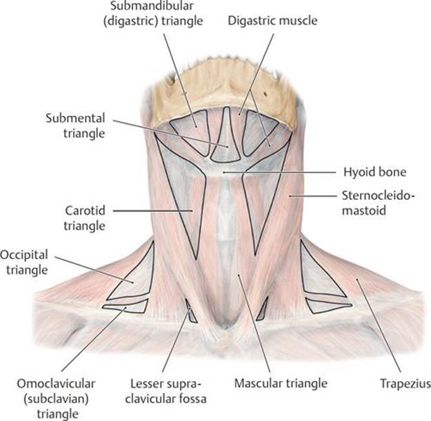
Fig. 21.20 Cervical regions
A Anterior view.

B Left lateral view.

Fig. 21.21 Topography of the anterior cervical region
Anterior view.
A Superficial dissection.
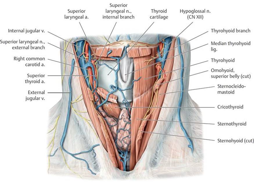
B Deep dissection.
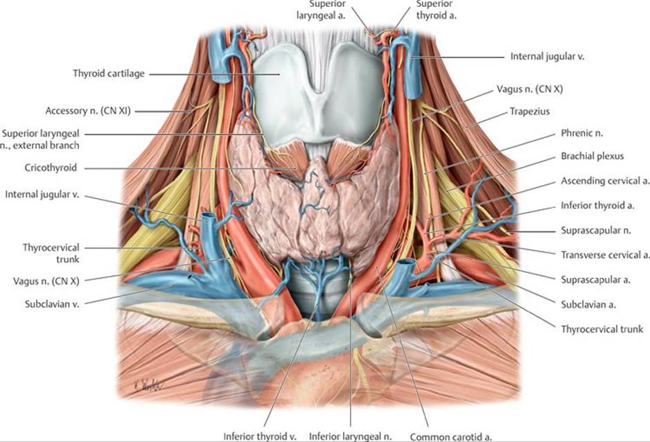
C Root of the neck.
 Investing (superficial) layer
 Muscular
 Visceral
 Prevertebral layer
 Carotid sheath
 Thyroarytenoid m.
 Lateral cricoarytenoid m.