
The eye, the organ of vision, and the ear, which contains the organs for hearing and balance, are anatomically the most complex of the sense organs. The eyes, enclosed within the bony orbits, are prominent features of the face. The ears have superficial and deep components that are related to the temporal bones on the sides of the head.
20.1 The Eye
The anatomy of the eye includes the bony orbit, the eyelids and lacrimal apparatus, the eyeball, and six extraocular muscles.
20.1a The Bony Orbit
The paired orbits lie on either side of the superior part of the nasal cavity, above the maxillary sinuses and below the anterior cranial fossae. These cavities are shaped like quadrangular pyramids with the apex directed posteriorly and the base opening onto the face (Fig. 20.1A and B).
– Seven bones of the skull form the bony orbit.
• The frontal bone forms the roof.
• The maxilla forms the floor.
• The ethmoid, lacrimal, nasal, and sphenoid bones form the medial wall.
• The zygomatic and sphenoid bones form the lateral wall.
– Numerous openings allow nerves and vessels to pass between the orbit and the middle cranial fossa, the nasal cavity, the pterygopalatine fossa, and the face (Table 20.1).
– The eyeball occupies the anterior part of the orbit, accompanied by six extraocular muscles, ophthalmic vessels, and five cranial nerves (optic, oculomotor, trochlear, trigeminal, and abducent). Periorbital fat supports and surrounds these structures.
TABLE 20.1  Openings in the Orbit for Neurovascular Structures
|
Opening* |
Nerves |
Vessels |
|
Optic canal |
Optic n. (CN II) |
Ophthalmic a. |
|
Superior orbital fissure |
Oculomotor n. (CN III) Trochlear n. (CN IV) Abducent n. (CN VI) Trigeminal n., ophthalmic division (CN V1) • Lacrimal n. • Frontal n. • Nasociliary n. |
Superior ophthalmic v. |
|
Inferior orbital fissure |
Infraorbital n. (CN V2) Zygomatic n. (CN V2) |
Infraorbital a. and v., inferior ophthalmic v. |
|
Infraorbital canal |
Infraorbital n. (CN V2), a., and v. |
|
|
Supraorbital foramen |
Supraorbital n. (lateral branch) |
Supraorbital a. |
|
Frontal incisure |
Supraorbital n. (medial branch) |
Supratrochlear a. |
|
Anterior ethmoidal foramen |
Anterior ethmoidal n., a., and v. |
|
|
Posterior ethmoidal foramen |
Posterior ethmoidal n., a., and v. |
* The nasolacrimal canal transmits the nasolacrimal duct.

Fig. 20.1  Bones of the orbit
A Anterior view.

B Lateral view of right orbit.
20.1b Eyelids and Lacrimal Apparatus (Figs. 20.2 and 20.3)
The upper and lower eyelids are moveable folds of skin that protect the eyeball from injury, irritation, and light.
– The eyelids are covered externally by skin and internally by the palpebral conjunctiva, a thin inner membrane that reflects onto the anterior eyeball as the bulbar conjunctiva. When the eyes are closed, the palpebral and bulbar conjunctivae form the conjunctival sac.
– Tarsal plates or tarsus, bands of dense connective tissue that provide support for both the upper and lower eyelids, are attached to medial and lateral palpebral ligaments, which connect to the medial and lateral margins of the orbit, respectively. Tarsal glands within the tarsal plates lubricate the edges of the eyelids to prevent them from sticking together.
– The orbicularis oculi muscle (see Fig. 19.1), innervated by the facial nerve (CN VII), closes the eye in a sphincter-like fashion. The levator palpebrae superioris muscle, innervated by the oculomotor nerve (CN III) and attached to the superior tarsal plate, opens the eye by lifting the upper eyelid.
– An orbital septum, which connects the tarsal plates to the margins of the orbit, holds the orbital fat within the orbit and helps to limit the spread of infection to and from the orbit.

Fig. 20.2 Eyelids and conjunctiva
Sagittal section through the anterior orbital cavity.
The lacrimal apparatus produces and drains tears that cleanse and lubricate the outer surface of the eye.
– The lacrimal gland, which produces and secretes tears, resides in the lacrimal fossa in the supralateral aspect of the orbit. Secretomotor parasympathetic fibers from the facial nerve (CN VII) stimulate the gland (see Fig. 18.19).
– Blinking of the eye sweeps the tears across the eye toward the medial angle, where they drain via superior and inferior puncta (openings) into lacrimal canaliculi and the lacrimal sac, the dilated superior part of the nasolacrimal duct.
– The nasolacrimal duct is a membranous structure that begins in the medial angle of the eye and terminates in the inferior meatus of the nasal cavity. Tears drain into the nasal cavity via this duct.
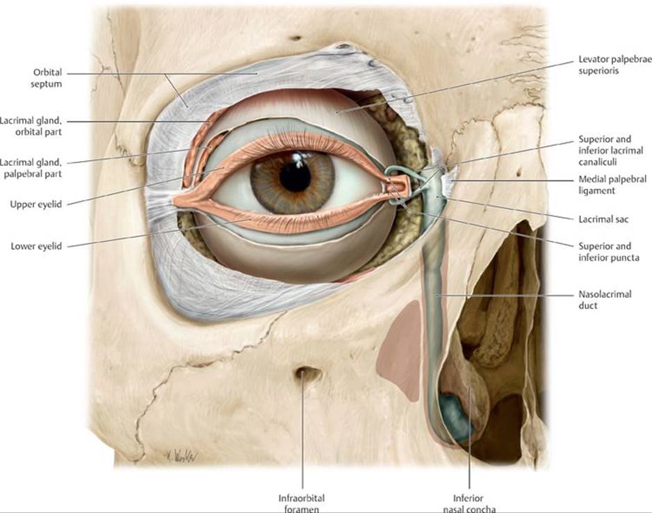
Fig. 20.3 Lacrimal apparatus
Right eye, anterior view. Removed: Orbital septum (partial). Divided: Levator palpebrae superioris (tendon of insertion).
20.1c The Eyeball
The eyeball, the organ of vision, has three concentric layers that form its outer walls: the sclera, the choroid, and the retina (Fig. 20.4).
– The sclera, the white part of the eye, forms the posterior five sixths of the outer fibrous layer of the eye; the cornea, the transparent part of the sclera, forms the anterior sixth. This outer layer is largely avascular but provides structure to the eyeball.
– The choroid, the middle vascular layer, provides oxygen and nutrients to the underlying retina (Fig. 20.5).
• The ciliary body connects the choroid with the circumference of the iris. Short, smooth muscle fibers, zonular fibers, which attach the ciliary body to the lens, control the thickness and refractive power of the lens and therefore the focus of the eye.
• The iris, an adjustable muscular diaphragm, surrounds a central aperture, the pupil.
○ The pupillary sphincter muscle of the iris responds to parasympathetic stimulation to constrict the pupil.
○ The pupillary dilator muscle of the iris responds to sympathetic stimulation to dilate the pupil.
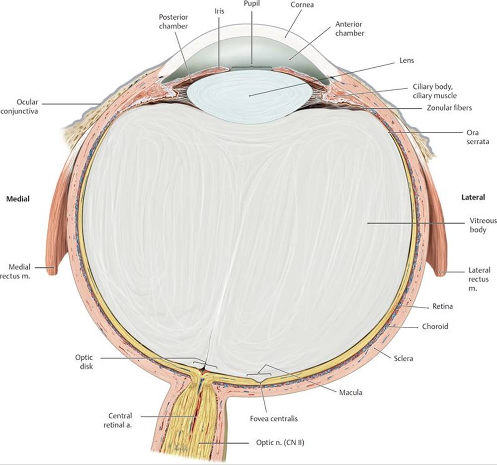
Fig. 20.4 Structure of the eyeball
Transverse section through the right eyeball, superior view.

Fig. 20.5 Cornea, iris, and lens
Transverse section through the anterior segment of the eye, anterosuperior view.
– The retina, the inner sensory layer, has a posterior optic part that is sensitive to light and a nonvisual part that continues anteriorly over the ciliary body and iris.
• The optic disc, a point on the retina where the optic nerve exits the eyeball, lacks photoreceptors and therefore is insensitive to light and is known as the blind spot.
• The macula of the retina, a spot lateral to the optic disk, is an area of intense visual acuity.
• The fovea centralis, a depression in the macula, is the area of greatest visual acuity.
– Light passes through four refractive media before focusing on the retina of the eye:
1. The cornea, the primary refractive medium for light entering the eye
2. The aqueous humor, a watery solution that fills the anterior and posterior chambers of the eye that lie anterior to the lens and ciliary body. The balance between its production and drainage determines the intraocular pressure.
3. The lens, a transparent biconcave disk that focuses objects on the retina by changing its thickness. In the process of accommodation, which is mediated by parasympathetic stimulation, the ciliary muscle contracts, causing the lens to thicken and bring near objects into focus.
4. The vitreous body, a jelly-like substance that fills the chamber of the eye posterior to the lens.
Corneal reflex
A positive corneal reflex is the bilateral contraction of the orbicularis oculi muscles (a blink) when the cornea is touched or exposed to bright light. The afferent limb is the nasociliary nerve of the ophthalmic division of the trigeminal nerve (CN V1); the efferent limb is the facial nerve (CN VII). The reflex protects the eye from foreign bodies and bright light.
Pupillary light reflex
The pupillary light reflex is rapid constriction (miosis) of both pupils (via the constrictor pupillae muscle) in response to shining a light into the eye. The afferent limb of the reflex is the optic nerve (CN II). The efferent limb is the parasympathetic fibers of the oculomotor nerve (CN III). Both pupils constrict because each retina sends fibers into the optic tracts of both sides. If the parasympathetic fibers are interrupted, both pupils dilate (mydriasis) as a result of the unopposed sympathetic outflow to the dilator pupillae muscle. This reflex mediates papillary aperture variation and is therefore one of the means by which the eye adapts to changes in light intensity.
Presbyopia and cataracts
The lens of the eye undergoes age-related changes that affect vision in the older patient. The loss of elasticity of the lens, and the subsequent loss of accommodation, diminishes a patient’s ability to focus on near objects, a condition known as presbyopia. Opacities of the lens or its capsule, known as cataracts, allow less light to reach the retina, resulting in blurred, cloudy vision. Treatment involves surgical removal of the affected lens and replacement with a plastic lens implant.
Glaucoma
Glaucoma refers to a group of eye diseases that involve increased intraocular pressure and atrophy of the optic nerve. In primary open-angle glaucoma, the most common form, venous channels in the angle between the cornea and the iris that allow drainage of the aqueous humor from the anterior and posterior chambers are blocked. The subsequent buildup of aqueous humor results in raised intraocular pressure and eventual damage to the optic nerve. This leads to a gradual loss of peripheral vision that progresses to tunnel vision. Pressure on the retina can lead to blindness.
20.1d Extraocular Muscles
– The six extraocular muscles that control the movement of the eyeball (Fig. 20.6A and B; Table 20.2) include
• four rectus muscles, the superior rectus, medial rectus, inferior rectus, and lateral rectus, that originate from a common tendinous ring at the apex of the orbit; and
• two oblique muscles, the superior oblique that arises near the apex and reflects back via the trochlea to attach to the eyeball, and the inferior oblique that arises from the medial aspect of the orbital floor.

Fig. 20.6 Extraocular muscles
Right eye.
A Anterior view.
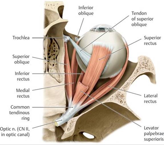
B Opened orbit, superior view.

A Superior rectus.

B Medial rectus.
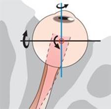
C Inferior rectus.

D Lateral rectus.

E Superior oblique.
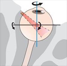
F Inferior oblique.

– The muscles allow six cardinal directions of gaze, which are the normal movements of the eyeball tested during clinical evaluation of ocular mobility (Fig. 20.7A and B).
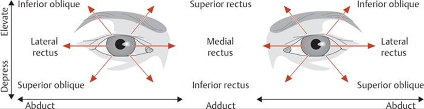
Fig. 20.7 Testing the extraocular muscles
A Starting with the eyes directed anteriorly, movement to any of the cardinal directions of gaze (arrows) requires activation of two extraocular muscles, each of which is innervated by a different cranial nerve, thus testing the function of those pairs of muscles.
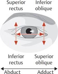
B Starting with the eyes adducted or abducted, elevating or lowering the eyes activates only the oblique or rectus muscles, respectively, allowing for the testing of the function of individual muscles.
20.1e Neurovasculature of the Orbit
– The ophthalmic artery supplies most structures of the orbit. One of its branches, the central retinal artery, runs within the optic nerve and is the sole arterial supply to the retina through its terminal branches (Fig. 20.8).
• The ophthalmic artery anastomoses with the facial artery through its supratrochlear branch and with the maxillary artery through its anterior and posterior ethmoidal and middle meningeal branches.

Fig. 20.8 Arteries of the orbit
Right orbit, superior view. Opened: optic canal and orbital roof.
– The superior and inferior ophthalmic veins, which drain structures in the orbit, primarily drain into the cavernous sinus but also communicate with the facial vein and pterygoid venous plexus (Fig. 20.9).
– Six cranial nerves (optic, oculomotor, trochlear, trigeminal, abducent, and facial) innervate structures in the orbit (Table 20.3; Fig. 20.10).
• The optic nerve (CN I) transmits images from the retina.
• The oculomotor nerve (CN III), trochlear nerve (CN IV), and abducent nerve (CN VI) innervate the extraocular muscles.
• The ophthalmic division of the trigeminal nerve (CN V1) carries general sensory fibers from structures in the orbit and distributes postganglionic autonomic fibers to target organs of the orbit and face.
• The facial nerve (CN VII) provides secretomotor (parasympathetic) innervation to the lacrimal gland.
Oculomotor nerve injury
The oculomotor nerve innervates most of the extraocular muscles. With paralysis of these muscles, the eye is directed downward and outward because of the unopposed action of the lateral rectus (innervated by the abducent nerve) and superior oblique (innervated by the trochlear nerve). The dilator pupillae is also unopposed, so the pupil remains fully dilated. Paralysis of the levator palpebrae superior allows the superior eyelid to droop.
– Autonomic innervation of structures in the orbit includes the following:
• Sympathetic fibers from the carotid plexus innervate the ciliary body and pupillary dilator muscle (responsible for pupil dilation).
• Parasympathetic fibers from the oculomotor nerve (CN III) synapse in the ciliary ganglion and travel via the short ciliary nerves to innervate the ciliary body and pupillary sphincter muscles (responsible for pupil constriction).
• Parasympathetic fibers from the facial nerve (CN VII) synapse in the pterygopalatine ganglion and travel via the zygomatic nerve (CN V2) to innervate the lacrimal gland (responsible for tear secretion).
Horner’s syndrome
Horner’s syndrome is a kaleidoscope of symptoms resulting from disruption of the cervical sympathetic trunk in the neck. The absence of sympathetic innervation is manifested on the affected side of the face as pupillary constriction (miosis), a sunken eye (enophthalmos), drooping of the upper eyelid (ptosis), loss of sweating (anhydrosis), and vasodilation.

Fig. 20.9 Veins of the orbit
Right orbit, lateral view. Removed: lateral orbital wall. Opened: maxillary sinus.
TABLE 20.3 Nerves of the Orbit
|
Nerve |
Nerve Fibers |
Distribution |
|
Optic (CN II) |
Special sensory for vision |
Retina |
|
Oculomotor (CN III) |
Somatic motor Parasympathetic: synapse in ciliary ganglion; post-synaptic fibers travel with nasociliary n. (CN V1) |
Muscles of the orbit, except the lateral rectus and superior oblique muscles Pupilary sphincter muscle and ciliary body |
|
Trochlear (CN IV) |
Somatic motor |
Superior oblique muscle |
|
Ophthalmic (CN V1) Lacrimal Frontal - Supratrochlear - Supraorbital Short ciliary Nasociliary - Anterior and posterior ethmoidal - Infratrochlear - Long ciliary |
General sensory General sensory General sensory General sensory Parasympathetic (CN III) and sympathetic General sensory General sensory General sensory Sympathetic (carotid plexus) |
Lacrimal gland, upper lateral eyeball Anterior scalp Anterior scalp Ciliary body and iris Nasal cavity, ethmoid and sphenoid sinuses External nose, conjunctiva, lacrimal sac Iris and cornea Pupil dilator |
|
Abducent (CN VI) |
Somatic motor |
Lateral rectus muscle |
|
Facial (CN VII) |
Parasympathetic: synapse in pterygopalatine ganglion; postsynaptic fibers travel with zygomatic n. (CN V2) |
Lacrimal gland |

Fig. 20.10 Innervation of the orbit
Right orbit, lateral view. Removed: Temporal bony wall.
20.2 The Ear
The ear, which contains the organs for hearing and equilibrium, is divided into external, middle, and internal parts (Fig. 20.11).
20.2a The External Ear
The external ear collects and conducts sound.
– The auricle, the visible external part of the ear, has a skeleton composed of elastic cartilage that is covered by skin.
– The external acoustic meatus (external auditory canal), a canal that extends 2 to 3 cm from the auricle to the tympanic membrane, conducts sound waves toward the middle ear. The outer third of the canal is cartilaginous, and the inner two thirds are formed by the temporal bone. Ceruminous and sebaceous glands in the subcutaneous tissue lining the cartilaginous part secrete earwax.
– The tympanic membrane, a thin, transparent membrane, separates the external and middle ear.
• Skin covers the tympanic membrane externally, and a mucous membrane lines it internally.
• The concave outer surface of the membrane has a central conelike depression, the umbo.
• A thin, superior portion of the membrane, the flaccid part (pars flaccida), is distinct from the rest of the membrane, the tense part (pars tensa).
– Posterior auricular and anterior auricular arteries, branches of the superficial temporal artery, supply the external ear.
– Sensation from the external ear is transmitted by
• the great auricular nerve (cervical plexus) from the auricle;
• the auriculotemporal nerve, a branch of CN V3, from the auricle and external surface of the tympanic membrane;
• the auricular branch of the vagus nerve (CN X) from the external surface of the tympanic membrane; and
• the glossopharyngeal nerve (CN IX) from the internal surface of the tympanic membrane.

Fig. 20.11 Ear
Coronal section through the right ear, anterior view.
20.2b The Middle Ear
– The middle ear, also known as the tympanic cavity, is an air-filled chamber housed in the petrous portion of the temporal bone (Figs. 20.12 and 20.13).
• Anteriorly, a pharyngotympanic tube, which connects the tympanic cavity to the nasopharynx, helps equalize pressure in the middle ear.
• Posteriorly, the aditus (inlet) to the mastoid antrum (a cavity in the mastoid process of the temporal bone) connects the tympanic cavity to the bony meshwork of mastoid air cells.
• A thin bony plate, the tegmen tympani, forms the roof of the tympanic cavity and separates it from the middle cranial fossa.
• The medial wall, which separates the tympanic cavity from the internal ear, has a promontory, covered by the tympanic nerve plexus, and two openings, the oval and round windows (see Fig. 20.15).
– The auditory ossicles, the malleus, incus, and stapes bones of the middle ear, articulate with each other through synovial joints and form a bony chain between the tympanic membrane and the oval window of the internal ear.
• The handle of the malleus is embedded in the tympanic membrane, and its head articulates with the incus.
• The incus articulates with the malleus and stapes.
• The head of the stapes articulates with the incus, and its base fits into the oval window of the bony labyrinth of the inner ear.
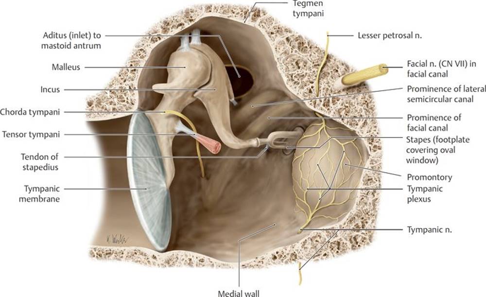
Fig. 20.12 Tympanic cavity
Right tympanic cavity, anterior view. Removed: Anterior wall.
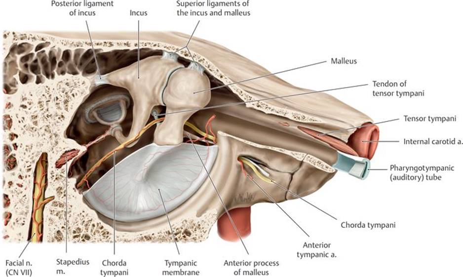
Fig. 20.13 Muscles and neurovascular relations in the tympanic cavity
Right middle ear, lateral view.
– Muscles of the middle ear dampen the movements of the auditory ossicles, thereby lessening the sound transmitted from the external ear.
• The tensor tympani, which is innervated by a branch of the mandibular nerve (CN V3), lessens the damage from loud sounds by tensing the tympanic membrane.
• The stapedius, which is innervated by a branch of the facial nerve (CN VII), dampens the vibrations of the stapes on the oval window.
– The ascending pharyngeal, maxillary, and posterior auricular branches of the external carotid artery and a small branch of the internal carotid artery supply the middle ear.
– The chorda tympani, a branch of the facial nerve (CN VII), has no branches in the middle ear but passes between the malleus and incus to exit the cavity through a small opening in the temporal bone.
– The glossopharyngeal nerve (CN IX) transmits sensation from the tympanic cavity and pharyngotympanic tube. Preganglionic parasympathetic fibers carried in the tympanic nerve (a branch of the glossopharyngeal nerve) synapse in the otic ganglion. The postganglionic fibers join with sympathetic fibers of the internal carotid plexus to form the tympanic plexus (see Fig. 18.22).
Otitis media
Otitis media is an infection of the middle ear that occurs commonly in children often following an upper respiratory tract infection. Fluid that accumulates in the middle ear can temporarily diminish hearing, and inflammation of the lining of the tympanic cavity can block the pharyngotympanic tube.
Hyperacusis
The stapedius muscle protects the delicate inner ear by modifying the vibrations of very loud sounds as they are transmitted through the middle ear to the stapes. Paralysis of the muscle resulting from a lesion of the facial nerve causes an extreme sensitivity to sound, a condition known as hyperacusis.
20.2c The Internal Ear
– The internal ear, which contains the organ for hearing, the auditory apparatus, and the organ for balance, the vestibular apparatus, is encased within the petrous part of the temporal bone and consists of (Figs. 20.14 and 20.15) the following:
• A bony otic capsule, which forms the walls of the bony labyrinth
• A bony labyrinth, a series of chambers and canals within the otic capsule containing the fluid perilymph. It includes the cochlea, the vestibule, and the semicircular canals.
• A membranous labyrinth, a series of sacs and ducts suspended within the bony labyrinth, and filled with a fluid, endolymph. The membranous labyrinth is made up of
○ the cochlear duct contained within the cochlea,
○ the utricle and saccule contained within the vestibule, and
○ the semicircular ducts contained within the semicircular canals.

Fig. 20.14 Projection of the otic capsule of the inner ear onto the skull
Petrous part of the temporal bone, superior view.
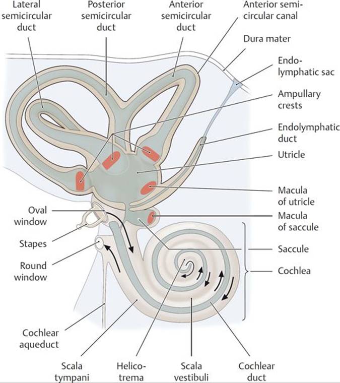
Fig. 20.15 Schematic of the inner ear
Right lateral view. The inner ear is embedded within the petrous part of the temporal bone. It is composed of a membranous labyrinth filled with endolymph floating within a similarly shaped bony labyrinth filled with perilymph.
– The auditory apparatus consists of the following:
• The cochlea, a space within the bony labyrinth that includes the bony cochlear (spiral) canal, which makes 2.5 turns around its axis, the modiolus (Fig. 20.16A). The basal turn of the cochlea forms the promontory on the medial wall of the middle ear and contains the round window, which is closed by a membrane.
• The cochlear duct, part of the membranous labyrinth, which is a blind-ended duct filled with endolymph and suspended within the cochlear canal (Fig. 20.16B):
○ The cochlear duct divides the cochlear canal into two channels, the scala vestibuli and scala tympani, which are continuous with each other at the helicotrema, a space at the apex of the canal.
○ At the base of the cochlea, the scala vestibuli lies against the oval window, and the scala tympani lies against the round window.
• The spiral organ (of Corti), which contains the sensory receptors for hearing, and is embedded in the basement membrane on the floor of the cochlear duct
– The sequence of sound transmission through the ear involves (Fig. 20.17) the following:
1. Transmission of sound waves from the external ear and external acoustic meatus to the tympanic membrane of the middle ear. The waves vibrate the auditory ossicles and, in turn, the oval window that is attached to the base of the stapes.
2. Transmission of vibrations of the oval window to the perilymph of the scala vestibuli, which creates pressure waves that displace the basement membrane and spiral organ of the cochlear duct. Nerve endings in the spiral organ transmit impulses to the brain along the cochlear nerve.
3. Transmission of the pressure waves of the perilymph from the scala vestibuli along the scala tympani to the round window, with dissipation into the tympanic cavity.
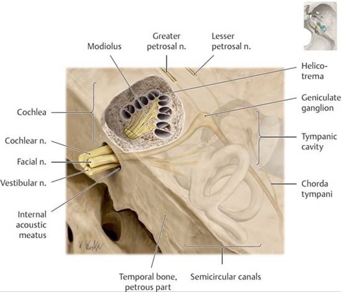
Fig. 20.16 Auditory apparatus
A Location of the cochlea. Superior view of the petrous part of the temporal bone with the cochlea sectioned transversely.
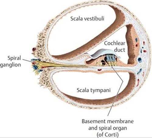
B Compartments of the cochlear canal, cross section.

Fig. 20.17 Propagation of sound waves by the ossicular chain
– The vestibular apparatus consists of the following (see Fig. 20.15):
• The vestibule of the bony labyrinth that communicates with the cochlea and semicircular canals
○ A small extension, the vestibular aqueduct, communicates with the posterior cranial fossa and contains the endolymphatic sac, a membranous storage space for excess endolymph.
• The utricle and saccule, part of the membranous labyrinth, which lie within the vestibule
○ The utricle communicates with the semicircular ducts; the saccule communicates with the cochlear duct.
○ Both the uticle and saccule contain specialized sensory fields called maculae, which occupy different positions in space and are sensitive to movement of the endolymph in the horizontal and vertical planes.
• Three semicircular canals of the bony labyrinth, which are arranged perpendicular to one another and communicate with the vestibule. Each canal has a swelling at one end, the bony ampulla.
• Three semicircular ducts, parts of the membranous labyrinth contained within the semicircular canals, that communicate with the utricle
○ An ampulla at one end of each semicircular duct contains the ampullary crest, an area of sensory epithelium. The ampullary crests respond to motion of the endolymph within the ducts caused by rotation of the head.
– The membranous labyrinth receives its blood supply from the internal auditory artery, a branch of the basilar artery via its anterior inferior cerebellar artery.
– The vestibular and cochlear nerves within the internal acoustic meatus form the vestibulocochlear nerve (CN VIII) (see Fig. 18.21)
• The vestibular nerve innervates organs of the vestibular apparatus: the maculae of the utricle and saccule and the ampullary crests of the semicircular canals. Neuron cell bodies lie within the vestibular ganglion in the internal acoustic meatus.
• The cochlear nerve innervates the spiral organ (of Corti) in the cochlea. Neuron cell bodies lie in the spiral ganglion of the cochlea at the modiolus.
Meniere’s disease
Meniere’s disease is an inner ear disorder resulting from blockage of the cochlear duct. Recurring episodes are characterized by tinnitus (ringing or buzzing in the ear), vertigo (the illusion of movement), and hearing loss. The hearing loss may fluctuate in intensity and from ear to ear but eventually becomes permanent.
Vertigo, tinnitus, and hearing loss
Trauma to the ear can cause three types of symptoms: vertigo, tinnitus, and hearing loss. Vertigo refers to the illusion of movement, or dizziness, and results from injury to the semicircular canals. Tinnitus refers to a ringing in the ears and is a disorder involving the cochlear duct. Causes of hearing loss can be either peripheral or centrally located. Conductive hearing loss occurs when there is impaired transmission of sound waves through the auditory canal to the auditory ossicles. Sensorineural hearing loss occurs when there is damage to the pathway between the cochlea and the brain.