|
|
|
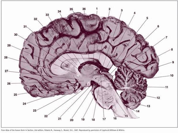
Plate 72 Gross Midsagittal Section of the Brain and Brain Stem with Meninges and Blood Vessels Intact |
Gross midsagittal section of the brain and brain stem with meninges and blood vessels intact. Arachnoid granulations are seen along the crest of the hemisphere. The posterior commissure, decussation of the superior cerebellar peduncles, and medial longitudinal fasciculus (MLF) are well demonstrated.
1. Stria medullaris of thalamus
2. Third ventricle
3. Habenular commissure
4. Pineal gland
5. Stratum opticum
6. Cerebral aqueduct
7. Inferior colliculus
8. Central gray substance
9. Superior medullary velum
10. Decussation of superior cerebellar peduncle
11. Medial longitudinal fasciculus (bundle)
12. Superior central nucleus
13. Choroid plexus of fourth ventricle
14. Obex
15. Inferior central nucleus
16. Medulla oblongata
17. Posterior commissure
18. Interpeduncular nucleus
19. Basilar artery
20. Central rami of basilar artery
21. Posterior perforated substance
22. Mamillary body
23. Infundibular recess
24. Supraoptic recess
25. Lamina terminalis
26. Hypothalamus
27. Preoptic area
28. Pericallosal artery
29. Anterior commissure
30. Septum pellucidum
31. Interventricular foramen
32. Column of fornix
33. Paraventricular nucleus of thalamus
34. Body of fornix
35. Arachnoidal granulations
36. Velum interpositum and tela choroidea
|
|
|
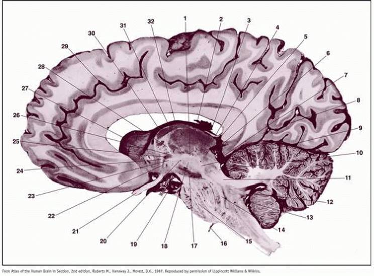
Plate 73 Gross Parasagittal Section through the Red Nucleus, Medial Lemniscus, and Inferior Olivary Nucleus |
Gross parasagittal section through the red nucleus, medial lemniscus, and inferior olivary nucleus. The corticospinal fibers can be traced from the crus cerebri to the spinal cord. The abducent nerve (CN VI), is seen exiting from the pontobullar sulcus.
1. Prerubral tract
2. Dorsal medial nucleus of thalamus
3. Crus of fornix
4. Medial pulvinar nucleus of thalamus
5. Pretectal area
6. Fasciolar gyrus
7. Posterior cerebral artery
8. Calcarine sulcus
9. Medial lemniscus
10. Lateral lemniscus
11. Superior cerebellar peduncle
12. Dentate nucleus
13. Striae medullares of fourth ventricle
14. Medial lemniscus
15. Corticospinal tract
16. Abducent nerve (VI)
17. Substantia nigra
18. Medullary lamina of red nucleus
19. Posterior cerebral artery
20. Tegmental area H
21. Optic nerve (II)
22. Ansa lenticularis
23. Tegmental area H2
24. Inferior thalamic peduncle
25. Zona incerta
26. Tegmental area H1
27. Ventricular surface of head of caudate nucleus
28. Mamillothalamic tract
29. Cingulate sulcus
30. Anteroventral nucleus of thalamus
31. Posteromedial ventral nucleus of thalamus
32. Internal medullary lamina of thalamus
|
|
|
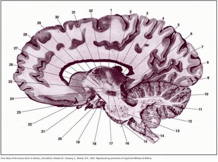
Plate 74 Gross Parasagittal Section through the Caudate Nucleus, Subthalamic Nucleus, Substantia Nigra, and Dentate Nucleus |
Gross parasagittal section through the caudate nucleus, subthalamic nucleus, substantia nigra, and dentate nucleus. The abducent nerve (CN VI) is seen exiting the pontobulbar sulcus. Damage to the subthalamic nucleus results in hemiballism. Parkinson disease results from a cell loss of the pigmented neurons in the substantia nigra.
1. Stratum zonale of thalamus
2. Centromedian nucleus of thalamus
3. Posteromedial ventral nucleus of thalamus
4. Medial pulvinar nucleus of thalamus
5. Medial geniculate body
6. Cingulum
7. Parahippocampal gyrus
8. Medial lemniscus
9. Peduncle of substantia nigra
10. Lateral corticobulbar fibers
11. Dentate nucleus
12. Inferior cerebellar peduncle
13. Pontine nuclei
14. Olive
15. Abducent nerve (VI)
16. Substantia nigra
17. Subthalamicotegmental tract
18. Posterior cerebral artery
19. Uncus
20. Oculomotor nerve (III)
21. Optic nerve (II)
22. Olfactory tract
23. Anterior cerebral artery
24. Capsule of subthalamic nucleus
25. Subthalamic nucleus
26. Anterior limb of internal capsule
27. Head of caudate nucleus
28. Genu of internal capsule
29. Tegmental area H2
30. Tegmental area H1
31. Zona inserta
32. Posterior lateral nucleus of thalamus
|
|
|
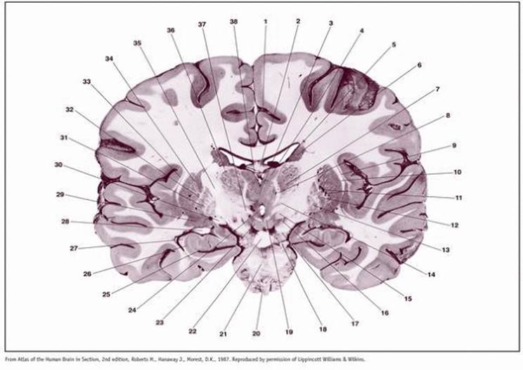
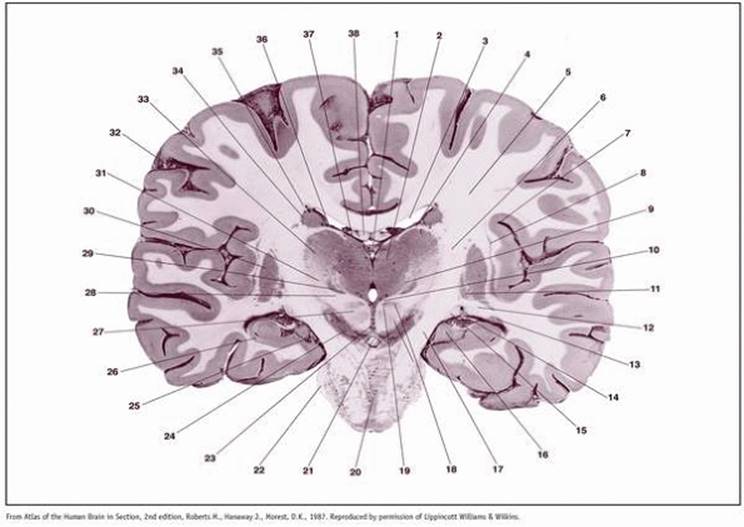
Plate 75 Coronal Section through the Anterior Commissure, Amygdala, Septal Nuclei, and Optic Chiasm |
Coronal section through the anterior commissure, amygdala, septal nuclei, and optic chiasm. The septal nuclei have reciprocal connections with the hippocampal formation. Bilateral destruction of the amygdaloid nuclei results in the Klüver-Busey syndrome (psychic blindness, hyperphagia, and hypersexuality.) An aneurism of the internal carotid artery within the cavernous sinus (CS) may compress the following nerves: CN III, CN IV, V-1, V-2, VI and postganglionic sympathetic fibers. Compression of postganglionic sympathetic fibers in the CS results in oculosympathetic paralysis of miosis, hemihidrosis, mild ptosis, and apparent enophthalmos.
1. Pericallosal artery
2. Body of corpus callosum
3. Lateral septal nucleus
4. Head of caudate nucleus
5. Anterior limb of internal capsule
6. Globus pallidus II
7. Extreme capsule
8. Insula
9. Putamen
10. Lateral cerebral fissure
11. Uncinate fasciculus (uncinate bundle)
12. Lenticulostriate branches of middle cerebral artery
13. Olfactory part of anterior commissure
14. Middle cerebral artery
15. Anterior cerebral artery
16. Internal carotid artery
17. Optic chiasm
18. Nucleus of diagonal band
19. Anterior perforated substance (substantia innominata)
20. Amygdala
21. Anterior commissure
22. Orbitofrontal fibers
23. Inferior occipitofrontal fasciculus (inferior occipitofrontal bundle)
24. Middle cerebral artery
25. Claustrum
26. External medullary lamina of globus pallidus
27. External capsule
28. Superior longitudinal fasciculus (bundle)
29. Corona radiata
30. Superior occipitofrontal fasciculus (superior occipitofrontal bundle)
31. Anterior horn of lateral ventricle
32. Septum pellucidum
33. Longitudinal cerebral fissure (interhemispheric fissure)
|
|
|
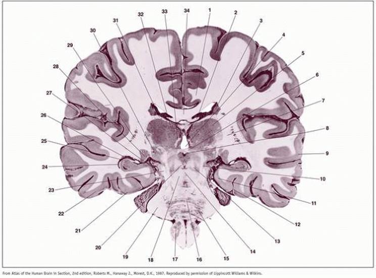
Plate 76 Coronal Section through the Posterior Limb of the Internal Capsule, Mamillothalamic Tract (MTT), Mamillary Body, and Hippocampal Formation |
Coronal section through the posterior limb of the internal capsule, mamillothalamic tract (MTT), mamillary body, and hippocampal formation. Note the MTT entering the anterior ventral nucleus. The optic tracts are visible bilaterally. Claustrum and third ventricle.
1. Cingulate artery
2. Body of fornix
3. Interthalamic adhesion (massa intermedia)
4. Anteroventral nucleus of thalamus
5. Stria terminalis and thalamostriate vein
6. Tail of caudate nucleus
7. Lateral ventral nucleus of thalamus
8. Mamillothalamic tract
9. Claustrum
10. External capsule
11. Lenticulostriate branches of middle cerebral artery
12. Globus pallidus II
13. Globus pallidus I
14. Zona incerta
15. Subthalamic nucleus
16. Substantia nigra
17. Cerebral peduncle
18. Posterior cerebral artery
19. Principal mamillary fasciculus (bundle)
20. Basilar artery
21. Pons
22. Interpeduncular fossa
23. Mamillary body
24. Third ventricle
25. Hippocampal sulcus
26. Hippocampus
27. Alveus of hippocampus
28. Inferior horn of lateral ventricle
29. Amygdala and nucleus basalis
30. Optic tract
31. Internal medullary lamina of globus pallidus
32. External medullary lamina of globus pallidus
33. Extreme capsule
34. Putamen
35. Posterior limb of internal capsule
36. Rostral peduncle of thalamus
37. Tegmental area H2
38. Velum interpositum
|
|
|
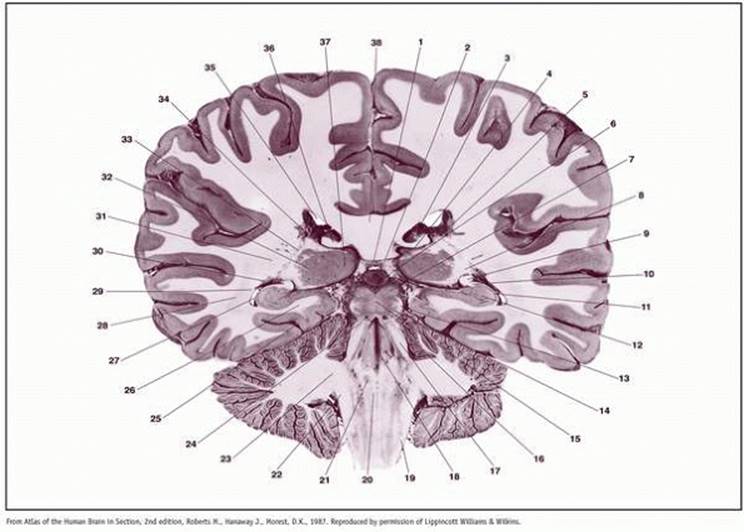
Plate 77 Coronal Section through the Thalamus, Ventral Posteromedial Nucleus (VPM) and the Ventral Posterolateral Nucleus (VPL), Posterior Limb of the Internal Capsule, Substantia Nigra, and Red Nucleus |
Coronal section through the thalamus, ventral posteromedial nucleus (VPM) and the ventral posterolateral nucleus (VPL), posterior limb of the internal capsule, substantia nigra, and red nucleus. The optic tract lies dorsal to the temporal horn of the lateral ventricle. A lesion of the red nucleus involves the ipsilaterial intra-axial oculomotor fibers and the contralateral dentatorubrothalamic fibers from the cerebellum. This is Benedikt syndrome, characterized by a third nerve palsy and a contralateral cerebellar ataxia.
1. Third ventricle
2. Dorsal medial nucleus of thalamus
3. Body of lateral ventricle
4. Rostral peduncle of thalamus
5. Corona radiata
6. Posterior limb of internal capsule
7. Circular sulcus of insula
8. Claustrum
9. Centromedian nucleus of thalamus
10. Medial longitudinal fasciculus (bundle)
11. Globus pallidus II
12. Optic radiation
13. Tail of caudate nucleus
14. Stria terminalis
15. Optic tract
16. Uncus
17. Cerebral peduncle
18. Red nucleus
19. Habenulo-interpeduncular tract (fasciculus retroflexus)
20. Medial nucleus of pons
21. Interpeduncular fossa
22. Middle cerebellar peduncle
23. Interpeduncular nucleus
24. Substantia nigra
25. Posterior cerebral artery
26. Hippocampus
27. Medullary lamina of red nucleus
28. Medial lemniscus
29. Posteromedial ventral nucleus of thalamus
30. Putamen
31. Posterolateral ventral nucleus of thalamus
32. Central sulcus
33. Posterior lateral nucleus of thalamus
34. Tail of caudate nucleus
35. Superior occipitofrontal fasciculus (superior occipitofrontal bundle)
36. Stria terminalis
37. Lateral dorsal nucleus of thalamus
38. Body of fornix
|
|
|
Plate 78 Coronal Section through the Lateral and Medial Lemnisci, Lateral and Medial Geniculate Nuclei, and Hippocampal Formation |
Coronal section through the lateral and medial lemnisci, lateral and medial geniculate nuclei, and hippocampal formation
1. Cingulum
2. Crus of fornix
3. Tail of caudate nucleus
4. Third ventricle
5. Posterior lateral nucleus of thalamus
6. Posterior commissure
7. Posterior limb of internal capsule
8. Cerebral aqueduct
9. Superior temporal gyrus
10. Medial lemniscus
11. Parahippocampal gyrus
12. Collateral sulcus (rhinal fissure)
13. Central tegmental bundle
14. Decussation of superior cerebellar peduncles
15. Pontine nuclei
16. Raphe of pons
17. Inferior olivary nucleus
18. Superior cerebellar peduncle
19. Middle cerebellar peduncle
20. Medial longitudinal fasciculus (bundle)
21. Lateral occipitotemporal gyrus
22. Inferior temporal gyrus
23. Lateral lemniscus
24. Inferior horn of lateral ventricle
25. Alveus of hippocampus
26. Tail of caudate nucleus
27. Posterior cerebral artery
28. Lateral geniculate body
29. Transcapsular caudatolenticular gray striae
30. Medial geniculate body
31. Rostral peduncle of thalamus
32. Dorsal medial nucleus of thalamus
33. Habenulo-interpeduncular tract (fasciculus retroflexus)
34. Body of corpus callosum
|
|
|
Plate 79 Coronal Section through the Pulvinar Nuclei, Pineal Gland (Epiphysis), Superior and Inferior Colliculi, and Trochlear Nerve (CN IV) |
Coronal section through the pulvinar nuclei, pineal gland (epiphysis), superior and inferior colliculi, and trochlear nerve (CN IV). The abducent nucleus is seen in the dorsomedial pontine tegmentum; it is surrounded by exiting root fibers of the fascial nerve (CN VII); the facial colliculus (FC) is found in the floor of the rostral pontine tegmentum. A lesion of the FC results in a central paralysis of the CN VII, a complete abducent paralysis, and a medial rectus palsy on attempted lateral conjugate gaze.
1. Velum interpositum
2. Internal cerebral vein
3. Choroid plexus of lateral ventricle
4. Choroidal fissure
5. Tail of caudate nucleus
6. Pineal gland
7. Superior colliculus
8. Triangular area
9. Fimbria of hippocampus
10. Alveus of hippocampus
11. Hippocampus (Sommer's sector)
12. Dentate gyrus
13. Brachium of inferior colliculus
14. Inferior colliculus
15. Commissure of inferior colliculus
16. Uncinate fasciculus (bundle)
17. Superior cerebellar peduncle
18. Median sulcus
19. Dorsal spinocerebellar tract
20. Medial longitudinal fasciculus (bundle)
21. Nucleus of abducent nerve (VI)
22. Inferior cerebellar peduncle
23. Middle cerebellar peduncle
24. Trochlear nerve (IV)
25. Collateral sulcus (rhinal fissure)
26. Cingulum
27. Inferior horn of lateral ventricle
28. Optic radiation
29. Tapetum
30. Tail of caudate nucleus
31. Retrolenticular part of internal capsule
32. Lateral pulvinar nucleus of thalamus
33. Transcapsular caudatolenticular gray striae
34. Thalamostriate vein
35. Body of lateral ventricle
36. Crus of fornix
37. Medial pulvinar nucleus of thalamus
38. Splenium of corpus callosum
|
|
|
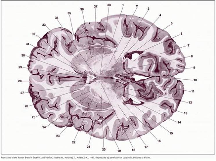
Plate 80 Axial Section through the Internal Capsule, Anterior Commissure, and Pulvinar Nuclei |
Axial section through the internal capsule, anterior commissure, and pulvinar nuclei. Locate the visual radiation and the basal ganglia: caudate nucleus, globus pallidus and putamen; caudate nucleus + putamen = lentiform nucleus, caudate nucleus + putamen = striatum.
1. Circular sulcus of insula
2. Lateral ventral nucleus of thalamus
3. Dorsal medial nucleus of thalamus
4. Tail of caudate nucleus
5. Fimbria of hippocampus
6. Tapetum
7. Optic radiation
8. Retrosplenial gyrus of hippocampus
9. Stria medullaris of thalamus
10. Longitudinal cerebral fissure (interhemispheric fissure)
11. Great cerebral vein
12. Medial pulvinar nucleus of thalamus
13. Choroidal fissure
14. Lateral pulvinar nucleus of thalamus
15. Choroid plexus of lateral ventricle
16. Triangular area
17. Retrolenticular part of internal capsule
18. Mamillothalamic tract
19. Branch of middle cerebral artery
20. Posterior limb of internal capsule
21. Long gyrus of insula
22. Globus pallidus I
23. Short gyri of insula
24. Globus pallidus II
25. Internal medullary lamina of globus pallidus
26. Anterior limb of internal capsule
27. Lateral preoptic nucleus
28. Column of fornix
29. Cingulate gyrus
30. Anterior commissure
31. Third ventricle
32. Head of caudate nucleus
33. Putamen
34. Paraventricular nucleus of hypothalamus
35. Lateral cerebral fissure
36. External medullary lamina of globus pallidus
37. Temporal operculum
38. Interthalamic adhesion (massa intermedia)
|
|
|
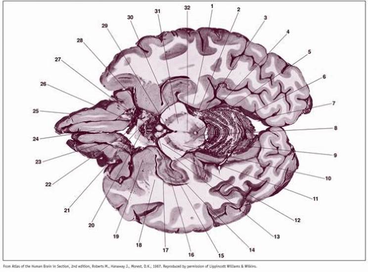
Plate 81 Axial Section through the Mamillary Nuclei and the Superior Colliculi |
Axial section through the mamillary nuclei, and the superior colliculi. In Wernicke's encephalopathy, one sees petechial hemorrhages in the mamillary nuclei. In Parkinson disease, the substantia nigra shows a loss of pigmented neurons. In Benedikt syndrome, a lesion of the red nucleus, the following structures are lesioned: root fibers of the oculomotor nerve (CN III), dentatorubrothalamic fibers, and the medial lemniscus.
1. Inferior horn of lateral ventricle
2. Choroid plexus of lateral ventricle, inferior horn
3. Medial geniculate body
4. Red nucleus
5. Medial longitudinal fasciculus (bundle)
6. Superior colliculus
7. Central gray matter
8. Superior cistern
9. Tegmental reticular formation (lateral mesencephalic nucleus)
10. Substantia nigra
11. Cerebral peduncle
12. Dentate gyrus
13. Tapetum
14. Hippocampus
15. Fimbria of hippocampus
16. Mamillary body
17. Principal mamillary fasciculus (bundle)
18. Branch of middle cerebral artery
19. Lateral cerebral fissure
20. Optic tract
21. Tuber cinereum
22. Medial orbital gyrus
23. Straight gyrus
24. Anterior cerebral artery
25. Anterior perforated substance
26. Mamillothalamic tract
27. Uncus
28. Amygdala
29. Anterior commissure
30. Lateral geniculate body
31. Tail of caudate nucleus
32. Alveus of hippocampus
|
|
|
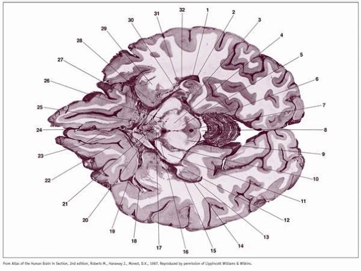
Plate 82 Axial Section through the Mamillary Nuclei, Inferior Colliculi, and Optic Chiasm |
Axial section through the mamillary nuclei, inferior colliculi, and optic chiasm. The amygdala and hippocampal formations are outstanding; lesions of the amygdala and hippocampal formation result in memory loss. Plates 72, 73, 74, 75, 76, 77, 78, 79, 80, 81 and 82 show normal brains with no neuropathology. Lead lines point to normal structures which in time will demonstrate pathology (e.g., lead line points to the normal substantia nigra). In Parkinson disease there is a depopulation of melanin-containing neurons in the substantia nigra and in the locus ceruleus.
1. Parahippocampal gyrus
2. Lateral lemniscus
3. Cisterna ambiens
4. Brachium of inferior colliculus
5. Cerebral aqueduct
6. Vermis of cerebellum
7. Calcarine sulcus
8. Central gray matter
9. Inferior colliculus
10. Medial lemniscus
11. Substantia nigra
12. Frontopontine tract
13. Parietotemporo-occipitopontine tract
14. Dentate gyrus
15. Hippocampus
16. Mamillary body
17. Alveus of hippocampus
18. Anterior commissure
19. Amygdala
20. Infundibulum
21. Optic chiasm
22. Anterior cerebral artery
23. Olfactory sulcus
24. Straight gyrus
25. Medial orbital gyrus
26. Olfactory tract
27. Optic tract
28. Temporal pole
29. Interpeduncular fossa
30. Corticospinal tract