
The cerebrum is the largest part of the human brain that fills most of the cranial cavity. Its large size is the result of a progressive (telencephalization) centralization of the various higher sensory and motor centres of the brain during evolution.
The cerebrum is a heavily, convoluted bilobed structure (Fig. 12.1). The two lateral halves are called cerebral hemispheres. When the two cerebral, hemispheres are viewed together from above, they assume the shape of an ovoid mass, which is broader behind than in front. The widest transverse diameter corresponds with a line connecting the two parietal tuberosities.
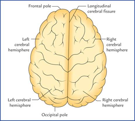
FIG. 12.1 Superior view of the cerebrum.
A deep median cleft, the longitudinal cerebral fissure, incompletely separates the two cerebral hemispheres. Both in front and behind, the cleft is complete, but in the central part the cleft extends downwards up to the corpus callosum which is a large mass of white fibres joining the two cerebral hemispheres across the median plane.
The longitudinal cerebral fissure is occupied by the following structures:
1. Falx cerebri (a sickle-shaped fold of dura mater).
2. Fold of arachnoid that follows the surfaces of the falx cerebri.
3. Pia mater covering the medial surface of the falx cerebri.
4. Anterior cerebral arteries and veins (which lie in the subarachnoid space between the arachnoid and the pia).
Each cerebral hemisphere consists of: (a) an outer layer of grey matter called cerebral cortex, (b) an inner mass of white matter, (c) large masses of grey matter embedded in the basal part of the white matter called basal ganglia/basal nuclei, and (d) a cavity within it called lateral ventricle (Fig. 12.2).

FIG. 12.2 Coronal section of the cerebral hemisphere showing its structure. Also note the borders and surfaces of the cerebral hemisphere. (CN = caudate nucleus, T = thalamus, P = putamen, C = claustrum, A = amygdaloid body.)
External Features of the Cerebral Hemisphere
The external features of the cerebral hemisphere include poles, surfaces, borders, sulci, and gyri.
Poles (Figs 12.2 and 12.3)
Each cerebral hemisphere presents three poles – frontal, occipital, and temporal. The anterior end of the hemisphere is the frontal pole and the posterior end the occipital pole. The temporal pole is below and in front at the junction of orbital and tentorial surfaces.
• The frontal pole at the anterior end of the hemisphere is more rounded than the occipital pole. It lies opposite the medial part of the superciliary arch.
• The occipital pole at the posterior end of the hemisphere is more pointed than the frontal pole. It lies at a short distance superolateral to the external occipital protuberance.
• The temporal pole between frontal and temporal poles points forwards. It fits into the anterior part of the middle cranial fossa and is overhung by the lesser wing of the sphenoid.
N.B. The temporal pole is the primitive posterior pole that has curved ventrocaudally for better accommodation during the growth of the cerebrum.
Surfaces
Each cerebral hemisphere has three surfaces – superolat-eral, medial, and inferior (Fig. 12.2).
1. The superolateral surface is most convex and most extensive. It faces upwards and laterally and conforms to the corresponding half of the cranial vault.
2. The medial surface is flat and vertical. It presents a thick C-shaped cut surface of the corpus callosum.
3. The inferior surface is irregular to adopt the floors of anterior and middle cranial fossae. It is divided into two parts by a deep horizontal groove or sulcus, the stem of lateral sulcus, viz. (a) a small anterior part, the orbital surface, and (b) a large posterior part, the tento-rial surface.
Borders
Each cerebral hemisphere presents six borders (Figs 12.2 and 12.3), viz. superomedial, superciliary, inferolateral, medial orbital, medial occipital and inferomedial.

FIG. 12.3 Inferior aspect of cerebral hemisphere showing borders, surfaces, and poles.
1. The superomedial border separates the superolateral surface from the medial surface.
2. The superciliary border is at the junction of superolateral and orbital surfaces. It lies just behind the superciliary arch hence its name strictly speaking, it is the orbital part of the inferolateral border.
3. The inferolateral border separates the superolateral surface from the tentorial surface. Posteriorly this border exhibits a notch, the preoccipital notch about 3 cm in front of the occipital pole. This notch is used as a useful surface landmark.
4. The medial orbital border (Fig. 12.3) separates the medial surface from the orbital surface.
5. The inferomedial/hippocampal border (Fig. 12.3) surrounds the cerebral peduncle. It is formed by the medial aspect of the uncus and parahippocampal gyrus.
6. The medial occipital border (Fig. 12.3) separates the medial surface from the tentorial surface.
Sulci and Gyri
The cerebral cortex (the surface layer of grey matter) is highly extensive in man. To accommodate it in the limited space available within the rigid cranial cavity, the surface of cerebral hemisphere becomes folded, producing numerous convolutions separated by fissures. These convolutions and fissures are termed gyri and sulci respectively. In human brain the total surface area of cerebral hemisphere is about 2000 cm but approximately two-third of this is hidden from the surface view within the walls of the sulci.
A brain with convoluted cerebral cortex is termed gyrencephalic while the one with smooth cortex, lissen-cephalic (Gk. lissos = smooth).
In general, man and other higher mammals have gyren-cephalic brain while the reptiles, birds and lower mammals have lissencephalic brain.
The sulci vary in depth from slight grooves to deep fissures and some of them are sufficiently deep to indent the wall of the lateral ventricle in the depth of the hemisphere.
The gyri consist of a central core of white matter (nerve fibres running to and from the overlying cortex) covered by a layer of grey matter, the cerebral cortex.
There is a great deal of individual variations in the details of sulci and gyri. Therefore, the following account deals with only some important sulci and gyri.
Main cerebral sulci
Main cerebral sulci are fairly constant in position and shape and include lateral, central, parieto-occipital and calcarine sulci.
Lateral sulcus (of Sylvius) (Figs 12.4, 12.6)
Lateral sulcus is the most conspicuous of all the cerebral sulci and has a stem and three rami. The stem of the sulcus begins as a deep cleft on the inferior surface of the cerebral hemisphere at the anterior perforated substance and extends laterally between the temporal pole and the posterior part of the orbital surface of the hemisphere. On reaching the superolateral surface it divides into three rami: (a) anterior horizontal, (b) anterior ascending, and (c) posterior.
The anterior horizontal ramus is about 2.5 cm long and passes forwards into the inferior frontal gyrus. The anterior ascending ramus runs upwards for about 2.5 cm in the same gyrus. The posterior ramus (the main part of the sulcus) is about 7.5 cm long and runs posteriorly and slightly upwards across the lateral surface and ends in the inferior parietal lobule by an upturned posterior end.
N.B. The three rami of lateral sulcus diverge from each other at a point called Sylvian point.
Central sulcus (of Rolando) (Fig. 12.4)
Central sulcus begins by cutting the superomedial border of the hemisphere about 1 cm behind the midpoint between the frontal and occipital poles, runs sinuously downwards and forwards at an angle of 70° and ends just above the posterior ramus of the lateral sulcus. Its upper end usually extends into the medial surface. The central sulcus forms the boundary between the motor area of the cerebral hemisphere in front and the sensory area behind.
N.B. It is the only sulcus of any length that indents the superomedial border of the hemisphere. This sulcus serves as a key to localize the other sulci and gyri for the pathologist while performing an autopsy.
Calcarine sulcus
Calcarine sulcus is present on the medial surface of the cerebral hemisphere. It begins as a deep fissure, a little below the posterior end of the corpus callosum, the sple-nium and follows an arched course with a convexity upwards to the occipital pole and may extend slightly on to the superolateral surface.
Parieto-occipital sulcus
Parieto-occipital sulcus is present on the medial surface of the hemisphere. It begins at the midpoint of the calcarine sulcus and courses upwards and slightly backwards to cut the superomedial border of the hemisphere about 5 cm in front of the occipital pole, and may extend slightly on to the superolateral surface.
Lobes of Cerebral Hemisphere (Fig. 12.4)
To discuss further about sulci and gyri and other aspects of the cerebral hemisphere, the superolateral surface of the hemisphere is arbitrarily divided into four lobes – frontal, parietal, temporal and occipital with the help of: (a) three main sulci, central, lateral and parieto-occipital, and (b) two imaginary lines. The first imaginary line is a vertical line joining the parieto-occipital sulcus to the preoccipital notch, and the second line is a backward continuation of the horizontal part of the posterior ramus of the lateral sulcus till it joins the first line (Fig. 12.4).

FIG. 12.4 Division of superolateral surface of the left cerebral hemisphere into four lobes.
The frontal lobe lies anterior to the central sulcus, and above the posterior ramus of the lateral sulcus.
The parietal lobe lies behind the central sulcus and in front of the upper part of the first imaginary line. Below it is bounded by the posterior ramus of lateral sulcus and the second imaginary line.
The temporal lobe lies below the posterior ramus of lateral sulcus and second imaginary line. It is separated from the occipital lobe by the lower part of the first imaginary line.
The occipital lobe lies behind the vertical line joining the parieto-occipital sulcus and preoccipital notch.
Insula/island of Reil (also called central lobe)
It is customary to consider the insula separately from the four main lobes (vide supra) of the cerebral hemisphere.
The insula is the submerged (hidden) portion of the cerebral cortex in the floor of the lateral sulcus (Fig. 12.5). It has been submerged from the surface during development of brain due to the overgrowth of the surrounding cortical areas and can be seen only when the lips of the lateral sulcus are widely pulled apart. It is triangular in shape and surrounded all around by a sulcus, the circular sulcus except anteroinferiorly at its apex called limen insulae which is continuous with the anterior perforated substance.
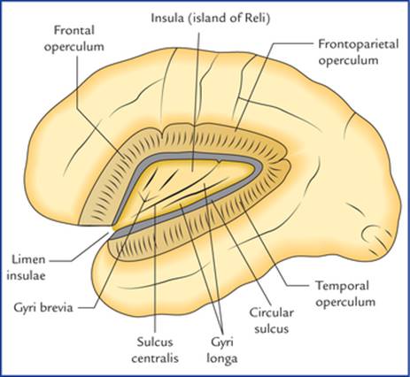
FIG. 12.5 The insula (island of Reil) exposed by removing the opercula. Note: Insula is also called ‘central lobe.’

FIG. 12.6 Superolateral surface of the left cerebral hemisphere showing lobes, sulci and gyri. (SFG = superior frontal gyrus, MFG = middle frontal gyrus, IFG = inferior frontal gyrus (a. pars orbitalis, b. pars triangularis, c. pars opercularis), STG = superior temporal gyrus, MTG = middle temporal gyrus, ITG = inferior temporal gyrus, IPL = inferior parietal lobule.)
The insula is divided into two regions – anterior and posterior by a central sulcus. The anterior region presents 3 or 4 short gyri called gyri brevia and the posterior region presents 1 or 2 long gyri called gyri longa.
The insula is hidden from the surface view by the overgrown cortical areas of frontal, parietal and temporal lobes. These areas are termed frontal, frontoparietal and temporal opercula (operculum = lid). The superior surface of the temporal operculum presents anterior and posterior transverse temporal gyri.
The middle cerebral artery and deep middle cerebral vein lie on the surface of the insula.
Sulci and Gyri on the Superolateral Surface of the Cerebral Hemisphere (Fig. 12.6)
In the frontal lobe
• The prefrontal sulcus often broken into two or three parts, runs downwards and forwards parallel and little anterior to the central sulcus. The area between the central and precentral sulci is called precentral gyrus.
• Anterior to the precentral sulcus there are two sulci called superior and inferior frontal sulci which run horizontally. These sulci divide the region of frontal lobe in front of precentral sulcus into superior, middle, and inferior frontal gyri.
• The anterior and ascending rami of lateral sulcus divide the inferior frontal gyrus into three parts. The part below the anterior ramus is called pars orbitalis, the part between the anterior and ascending rami the pars trian-gularis and the part posterior to the ascending ramus, the pars opercularis.
In the parietal lobe
• The postcentral sulcus runs downwards and forwards, a little behind and parallel to the central sulcus. The area between these two sulci is called the postcentral gyrus.
• The rest of the parietal lobe is divided into a superior and inferior parietal lobules by an intraparietal sulcus which runs horizontally backwards from the postcentral sulcus.
• The upturned posterior end of the posterior ramus of lateral sulcus, and the posterior ends of superior and inferior temporal sulci extends into the inferior parietal lobule to divide it into three parts: (a) the part that surrounds the posterior ramus of lateral sulcus is called supra marginal gyrus, (b) the part surrounding the superior temporal sulcus, the angular gyrus, and (c) the part surrounding the inferior temporal sulcus, the arcus temporo-occipitalis.
In the temporal lobe
There are two sulci in this lobe that run parallel to the posterior ramus of the lateral sulcus. These are termed superior and inferior temporal sulci, and divide the temporal lobe into superior, middle and inferior temporal gyri.
The superior surface of superior temporal gyrus presents two transverse temporal gyri. The anterior transverse temporal gyrus also called Heschl's gyrus forms the primary auditory area of the cortex.
In the occipital lobe
The occipital lobe possesses rather three short sulci, lateral and transverse occipital sulci and lunate sulcus.
1. Lateral occipital sulcus runs horizontally and divides this lobe into superior and inferior occipital gyri.
2. Lunate sulcus is C-shaped sulcus with forward convexity just in front of the occipital pole.
3. Transverse occipital sulcus runs downwards into the uppermost part of the occipital lobe from the supero-medial border of hemisphere, a little behind the parieto-occipital sulcus.
N.B. The cortex surrounding the parieto-occipital sulcus on the superolateral surface is termed arcus parieto-occip-italis. It lies in both parietal and occipital lobes.
Sulci and Gyri on the Medial Surface of the Cerebral Hemisphere (Fig. 12.7)
The presence of corpus callosum is the most conspicuous feature seen on the medial surface of the cerebral hemisphere. It is C-shaped thick bundle of commissural fibres. It consists of a central part, the trunk, a thick posterior end, the splenium and curved anterior end, the genu.
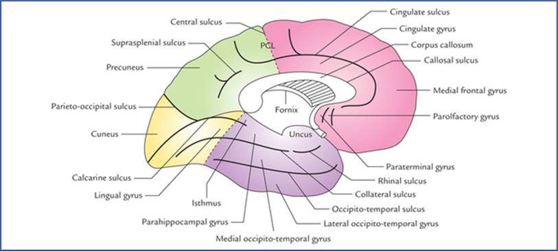
FIG. 12.7 Medial surface of the left cerebral hemisphere showing lobes, sulci and gyri. (Note: In this figure tentorial surface of hemisphere is also seen.) The interrupted lines indicate the approximate boundaries of the lobes. (PCL = paracentral lobule.)
The sulci and gyri on the medial surface are located above, in front and behind the corpus callosum.
These are as follows:
• Cingulate sulcus: It is the most prominent sulcus which follows a curved course about 1 cm above and parallel to the upper convex margin of the corpus callosum. Anteriorly it ends below the genu of corpus callosum, posteriorly it turns upwards to reach the superomedial border of the hemisphere a little behind the upper end of the central sulcus.
The area between the cingulate sulcus and the corpus callosum is termed cingulate gyrus.
• Just in front of lamina terminalis, there are paraterminal and parolfactory gyri and anterior and posterior parol-factory sulci.
• Callosal sulcus. It separates the cingulate gyrus from the corpus callosum.
• The part of medial surface between the cingulate sulcus and the superomedial border of hemisphere is divided by a short offshoot sulcus ascending from the cingulate sulcus above the middle of the trunk of corpus callosum into two parts:
– a small part around the upper part of the central sulcus, the paracentral lobule, and
– a larger medial part the medial frontal gyrus.
The boundaries of paracentral lobule needs to be elaborated, as it is the cortical (highest) centre of micturition and defecation. The paracentral lobule is bounded above, by the superomedial border of the hemisphere, below by the cingulate sulcus, and posteriorly by the upturned posterior end of the cingulate sulcus. The paracentral lobule is invaded by the downturned upper end of the central sulcus.
• The posterior part of medial surface behind the para-central lobule has two main sulci: the calcarine sulcus, and the parieto-occipital sulcus.
(a) Calcarine sulcus. It is already described on page 142. A small region between the splenium and calcarine sulcus is termed isthmus.
(b) Parieto-occipital sulcus. It is already described on page 142.
The triangular area between the posterior part of the cal-carine sulcus (also called postcalcarine sulcus) and the parieto-occipital sulcus is called cuneus.
The quadrangular area between the parieto-occipital sul-cus and paracentral lobule is termed precuneus.
A small sulcus a little above and parallel to the splenium is called suprasplenial sulcus. It separates the precuneus from the cingulate gyrus.
Sulci and Gyri on the Inferior Surface of the Cerebral Hemisphere (Fig. 12.8)
On the orbital part of inferior surface (orbital surface)

FIG. 12.8 Inferior surface of the left cerebral hemisphere showing various sulci and gyri. Note: Inferior surface is divided into small anterior part, the orbital surface and large posterior part, the tentorial surface. The midbrain has been cut across.
• Olfactory sulcus: It is a straight sulcus which runs antero-posteriorly close to the medial border of the orbital surface. It is called olfactory sulcus because it lodges the olfactory bulb and tract.
The area medial to this sulcus is called gyrus rectus.
• Orbital sulcus: It is an irregular H-shaped sulcus and divides the rest of the orbital surface into anterior, posterior, medial and lateral orbital gyri.
On the tentorial part of inferior surface (tentorial surface)
The tentorial surface is marked by two major sulci that run anteroposteriorly. The medial one is called collateral sulcus and the lateral one, the occipito-temporal sulcus. The latter is continuous around the inferolateral margin with the inferior temporal gyrus.
Posteriorly the collateral sulcus is parallel to the calcar-ine sulcus and here the area between these two sulci is termed lingual gyrus. Anteriorly the lingual gyrus is continuous with the parahippocampal gyrus. Anterior end of parahippocampal gyrus hooks sharply backwards and is limited laterally by a short rhinal sulcus. This hook-like anterior end of parahippocampal gyrus is called uncus.
Posteriorly the parahippocampal gyrus is continuous with the cingulate gyrus through the isthmus.
The area between the occipito-temporal sulcus laterally and the collateral and rhinal sulci medially is known as medial occipito-temporal gyrus. The area lateral to the occipito-temporal sulcus is termed lateral occipito-temporal gyrus. This gyrus is continuous around the infero-lateral margin of the hemisphere with the inferior temporal gyrus.
The Cerebral Cortex
The cerebral cortex is the surface layer of grey matter covering the cerebral hemisphere. The cortex represents the highest degree of evolutionary development of the human brain both in its relative size to other parts of the brain and its range of functions. It is principally responsible for three basic brain functions, viz. perception, adaptation responses, and movement which are common in all animals. In addition it is the site for various types of mental activities like memory, learning, speech and language abilities, intelligence and creative thinking.
In general each cerebral hemisphere controls the opposite half of the body, i.e. it sends motor commands to, and receives sensory information from, the contralateral half of the body. The cortex is highly folded into intricate convexities and grooves to increase its surface area in order to accommodate the large volume of grey matter in the limited space within the cranium which provides for the wide range of mental activities (vide supra) and forms the basis for man's superiority over other animals.
Types of Cerebral Cortex
The cortical areas vary in their phylogenetic scale. The oldest parts form archicortex, the recent parts neocortex, and those of intermediate origin paleocortex.
The archicortex and paleocortex together constitute the allocortex (= other cortex).
• Archicortex: Phylogenetically it is the oldest and constitutes the sizeable portion of the cerebrum of lower vertebrates. In man this rather primitive cortex is represented by the hippocampus and parts of rhinencephalic regions. Structurally it is simple and made up of three layers.
• Paleocortex: Phylogenetically it is intermediate in development and in man it is represented by the cingulate gyrus.
• Neocortex: Phylogenetically it is most recent in development and in man it comprises about 90% of the total area of the cerebral cortex. Structurally it is thick and consists of six layers.
Structure of Cerebral Cortex
The cerebral cortex mainly consists of nerve cells, synapses, and neuroglia. The human cerebral cortex contains enormous number (about 14000 millions) of neurons.
Types of Neurons in the Cerebral Cortex
The following five most conspicuous types of neurons present in the cerebral cortex are: (a) pyramidal cells, (b) stellate/granule cells, (c) horizontal cells of Cajal, (d) cells of Martinotti, and (e) fusiform cells (Fig. 12.9). The first two types of cells are the main ones.

FIG. 12.9 Five main types of neurons in the cerebral cortex.
1. The pyramidal cells are pyramidal in shape with their apices directed towards the surface. The axon arises from the base of the cell body while dendrites arise from its apex and basal angles. The axons enter the white matter as projection fibres.
According to the size of their cell bodies, they are called small (10 (μm), medium (50 (μm) and large (100–125 (μm) pyramidal cells. The latter are also termed large pyramidal cells of Betz (or only Betz cells).
2. The stellate/granule cells are much smaller than pyramidal cells and their cell bodies measure about 8 xm in diameter. They have star-shaped bodies with short axons and many dendrites. These cells are so small that they appear like granules in Nissl-stained material, hence the name, granule cells.
In certain areas of the cerebral cortex, they are so numerous that they resemble a cloud of dust particles and cerebral cortex in these areas is called koniocortex (Gk. Konios = cloud).
3. The fusiform cells have fusiform cell body with their long axis being vertical to the surface. They are concentrated in the deepest cortical layers.
4. The horizontal cells of Cajal are fusiform and oriented horizontally. They are found in the most superficial layer of the cortex.
5. The cells of Martinotti are small multipolar cells that are present throughout the layers of cerebral cortex.
The cortical neurons connect with other neurons in following three ways:
1. Projection neurons transmit impulses to the subcortical centres, viz. corpus striatum, thalamus, brainstem, or spinal cord.
2. Association neurons establish connections with the cortical nerve cells elsewhere in the same cerebral hemisphere.
3. Commissural neurons proceed to the cortex of the opposite cerebral hemisphere and establish connections between the cortical nerve cells of two cerebral hemispheres.
Layers of the Cerebral Cortex (Fig. 12.10)
The neocortex consists of six layers or laminae. From superficial to deep these are as follows:
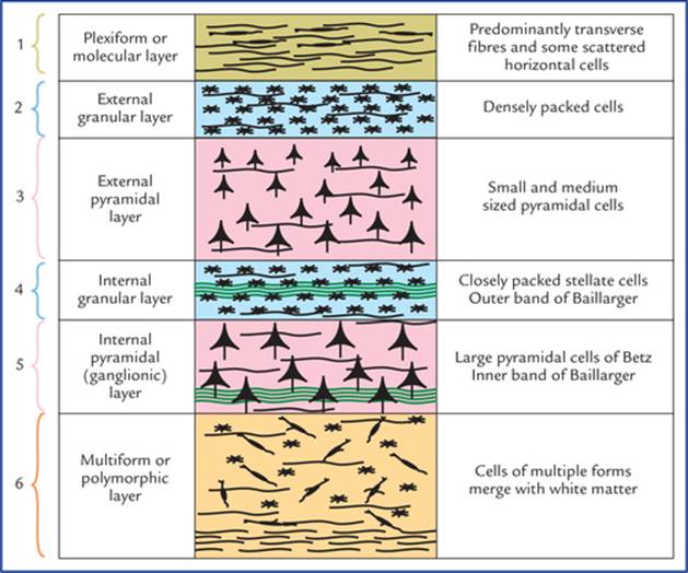
FIG. 12.10 The six layers of the cerebral cortex.
1. Molecular (plexiform) layer is made up predominantly of nerve fibres and few scattered horizontal cells of Cajal.
2. External granular layer is made up mainly of densely packed stellate (granule) cells.
3. External pyramidal layer is made up of small and medium sized pyramidal cells. The sizes of cells increase from superficial to deeper borders of the layer.
4. Internal granular layer is made up of closely packed stellate (granule) cells. In the middle of this layer there is a band of horizontally arranged white fibres called white stria or external band of Baillarger. The white stria is most marked in the visual cortex, hence visual cortex is also called striate cortex.
5. Internal pyramidal (ganglionic) layer is made up mainly of large pyramidal cells (of Betz). The basal part of this layer contains a thin band of horizontally arranged fibres called inner band of Baillarger. These cells account for about 3% of the projection fibres of the corticospinal (pyramidal) tract.
6. Multiform layer (or layer of polymorphic cells) is made up of cells of multiple forms (i.e. neurons of various sizes and shapes). This layer fuses with the white matter.
Variations in the Cortical Structure
The cerebral cortex shows considerable variation in its structure from region to region both in terms of thickness and prominence of various layers described above. Based on structural variation, the cerebral cortex is classified into following types:
1. Homotypical cortex: In this type, all the six layers of cortex are well defined.
2. Heterotypical cortex: In this type, all the six layers are not well defined (i.e. there are less than six layers). The heterotypical cortex is further divided into following two types:
(a) Granular cortex: In this type, the granular layers are well developed while the pyramidal layers are poorly developed. Since the density of granule cells is very high, it is termed granular cortex. The granular cortex is the characteristic feature of the sensory areas, viz. primary sensory, acoustic, and visual areas.
(b) Agranular cortex: In this type, the granular layers are poorly developed while pyramidal layers are well developed with densely packed pyramidal cells. The agranular cortex is the characteristic feature of the motor areas, viz. primary motor and other areas of the frontal lobe. The cortex is named agranular due to paucity or absence of granule cells.
Functional Areas of the Cerebral Cortex
The cerebral cortex is demarcated into large number of areas which differ from each other in structure as well as in function. Brodmann (1909) had divided the cerebral cortex into 47 such areas and indicated each of them by a number.
Types of cortical areas: According to the classical teaching, the cerebral cortex possesses three types of functional areas.
1. Motor areas: These areas are primarily concerned with the motor functions and give origin to the projection fibres which form corticospinal and corticonuclear tracts.
2. Sensory areas: These areas are primarily concerned with the sensory functions and receive afferent fibres from the thalamic nuclei in which major sensory pathways terminate.
3. Association areas: These areas are not concerned with primary motor or sensory functions but have more important associative, integrative and cognitive functions. Association areas occupy over 75% of the total surface area of the cerebral cortex in man.
Functional areas in the frontal lobe (Figs 12.11 and 12.12)
Primary motor area (area 4 of Brodmann)
Primary motor area is located in the precentral gyrus on the superolateral surface and extends to the anterior part of paracentral lobule on the medial surface of the cerebral hemisphere. It contains large number of pyramidal cells including large pyramidal cells (of Betz). About 40% pyramidal (corticospinal and corticonuclear) fibres arise from this area.
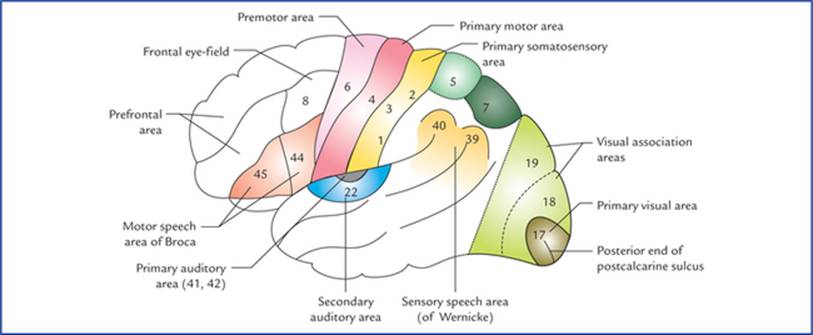
FIG. 12.11 The functional areas on the superolateral surface of the left cerebral hemisphere.
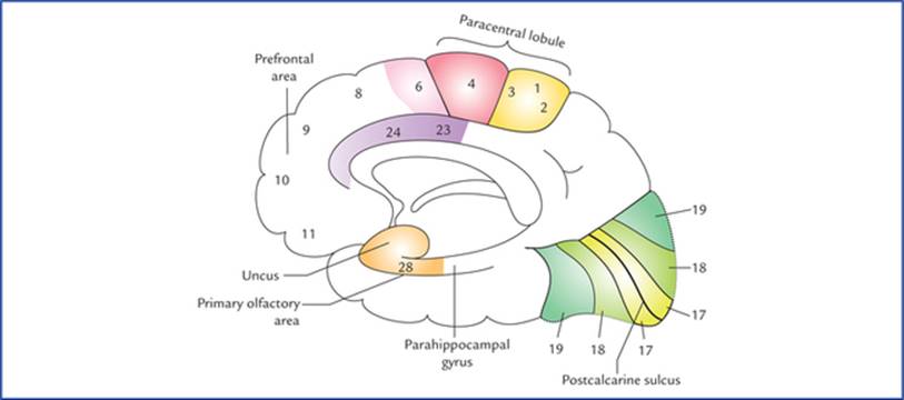
FIG. 12.12 The functional areas on the inferomedial surface of the right cerebral hemisphere.
The electrical stimulation of primary motor area produces contraction of muscles of (mainly) opposite half of the body, i.e. it controls voluntary motor activities of the opposite half of the body.
Although the cortical control of musculature of body is mainly contralateral, there is significant bilateral control of the muscles of the upper part of the face, tongue (genio-glossus), mandible, larynx, pharynx and axial musculature.
Specific regions within the area are responsible for movements in the specific parts of the body. Only movements are represented in this area and not the muscles.
The human body is represented in an upside down manner in the precentral gyrus (inverted homunculus) as shown in Figure 12.13. The pharyngeal region and tongue are represented in the lowermost part, followed by face, hand, trunk and thigh. The legs, feet and perineum are represented on the medial surface of the hemisphere in the paracentral lobule.

FIG. 12.13 The motor (A) and sensory (B) homunculi showing proportional somatotopical representation in the main motor and sensory cortex (after W. Penfield and T. Rasmussen, 1950).
Further, there is disproportionate registration of the individual parts. The area of cortex controlling a particular movement is proportional to the skill involved in performing that movement and not to the bulk of muscle participating in the movement. Thus, face especially the lips, tongue, larynx and hand have disproportionately larger areas while the trunk and lower limb have smaller areas.
Clinical Correlation
The lesions of primary motor area in one hemisphere produce flaccid paralysis of the extremities of the opposite half of the body (hemiplegia). The masticatory, laryngeal, pharyngeal, upper facial and extraocular muscles are spared for being represented bilaterally.
Premotor area (area 6 of Brodmann)
Premotor area is located anterior to the primary motor area in the posterior parts of superior, middle and inferior frontal gyri and extends on to the medial surface of the hemisphere. The premotor area is wider above than below and lacks the giant pyramidal cells (of Betz). It is the main site for the cortical origin of extrapyramidal (corticorubral, cortico-olivary, corticonigral, etc.) fibres.
The premotor area is responsible for successful performance of the voluntary motor activities. In fact, the primary motor area receives numerous inputs from the sensory cortex, the thalamus and the basal ganglia. It stores the programmes of motor activity assembled as a result of past experience. The premotor area thus appears to be responsible for programming the intended movements of the primary motor area, and controlling the movements in progress.
Clinical Correlation
The lesions of premotor (secondary motor) area produce difficulty in the performance of skilled movements.
N.B. The premotor and primary motor areas together are referred to as the primary somatomotor area (MsI) (Fig. 12.14).

FIG. 12.14 The main sensorimotor areas are projected diagram-matically upon the superolateral (B) and medial (A) surfaces of the cerebral hemisphere. Note: Primary (or first) somatomotor area includes both primary motor area and premotor area (MsI), the supplementary motor area is located in the medial frontal gyrus on the medial surface (MsII). Primary somatosensory area is located in postcentral gyrus (SmI) and second somatosensory area is located in the upper lip of the posterior ramus of lateral sulcus (SmII).
Supplementary motor area (MsII)
Supplementary motor area is located in the medial frontal gyrus on the medial surface of the hemisphere anterior to the paracentral lobule (Fig. 12.14). The body is represented from before backwards in craniocaudal order.
The stimulation of supplementary motor area produces complex movements, some are described as ‘assumption of posture’ with bilateral effects, including turning the head, assuming positions of trunk and lower limb, etc.
Clinical Correlation
Lesions of supplementary motor area produce bilateral flexor hypotonia with no paresis or paralysis.
Frontal eye field (area 8 of Brodmann)
The frontal eye field is located in the posterior part of the middle frontal gyrus just anterior to the facial area of the precentral gyrus. The electrical stimulation of this region causes deviation of both the eyes especially to the opposite side (conjugate movements of the eyes). The frontal eye field controls voluntary scanning movements of the eyes and is independent of the visual stimuli. The frontal eye field is connected to the visual area of occipital cortex by association fibres.
N.B. The involuntary following of moving objects by the eyes involves the visual area of the occipital cortex.
Clinical Correlation
Lesions of the frontal eye field of one hemisphere cause the two eyes to deviate to the side of lesion and an inability to turn the eyes to the opposite side. The involuntary tracking movement of the eyes when following moving objects is unaffected since the lesion does not involve the visual cortex of the occipital lobe.
Motor speech area of Broca (areas 44 and 45 of Brodmann)
The motor speech area of Broca is located in the pars triangularis (area 45) and pars opercularis (area 44) of inferior frontal gyrus of frontal lobe of left hemisphere (dominant hemisphere) in most of the individuals, and the persons are right handed. However, in 30% cases it is present in the right hemisphere and persons are left handed. Broca's area is responsible for the production of expressive speech/vocalization. It brings about the formation of words by its connections with the adjacent primary motor area, which in turn appropriately stimulate the muscles of the larynx, mouth, tongue, soft palate, and the respiratory muscles.
Clinical Correlation
The lesions of motor speech area of Broca result in loss of ability to produce speech, i.e. expressive aphasia (also called motor aphasia). The patients, however, retain the ability to think about the words they wish to say, they can write the words, and they can understand their meaning when they see or hear them.
Thus, although the language is understood, it cannot be expressed in speech even though there is no paralysis of muscles of lips, tongue, and vocal cords, etc.
Prefrontal area
The part of the frontal lobe rostral to the motor and pre-motor areas is referred to as prefrontal area. This area is concerned with the individual's personality. It exerts its influence in determining the initiative and judgement of an individual. It is also concerned with depth of emotions, social, moral and ethical awareness, concentration, orientation, and foresightedness.
It is an area of the cerebral cortex that is capable of associating experiences that are necessary for the production of abstract ideas.
Clinical Correlation
Bilateral destruction of prefrontal areas due to trauma or tumours results in profound change in personality. There is loss of concentration, initiative and judgement. The individual no longer conforms to the accepted mode of social behaviour and becomes careless about his dress and appearance (i.e. inappropriate social behaviour), e.g. use of obscene language, urinating in public.
Functional areas in the parietal lobe
Primary sensory area (areas 3, 1 and 2 of Brodmann)
Primary sensory area is located in the postcentral gyrus and extends into the posterior part of the paracentral lobule on the medial surface of the hemisphere. The opposite half of the body is represented up-side down exactly in same fashion as in the primary motor area (Fig. 12.13). Similarly, the area of cortex assigned for a particular part is not proportional to the size of that part but to its functional significance (i.e. to the intricacies of sensations received from it). Thus, the thumb, fingers, lips and tongue have a disproportionately large representation. The primary sensory area is concerned with the perception of extero-ceptive (pain, touch and temperature) and proprioceptive (vibration, muscle and joint sense) sensations from the opposite half of the body. However, sensations from pharynx, larynx and perineum go to both sides.
The primary sensory area receives projection fibres from ventral posterolateral (VPL) and ventral posteromedial (VPM) nuclei of the thalamus.
Clinical Correlation
Lesions of primary sensory area lead to the loss of appreciation of exteroceptive and proprioceptive sensations from the opposite half of the body. The crude pain, temperature and touch sensations often return, but this is believed to be due to functions of the thalamus.
Secondary sensory area (SmII)
Secondary sensory area is located in the upper lip of the posterior ramus of the lateral sulcus. The face area lies most anterior and the leg area is posterior. The whole body is represented bilaterally. This area relates more to the pain perception.
The ablation of this area may relieve intractable pain.
Sensory association area (Fig. 12.11)
Sensory association area occupies the superior parietal lobule corresponding to the Areas 5 and 7 of Brodmann. It is concerned with the perception of shape, size, roughness, and texture of the objects. Thus, it enables the individual to recognize the objects placed in his/her hand without seeing. Such ability is referred to as stereognosis.
Clinical Correlation
Lesions of this area result in inability to recognize or identify an object by its feel. This condition is called tactile agnosia or astereognosis.
Sensory speech area of Wernicke (Fig. 12.11)
Sensory speech area is located in the left dominant hemisphere occupying the posterior part of the superior temporal gyrus of temporal lobe and angular (area 39) and supramarginal (area 40) gyri of the inferior parietal lobule.
The Wernicke's area is concerned with the interpretation of language through visual and auditory input. It is also an essential zone for constant availability of the learned word patterns.
N.B. The sensory and motor speech areas exist together in one hemisphere only. The Wernicke's area is connected to the Broca's area by a bundle of nerve fibres called arcuate fasciculus.
Clinical Correlation
Lesions of Wernicke's area in the dominant hemisphere produce loss of ability to understand the spoken and written speech. This condition is called receptive sensory aphasia. Since Broca's area is unaffected, the expressive speech is unimpaired and the individual can produce a fluent speech. However, he is unaware of the meaning of the words he uses consequently he uses, incorrect words or even non-existent words. The person is unaware of his mistakes. To the others his speech sounds like an incomprehensive foreign language.
The angular and supramarginal gyri are essential for the process of learning such as reading, writing, and computing. The lesions of these areas produce wide variety of aphasic disorders, like disabilities in reading (alexia), writing (agraphia), computing (acalculia), and recognition of names of the objects (anomia).
Lesions involving both Broca's and Wernicke's speech areas result in loss of the production of speech as well as loss of understanding of the spoken and written speech. This condition is called global aphasia.
The motor and sensory aphasias are compared in the Table 12.1.
Table 12.1
Comparison between motor and sensory aphasias
|
Motor (Broca's) aphasia |
Sensory (Wernicke's) aphasia |
|
|
Site of lesion |
Posterior part of inferior frontal gyrus (areas 44, 45) |
Posterior part of superior temporal gyrus and adjoining part of inferior parietal lobule (areas 22, 39, and 40) |
|
Speech |
Effortful, dysarthric, telegraphic, and nonfluent |
Fluent |
|
Comprehension of spoken speech |
Good |
Poor |
|
Ability to read, write, and calculate |
Intact |
Lost |
|
World blindness (inability to comprehend written speech) |
Absent |
Present |
Further Consideration of Motor and Sensory Areas
Recently it has been found that both motor and sensory areas are not exclusively motor or sensory but sensorimotor in nature. The motor areas are predominantly motor while the sensory areas are predominantly sensory, and they are abbreviated as Ms and Sm respectively, according to relative significance of their functional attributes. Thus, primary somatomotor area is abbreviated as MsI, supplementary motor area as MsII, first somatosensory area as SmI and second somatosensory area as SmII (Fig. 12.11).
Functional areas in the temporal lobe
Primary auditory area (Brodmann's areas 41 and 42)
Primary auditory area is located in the inferior wall of the lateral sulcus, and to be very specific on the superior surface of the superior temporal gyrus occupying the anterior transverse temporal gyrus (Heschl's gyrus) and extends slightly to the adjacent part of the superior temporal gyrus (Fig. 12.11).
The primary auditory area receives input from the medial geniculate body through auditory radiations. The medial geniculate body receives input from organ of Corti in the cochlea of inner ear of both the sides but mainly from the opposite side. This area is concerned with the reception of isolated impressions of loudness, quality and pitch of the sound. In addition it also picks up the source of the sound.
Clinical Correlation
Unilateral lesions of the primary auditory area result in slight loss of hearing because it receives auditory input from the cochleae of both sides, but loss will be greater in the opposite ear. Bilateral lesions of the primary auditory areas cause complete cortical deafness.
Secondary auditory area/auditory association area (Brodmann's area 22)
Secondary auditory area is situated on the lateral surface of the superior temporal gyrus slightly posterior to the primary auditory area which it surrounds (Fig. 12.9). It receives auditory impulses from primary auditory area and correlates them with the past auditory experiences. Thus, this area is necessary for the interpretation of the sound heard.
Clinical Correlation
The lesions of secondary auditory area result in an inability to interpret the meaning of the sounds heard, and the patient may experience word deafness (auditory verbal agnosia).
Functional areas in the occipital lobe
Primary visual area/striate area (Brodmann's area 17)
Primary visual area is situated in the walls and floor of the posterior part of the calcarine sulcus (postcalcarine sul-cus) and may extend around the occipital pole on to the superolateral surface of the hemisphere.
The most marked structural feature of the visual cortex is the presence of white stria (visual stria of Gennari), hence the name, the striate area. The visual cortex is relatively thin and contains huge amount of granule cells.
The visual cortex receives afferent fibres from lateral geniculate body via geniculocalcarine tract/optic radiations. The visual cortex receives fibres from temporal half of the ipsilateral retina and the nasal half of the contralat-eral retina. Thus, right half of the field of vision is represented in the visual cortex of the left cerebral hemisphere and vice versa. It is also important to note that impulses from the superior retinal quadrants (inferior field of vision) pass to the superior wall of the calcarine sulcus, while the inferior retinal quadrants (superior field of vision) pass to the inferior wall of the calcarine sulcus.
The macular area which is the central area of retina and responsible for maximum visual acuity (keenest vision) has extensive cortical representation, occupying approximately posterior one-third of the visual cortex.
The primary visual area is concerned with reception and perception of isolated visual impressions like colour, size, form, motion, illumination and transparency.
Clinical Correlation
Lesions of the primary visual area result in the loss of vision in the opposite visual field (crossed homonymous hemianopia). The unilateral lesions of superior wall of postcalcarine sulcus result in inferior quadrantic hemianopia, whereas lesions involving inferior wall of postcalcarine sulcus result in superior quadrantic hemianopia.
The most common causes of these lesions are vascular accidents, tumours and injuries from gunshot wounds.
N.B. A common finding with most of the lesions of occipital cortex is loss of peripheral vision with normal macular vision, called macular sparing. This is because:
• The macular representation in the occipital cortex is separate and far greater than the peripheral part of the retina (the peripheral part of retina is represented anteriorly and macula posteriorly in the occipital cortex (mainly in the occipital pole). Therefore, occipital lesions must extend for a considerable distances to destroy macular as well as peripheral vision.
• The lesions of visual cortex usually occur due to thrombosis of posterior cerebral artery (calcarine branch). The visual cortex representing macula is spared because this is the site where the areas of distribution of posterior and middle cerebral arteries meet, hence this area may receive blood supply from middle cerebral artery even if the posterior cerebral artery is blocked.
• Patient's shifting of the eyes very slightly while the visual fields are being tested by an ophthalmologist.
Secondary visual area/visual association area (Brodmann's areas 18 and 19)
The secondary visual area surrounds the primary visual area and occupies most of the remaining visual cortex on the medial and superolateral surfaces of the cerebral hemisphere (Figs 12.11 and 12.12).
This area receives afferent fibres from primary visual area. It relates the visual information received from primary visual area to the past visual experiences, thus enabling the individual to recognize and appreciate what he is seeing. In other words, the secondary visual area is responsible for recognition of the objects seen.
Clinical Correlation
Lesions of the secondary visual area result in a loss of ability to recognize objects (visual agnosia) seen in the opposite field of vision.
Other functional areas in the cerebral cortex
• Taste area (gustatory area) is located in the inferior part of the parietal lobe, posterior to the general sensory area for the mouth or in the lower end of the postcentral gyrus in the superior wall of the lateral sulcus or in the adjoining area of the insula (Brodmann's area 43).
• Vestibular area is probably located near that part of the postcentral gyrus which is concerned with the sensations of the face.
• Olfactory area (Brodmann's area 28) is located in the anterior part of the parahippocampal gyrus and uncus.
Cerebral Hemispheric Dominance
In spite of an apparent congruity in size, shape and features of the left and right cerebral hemispheres, they are not ‘mirror images’ as far as certain neuronal activities are concerned. There are certain established functional differences between the two hemispheres (certain brain functions are lateralized. Each hemisphere has its own contributions, none is more important than the other).
The left hemisphere is more efficient as far as handed-ness, perception of language, speech, writing and calculation (numerical skills) are concerned. The right hemisphere is more efficient with spatial perception (geometrical and spatial relationships), recognition of faces, creative acts of arts and music, and non-verbal ideation (Fig. 12.15).
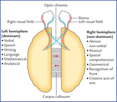
FIG. 12.15 Lateralization of functions in the dominant and non-dominant hemispheres.
The term dominant hemisphere refers to the side concerned with the perception and production of language/ speech. According to this concept, the left hemisphere is dominant in over 90% of people, in whom the right hemisphere is described as the minor or non-dominant hemisphere.
The left hemisphere controls the right side of the body, including the skilful right hand. Consequently over 90% of the adult population is right-handed.
During childhood, one hemisphere slowly comes to dominate over the other, and it is only after the first decade that the dominance becomes fixed.
Clinical Problems
1. What do you understand by cerebral hemispheric dominance? Mention its anatomical basis.
2. Explain why a 5-year-old child with damage in his dominant hemisphere can easily learn to become left handed and speak well, whereas in the case of an adult this is difficult.
3. A 55-year-old Dean of a medical institute received a severe blow on his head that caused a depressed fracture of his frontal bone. He was admitted in the hospital and discharged after 72 hours of observation. He joined his duty but in a few days the faculty members and students noticed that dean's social behaviour has changed dramatically. He no more dresses himself properly and lacks initiative and drive. He also lacked inhibition and one day he was seen urinating in the dustbin of his office. Explain the cause of his altered behaviour and personality?
4. A 62-year-old man on recovering from a cerebral stroke found that he has difficulty in understanding the spoken speech, although he understands written speech well. Mention which area of the cerebral cortex is involved?
5. An elderly patient has difficulty in understanding the written speech but he can easily understand the spoken speech. Mention which area of cerebral cortex is damaged?
6. Trace the neural pathway needed for a blind folded person to name an object placed in his right hand.
Clinical Problem Solving
1. Seepage 153.
2. In newborns both the hemispheres have equal potential to perceive and produce speech. But as they grow, one hemisphere slowly comes to dominate the other due to continuous learned behavioural stimulus to the left hemisphere, and it is only after the first decade that the dominance becomes fixed.
3. The abnormal behaviour and personality changes noticed in dean were due to a severe lesion involving the prefrontal areas of both frontal lobes of cerebrum secondary to the depressed fracture of the frontal bone (also seepage 151).
4. Secondary auditory area (Brodmann's area 22), because this area receives auditory impulses from primary auditory area and correlates them with the past experience, then this information is passed on to the sensory speech area of the Wernicke which permits the understanding of the spoken speech (see also pages 150 and 151).
5. Secondary visual area, because this area correlates the visual impulses received from the primary visual area to the past experiences. Then this information is passed on to the Wernicke's area for understanding the written speech.
6. Right hand → Spinal cord → Primary sensory area of dominant hemisphere → Wernicke's area (object is given name) → Broca's area → Premotor and motor areas → muscles involved in production of speech.