
The basal ganglia are the large masses of grey matter situated within the white core of each cerebral hemisphere and form essential constituents of the extrapyramidal system. The basal ganglia are now recognized as basal nuclei, but the former term is still commonly used.
Anatomically, the term basal ganglia include:
• Corpus striatum,
• Claustrum, and
• Amygdaloid body.
Functionally, the term basal ganglia also include substantia nigra and subthalamus. Some workers also include red nucleus.
The basal nuclei are important in organizing and coordinating somatic motor activities especially willed movements. They are also involved in automatic stereotyped postural and reflex motor activities such as normal individuals swing their arms when they walk.
Corpus Striatum
The corpus striatum is situated lateral to the thalamus. Topographically it is almost completely divided into the caudate nucleus and the lentiform nucleus by a band of nerve fibres, the internal capsule. However, anteroinferior ends of these nuclei remain connected by a few bands of grey matter across the anterior limb of internal capsule. These bands give it a striated appearance, hence the name corpus striatum (Figs 13.1–13.3).

FIG. 13.1 Lateral aspect of the left corpus striatum.

FIG. 13.2 Relationship of the corpus striatum to the internal capsule.
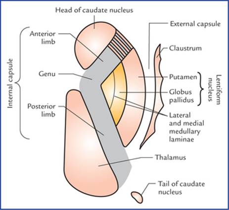
FIG. 13.3 Corpus striatum, thalamus, claustrum and internal capsule as seen in horizontal section of the cerebral hemisphere.
The lentiform nucleus consists of two parts: a darker lateral part putamen and a medial paler part globus pallidus.
Phylogenetically corpus striatum forms two distinct functional units, the paleostriatum and the neostriatum.
The globus pallidus is relatively ancient and termed paleostriatum/pallidum. The caudate nucleus and putamen being recent in development, together form the neostriatum/ striatum. The striatum is largely afferent whereas pallidum is largely efferent structure.
These features of corpus striatum may be summarized as follows:

Caudate Nucleus
Caudate nucleus is a large comma-shaped mass of grey matter, which surrounds the thalamus and is itself surrounded by the lateral ventricle (Fig. 13.4). Its whole length of convexity projects into the cavity of lateral ventricle.
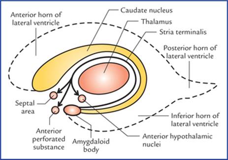
FIG. 13.4 Relationship of caudate nucleus with the cavity of the fourth ventricle and thalamus. Note that the stria terminalis, the main efferent tract of amygdaloid body projects to the septal area, anterior perforated substance, and anterior hypothalamus.
Its rounded anterior part in front of interventricular foramen is called its head. The head gradually and imperceptibly tapers caudally into the body and then into a tail which merges at its anterior extremity with an almond-shaped mass of grey matter called amygdaloid body (Fig. 13.2).
The head is large and rounded, and forms the floor and lateral wall of the anterior horn of lateral ventricle. The bands of grey matter connect it to the putamen across the anterior limb of internal capsule (Fig. 13.3).
The body is long and narrow, and forms the floor of the central part of lateral ventricle. It is separated from thalamus by stria terminalis and thalamostriate vein.
The tail is long and slender, and forms the roof of inferior horn of lateral ventricle. It terminates anteriorly (topographically) in the amygdaloid body.
Lentiform Nucleus
Lentiform nucleus is a large lens-shaped (biconvex) mass of grey matter beneath the insula forming the lateral boundary of internal capsule. In horizontal section of cerebrum, it appears wedge-shaped with broad convex base directed laterally.
It has three surfaces and divides into two parts:
Surfaces
• The lateral surface is convex and related to thin sheet of white matter, the external capsule. It is grooved by lateral striate arteries (the central branches of middle cerebral artery).
• Medial surface is more convex and related to internal capsule (limbs and genu). In transverse sections, the medial surface is angulated at the genu.
• Inferior surface is related to sublentiform part of internal capsule and lies close to the anterior perforated substance.
Parts
A vertical plate of white matter (external medullary lamina) divides the lentiform nucleus into two parts, the putamen and the globus pallidus. These are clearly seen on a cut section.
The putamen is larger lateral part and consists of densely packed small cells. It is darker in colour, and structurally similar to the caudate nucleus.
The globus pallidus is smaller medial part. It is lighter in colour and consists of large (motor) cells. It is also known as pallidum as it appears pale in section (pallid = pale). The globus pallidus is further subdivided by an internal medullary lamina of white matter into outer and inner segments (Fig. 13.3).
Connections of corpus striatum (Fig. 13.5)
The striatum (caudate nucleus and putamen) is the receptive part while globus pallidus is the efferent part (outflow centre) of the corpus striatum.
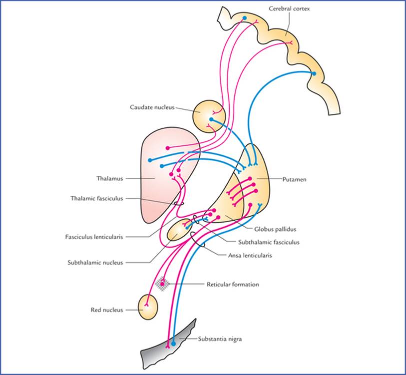
FIG. 13.5 Chief afferent and efferent connections of corpus striatum.
Afferent connections
The striatum receives afferents chiefly from cerebral cortex, thalamus and substantia nigra.
• Corticostriate fibres arise from a wide area of the ipsilateral cerebral cortex and reach the striatum through both internal and external capsules.
• Thalamostriate fibres arise from mediodorsal, intralaminar and midline nuclei of the thalamus. The majority of these fibres end in the caudate nucleus, the remaining pass through the internal capsule to reach the putamen.
• Nigrostriate fibres arise from substantia nigra and ascend up to terminate in the corpus striatum, mainly in putamen and caudate nucleus.
The nigrostriate fibres carry dopamine synthesized by nerve cells of substantia nigra to striatum (melanin normally present in the substantia nigra is a byproduct of dopamine metabolism). It is believed that these fibres have an inhibitory effect on corpus striatum.
Efferent connections
Most of the output of striatum goes to the globus pallidus, however, some efferents also go to substantia nigra.
The globus pallidus is the main efferent component of corpus striatum.
The outflow from globus pallidus goes mainly to the thalamus (ventrolateral and ventroanterior nuclei) (Figs 13.5, 13.6). This occurs by way of two fasciculi: (a) ansa lenticularis looping round the posterior limb of internal capsule, and (b) fasciculus lenticularis traversing the internal capsule.

FIG. 13.6 Outflow tracts from globus pallidus (paleostriatum). Also, note the interconnection between subthalamic nucleus and globus pallidus.
These fasciculi enter the region between the red nucleus and thalamus, where they unite with the dentatorubro-thalamic tract to form the thalamic fasciculus, which ends in the ventrolateral and ventroanterior nuclei of thalamus which in turn project to the motor and premotor areas of cerebral cortex.
The subthalamic fasciculus passes through the internal capsule and forms direct reciprocal connections between subthalamic nucleus and globus pallidus.
Some fibres from globus pallidus also pass to the substantia nigra (pallidonigral fibres).
Functions of corpus striatum
In submammalian vertebrates, the corpus striatum is the sensory and motor integrating and control centre. In later evolution, the activity of corpus striatum has become subordinate to those of cerebral cortex but remain essential for muscle tone, quality of movement, posture and locomotion.
• Corpus striatum controls the automatic associated movements like, swinging of arms during walking.
• It helps in smoothening the voluntary motor activity of the body.
Claustrum
Claustrum is a thin saucer-shaped mass of grey matter situated between the putamen and insula. It is considered as a detached part of the insula (Fig. 13.6).
Its connections and functions are not known.
Amygdaloid Body
Amygdaloid body is an almond-shaped mass of grey matter in the temporal lobe, lying anterosuperior to the tip of inferior horn of lateral ventricle. It is situated deep to uncus which serves as a surface landmark for its location.
Developmentally it is related to basal nuclei but functionally it is included in the limbic system and therefore shares its functions. For details see limbic system (page 229).
Substantia Nigra
Substantia nigra is described in detail in Chapter 8.
Subthalamus (Subthalamic Nucleus)
This small nucleus in the ventral part of the diencephalon looks like a biconvex lens in coronal section. It is located caudal to the lateral half of the thalamus and inferomedial to the globus pallidus. It is separated from thalamus by a smaller nucleus, called zona inserta. The subthalamic nucleus and globus pallidus are interconnected by the subthalamic fasciculus which traverses the internal capsule (Fig. 13.6).
Functions of Basal Nuclei
• Concerned with planning and programming of voluntary movements.
• Determine how rapidly a movement is to be performed and how large the movement must be.
• Decrease muscle tone and inhibit unwanted muscular activity.
• Regulate the muscle tone and thus helps in smoothening the voluntary motor activities of the body.
• Control automatic associated movements, like swinging of arms during walking.
• Control group of movements for emotional expression.
• Control reflex muscular activity.
Neural Circuitry Involved in Functioning of Basal Nuclei (Flowchart 13.1)
The striatum receives information from three major sources: (a) the cerebral cortex, (b) the thalamus, and (c) the substantia nigra. The striatum projects to the globus pallidus and substantia nigra. The globus pallidus (the outflow nucleus of striatum) projects to the thalamus and subthalamic nucleus. The substantia nigra also projects to the thalamus. The thalamus projects to the cerebral cortex. This information is integrated within the corpus striatum and outflow passes back to the above areas. This circular pathway functions as follows:
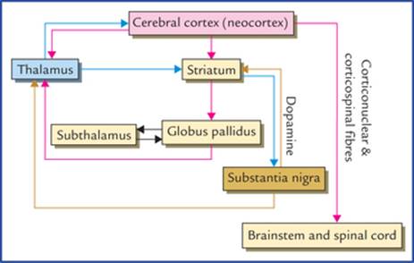
FLOWCHART 13.1 Neural circuitry of basal nuclei.
The activity of the basal ganglia is initiated by information received from the cerebral cortex, the thalamus, and the brainstem (substantia nigra and red nucleus). The outflow from the basal ganglia is channelled through the globus pallidus, which then influences the activities of the motor areas of the cerebral cortex. The motor cortex then controls the motor activities through corticospinal and corticonuclear fibres. Thus, basal ganglia can control muscular movements by influencing the cerebral cortex rather than through direct descending pathways to the brainstem and spinal cord.
Clinical Correlation
Disorders of the Basal Ganglia
The lesions of basal ganglia result in various forms of unwanted involuntary movements and disturbance in muscle tone. These disorders include parkinsonism, chorea, athetosis and ballismus.
• Parkinsonism (also called Parkinson's disease/paralysis agitans)
This disease usually occurs after 50 years of age due to deficiency of the neurotransmitter dopamine in the corpus striatum following a lesion in substantia nigra and/or its projections (i.e. nigrostriate fibres). Dopamine is synthesized in the melanin containing pigmented cells of substantia nigra and transported to the corpus striatum through nigrostriate fibres. Dopamine causes inhibition of cells within the corpus striatum. The neurological changes in the Parkinsonism appear to be a release phenomenon due to lack of inhibitory influences following dopamine deficiency.
Characteristic features of Parkinsonism (Fig. 13.7)
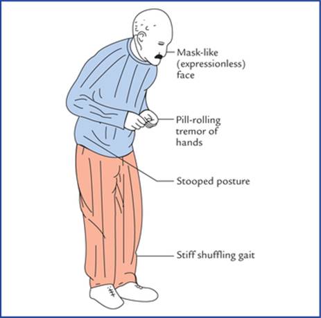
FIG. 13.7 Presenting features of patient suffering from Parkinsonism.
– Resting tremors, i.e. a slight shaking of hands when person is not performing a task. The tremors are diminished with movement and exaggerated by emotional excitation.
– Lead-pipe or cogwheel type of muscular rigidity due to increased muscle tone (in contrast to clasp-knife rigidity of upper motor neuron lesions).
– Pill-rolling movements of hands, consisting of circular movements of the opposed thumb and index fingertips is a kind of resting tremor).
– Mask-like face or loss of facial expression, due to lack of control of group of muscles of movements for emotional expression.
– Stiff, shuffling gait due to rigidity of joints.
– Stooped-posture. The stance of the patient is affected by rigidity. The back is flexed, the arms adducted and flexed, and knees bent.
– General slowing-down of movements and absence of associated movements, such as arm-swinging during walking.
To summarize the Parkinson's disease is characterized by a triad of symptoms, viz. tremor, rigidity, and akinesia.
Treatment
– The administration of L-dopa, a precursor of dopamine, or
– By placing small lesions (stereotactic surgery) in the globus pallidus and thalamus to diminish the cortical effects of abnormal discharges from corpus striatum, or
– By striatal implants of dopamine containing neurons of fetal origin.
• Chorea
This is characterized by quick, jerky, irregular purposeless involuntary movements, which involve primarily, the tongue, face and limbs. Swift grimaces and sudden movements of the head or limbs are good examples. The chorea is of two types: (a) Sydenham's chorea, and (b) Huntington's chorea (Table 13.1).
Table 13.1
Differences between Sydenham's chorea and Huntington's chorea
|
Sydenham's chorea (St. Vitus' dance) |
Huntington's chorea |
|
|
Age |
Occurs in children (between the ages of 5 and 15 years) primarily affecting girls and is associated with rheumatic fever |
Occurs in adults (between the ages of 30 and 45 years) and is an autosomal dominant inherited disease |
|
Pathology |
Antibodies produced by the antigens of streptococcal bacteria combine with the proteins of the membranes of neurons of striatum (caudate nucleus and putamen) producing inflammatory changes in the striatum |
Degeneration of GABA secreting neurons of striatum (caudate nucleus and putamen). Thus, striatonigral pathway is not able to inhibit the dopamine/secreting neurons of the substantia nigra. As a result, there occurs an over activity of nigrostriate fibres |
|
Involuntary movements |
Relatively less rapid and jerky |
Relatively more rapid and jerky |
|
CT scan of brain |
Shows no significant finding |
Shows enlarged lateral ventricle due to degeneration of caudate nucleus |
|
Recovery |
Full recovery (as a rule) |
No recovery |
• Athetosis
This consists of slow, sinuous, writhing movements (called athetoid movements) that commonly involve distal segments of the limbs, i.e. movements occur more in the muscles of the fingers and toes than in the proximal muscles of the limbs. The athetosis results due to lesions in neo-striatum and globus pallidus, breaking neuronal circuitry involving basal nuclei and cerebral cortex.
• Ballismus
It usually results due to vascular lesion of the subthalamic nucleus where normal smooth movements of different parts of the body are integrated.
The ballismus is characterized by violent burst of irregular movements in the trunk, girdles and proximal extremities. It usually involves the proximal musculature of opposite extremity, and limb suddenly flies about in all directions out of control, i.e. contralateral flinging (ballistic) movements of one or both extremities.
The disease may be restricted to one limb (monoballismus), but usually involves both upper and lower limbs on the contralateral side of the lesion (hemiballismus). This is because the subthalamic nucleus of one side projects mainly to the ipsilateral cortex through globus pallidus and thalamus.
Clinical Problems
1. A patient aged 65, presents with unintentional or resting tremors of hands (i.e. tremors occur at rest and disappear during voluntary movement), and slow voluntary movements. On examination, the following signs are elicited:
(a) Pill-rolling movements of hands, (b) face is expressionless (mask-like), (c) speech is slurred and monotonous, (d) cannot stand upright (a stooped posture), (e) short, shuffling gait, (f) increased muscle tone, and (g) cogwheel rigidity of joints. Make the clinical diagnosis.
2. What do you understand by the term ‘chorea’ and tell the differences between the Sydenham's chorea and Huntington's chorea?
3. A patient aged 55 has suddenly developed involuntary movements in his trunk and the left upper limb. His limb abruptly, vigorously and aimlessly would be thrown about in all directions, knocking over anything that comes in its path. Make the clinical diagnosis.
Clinical Problem Solving
1. This is a typical case of Parkinsonism (see page 159).
2. The term chorea stands for a syndrome in which the patient exhibits involuntary, quick, jerky, irregular movements, which are non-repetitive, viz. swift grimaces (grimace = distortion of the face). The differences between Sydenham's chorea and Huntington's chorea are presented in Table 13.1.
3. It is a typical case of left sided hemiballismus due to damage of right subthalamic nucleus (also seepage 160).