
The continuous blood supply to the brain is of utmost importance because of its high metabolic demands for oxygen and glucose. It is highly sensitive to hypoxia (inadequate O2) and hypoglycaemia (subnormal concentration of glucose in the blood). The consciousness is lost within 10 seconds of cessation of blood flow, and if the state continues, an irreversible brain damage starts to occur at about 4 minutes and is completed within 10 minutes.
The brain is one of the most metabolically active organs of the body as it depends on aerobic metabolism of glucose.
Although the brain constitutes only 2% (1/50) of the total body weight, it receives 20% (1/5) of the total cardiac output and consumes 20% of the total O2 used by the body.
The cerebrovascular diseases (thrombosis, embolism and haemorrhage) are the third most common cause of death and the neurological signs depend on the site of lesion. Therefore an adequate knowledge of the blood supply of the brain is essential for proper diagnosis and treatment of these diseases.
N.B. The arterial occlusion by a thrombus which often leads to infarction of the portion of the brain supplied by the affected artery is the most common type of the cerebrovascular disease.
Arteries of The Brain
The brain is supplied by the paired internal carotid and vertebral arteries via an extensive system of branches (Fig. 15.1):

FIG. 15.1 Four arteries supplying the brain.
The two vertebral arteries unite at the lower border of the pons to form the basilar artery which ascends in the midline on the ventral surface of the pons and at its upper border terminates by dividing into right and left posterior cerebral arteries.
Each internal carotid artery ends in the region of anterior perforated substance by dividing into a larger middle cerebral artery, and a smaller anterior cerebral artery.
N.B. Thus the brain is supplied by two systems of arteries: (a) vertebral system, consisting of a pair of vertebral arteries, and (b) carotid system, consisting of a pair of internal carotid arteries.
Circle of Willis (circulus arteriosus)
The major arteries supplying the cerebrum (i.e. branches of basilar and internal carotid arteries) get interconnected to one another at the base of the brain to form a six-sided polygon of arteries called circulus arteriosus or circle of Willis (Figs. 15.2 and 15.3). The circle of Willis is formed around the interpeduncular fossa and lies in the interpeduncular subarachnoid cistern. It contributes most of the arterial blood supply to the brain.

FIG. 15.2 Polygonal anastomoses between the branches of internal carotid and basilar arteries.

FIG. 15.3 Circle of Willis and the branches of arteries supplying the brain. The central branches of cerebral arteries are shown by abbreviations: AM = anteromedial group, PM = posteromedial group, AL = anterolateral group, PL = posterolateral group.
Circle of Willis is formed:
Anteriorly, by the anterior communicating and the anterior cerebral arteries.
Posteriorly, by the basilar artery dividing into two posterior cerebral arteries.
Laterally on each side, by the posterior communicating artery connecting the internal carotid artery with the posterior cerebral artery.
Functional significance of circle of Willis
Normally there is little or no mixing of blood streams: (a) of two vertebral arteries in the basilar artery, (b) of two anterior cerebral arteries in the anterior communicating artery, and (c) of internal carotid and posterior cerebral arteries in the posterior communicating artery. Therefore, right half of the brain is supplied by right vertebral and right internal carotid arteries and left half of the brain is supplied by left vertebral and left internal carotid arteries.
However, if one of the major arteries forming the circle of Willis is blocked, the circle of Willis provides the various alternative roots for collateral circulation like an arterial traffic circle.
Clinical Correlation
• Congenital cerebral aneurysms
These occur mostly at the sites where two arteries join in the formation of the circle of Willis. The basic abnormality at these points is the congenital deficiency of the tunica media (elastic tissue) in the arterial wall. The aneurysms are berry-shaped, hence they are generally termed berry aneurysms.
• Subarachnoid haemorrhage
The subarachnoid haemorrhage commonly but not exclusively results from rupture of congenital berry aneurysms in the interpeduncular cistern.
The subarachnoid haemorrhage produces a sudden severe pain in head followed by mental confusion. The death may quickly occur, or the patient may survive the first bleeding only to die few days or weeks later.
Vertebral System
Vertebral artery (Fig. 15.4)
The vertebral artery, a branch of subclavian artery, ascends in the foramina transversaria of upper six cervical vertebrae. On reaching the base of skull, it winds backwards and medially around the lateral mass of the atlas and pierces posterior atlanto-occipital membrane, to enter the posterior cranial fossa through the foramen magnum where it runs on the anterolateral aspect of the medulla. Here the two vertebral arteries converge, and unite at the lower border of the pons to form the basilar artery (Fig. 15.4).

FIG. 15.4 Origin and course of internal carotid and vertebral arteries.
Clinical Correlation
The loop of vertebral artery around the lateral mass of atlas may damp down the arterial pulsations within the cranial cavity.
Branches of the cranial part of the vertebral artery (Fig. 15.3)
• Anterior spinal artery is a small branch arising near the termination of the vertebral artery. It descends in front of the medulla and unites with its fellow of the opposite side at the level of the lower end of the olive to form a single median trunk that descends along the anterior longitudinal fissure of the spinal cord.
• Posterior spinal artery arises from vertebral artery and sometimes from posterior inferior cerebellar artery. It passes downwards on the posterior surface of the spinal cord, after dividing into two branches; one along the medial side, and the other along the lateral side of the dorsal roots of the spinal nerves.
• Posterior inferior cerebellar artery is the largest branch of the cranial (4th) part of the vertebral artery. It arises near the lower end of the olive, winds backwards around the medulla oblongata, and then ascends to the pon-tomedullary junction.
• Meningeal branches are small and supply the dura mater of the posterior cranial fossa.
• Medullary arteries are several minute vessels which supply the medulla oblongata.
Basilar artery (Figs 15.1–15.3)
Basilar artery is formed by the union of two vertebral arteries at the lower border of the pons. It ascends in the basilar sulcus on the ventral aspect of the pons in the cis-terna pontis and terminates at the upper border of the pons by dividing into right and left posterior cerebral arteries.
Branches of basilar artery
• Pontine branches are numerous short slender parame-dian vessels which pierce the pons to supply it.
• Anterior inferior cerebellar artery arises close to the lower border of the pons and runs backwards and laterally usually ventral to the VIIth and VIIIth cranial nerves. Then it forms a loop over the flocculus of the cerebellum and peeps into the internal acoustic meatus for a variable distance lying below the VIIth and VIIIth cranial nerves. After exit from the meatus it supplies the anterolateral portion of the inferior surface of the cerebellum.
• Labyrinthine artery is a long slender branch which arises either from basilar artery or from anterior inferior cere-bellar artery. It accompanies the vestibulocochlear nerve and enters the internal auditory meatus to supply the internal ear. It is an end artery.
• Superior cerebellar artery arises close to the superior border of the pons, runs laterally below the oculomotor nerve (which is interposed between this artery and the posterior cerebral artery), and winds round the cerebral peduncle below the trochlear nerve to reach the superior surface of the cerebellum which it supplies.
• Posterior cerebral artery passes laterally parallel to the superior cerebellar artery, curves around the midbrain to reach the medial surface of the cerebral hemisphere, beneath the splenium of corpus callosum (Figs 15.5 and 15.6). The artery gives off temporal branches which ramify over the inferior surface of the temporal lobe, and calcarine and parieto-occipital branches which run along the corresponding sulci.

FIG. 15.5 Arteries on the inferior surface of the cerebral hemisphere.
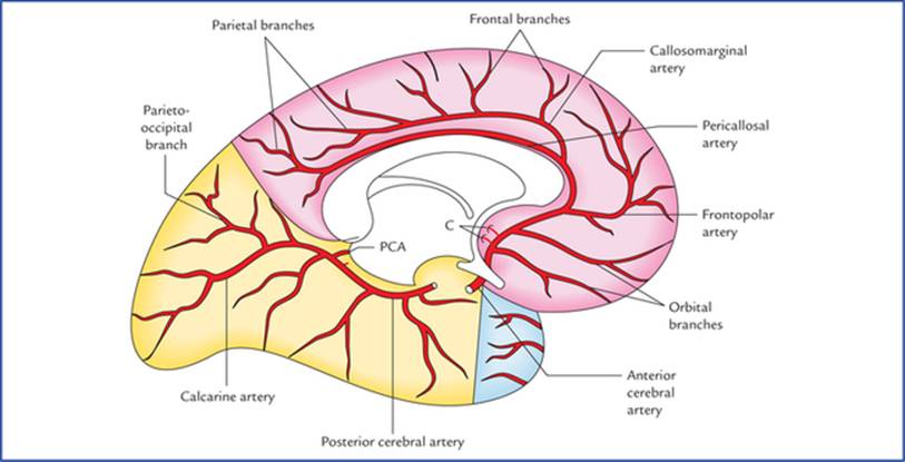
FIG. 15.6 Arteries on the inferomedial aspect of the cerebral hemisphere. (C = central branches, PCA = posterior choroidal artery.)
1. Anterior temporal branch passes forwards.
2. Posterior temporal branch passes posteriorly towards the occipital pole.
3. Parieto-occipital branch runs towards the parieto-occipital sulcus.
4. Calcarine branch (often called calcarine artery) passes posteriorly in the calcarine sulcus.
Carotid System
Internal carotid artery (Fig. 15.4)
The internal carotid artery, a terminal branch of the common carotid artery, traverses the carotid canal in the base of the skull and enters the middle cranial fossa beside the dorsum sellae of the sphenoid bone. Here it first runs forwards along the floor and medial wall of the cavernous sinus and then turns upwards on the medial side of the anterior clinoid process. At this point the artery pierces the dural roof of the cavernous sinus and also the arachnoid mater to enter the subarachnoid space. Now it first runs backwards and then upwards to come to lie lateral to the optic chiasma just underneath the anterior perforated substance of the brain, where it terminates by dividing into two branches, a larger middle cerebral artery and a smaller anterior cerebral artery (Figs 15.2 and 15.3).
Clinical Correlation
The intracavernous and supracavernous course of internal carotid artery forms a U-shaped bend (hair-pin bend) with convexity forward called carotid siphon. The siphon dampens the arterial pulsations to provide a more steady regular stream of blood flow to the brain.
The carotid siphon is an important part of the cerebral angiogram to inspect, for masses in the pituitary region will open out (widen) the curl in the artery.
Branches of the cerebral part of the internal carotid artery (Fig. 15.3)
• Ophthalmic artery arises from the ventral convexity of the carotid siphon and enters the optic canal to reach the orbital cavity to supply the structures of the orbit including eyeball.
• Posterior communicating artery arises close to the termination of the internal carotid artery. It runs backwards and anastomoses with the proximal part of the posterior cerebral artery.
• Anterior choroidal artery is a long slender branch, which arises just distal to the origin of the posterior communicating artery. It courses backwards above and along the optic tract, to enter the inferior horn of the lateral ventricle through the choroid fissure to end in the choroid plexus.
Due to its long subarachnoid course and a relatively small lumen, the anterior choroidal artery is most susceptible to thrombosis and is often referred to as artery of cerebral thrombosis.
• Anterior cerebral artery is a smaller terminal branch of the internal carotid artery. It runs forwards and medially above the optic nerve to the commencement of the median longitudinal cerebral fissure, where it comes very close to its fellow of the opposite side and gets joined with it by a short transverse anterior communicating artery. The anterior cerebral artery then curves around the genu of corpus callosum.
The branches given off just distal to the anterior communicating artery supply the medial part of the orbital surface of the frontal lobe.
The artery continues along the upper surface of the corpus collosum as the pericallosal artery and gives a large branch, the callosomarginal artery which runs in the cingulate sulcus. Near the splenium of corpus callosum, the artery ends by anastomosing with the branches of the posterior cerebral artery.
Clinical Correlation
The arched course of the anterior cerebral artery around the genu of corpus callosum makes it easy to identify it in a carotid angiogram.
• Middle cerebral artery is the larger terminal branch of the internal carotid artery. It appears to be the direct continuation of the internal carotid artery and carries about 30% of the carotid blood flow.
The middle cerebral artery first runs laterally in the stem of the lateral sulcus (Fig. 15.5) and then turns backwards and upwards in the posterior ramus of the lateral sulcus, where it breaks up into frontal, parietal and temporal branches which emerge from the lateral sulcus and run towards the areas of their supply. Some of these branches are named (Fig. 15.7).

FIG. 15.7 Arteries on the superolateral surface of the left cerebral hemisphere.
The branches of main arteries supplying the brain are summarized in Table 15.1
Table 15.1
Branches of the main arteries of the brain
|
Cerebral part of internal carotid artery |
Fourth part of vertebral artery |
Basilar artery |
|
• Ophthalmic artery |
• Meningeal arteries |
• Anterior inferior cerebellar artery |
|
• Anterior cerebral artery |
• Anterior spinal artery |
• Labyrinthine artery |
|
• Middle cerebral artery |
• Posterior spinal artery |
• Pontine arteries |
|
• Posterior communicating artery |
• Posterior inferior cerebellar artery |
• Superior cerebellar artery |
|
• Anterior choroidal artery |
• Medullary arteries |
• Posterior cerebral artery |
It is interesting to note from Table 15.1 that each main artery supplying the brain gives off five sets of branches thus making a total of 15 sets. Out of which there are: three pairs of cerebral arteries (anterior, middle, and posterior) and three pairs of cerebellar arteries (posteroinferior, anteroinferior, and superior).
Clinical Correlation
The cerebral thrombosis most commonly affects middle cerebral artery or its main branches because it is a direct continuation of internal carotid artery.
Arterial Supply of the Cerebrum
The cerebrum is supplied by three pairs of cerebral arteries, viz. anterior, middle and posterior.
Branching pattern of the cerebral arteries
The cerebral arteries give three types of branches: (a) cortical, (b) central, and (c) choroidal.
Cortical branches
Cortical branches supply the outer portion of the cerebrum. The cortical vessels freely anastomose and form a network in the pia mater on the surface of the cerebral cortex. From this network branches arise and pierce the cortex at right angle. Once they enter the cortex, they become the end arteries. There are two types of these end arteries: (a) short, which confine themselves only to the cortex, and (b) long, which pass through the cortex and reach the outer portion of the white matter.
Central branches (Fig. 15.3)
Central branches are numerous slender thin-walled perforating branches which supply the centrally located parts of the cerebrum, viz. corpus striatum, internal capsule, etc. They are end arteries. The central arteries arise in the region of arterial circle of Willis, and are arranged into following four groups:
• Anteromedial group: They arise from anterior cerebral and anterior communicating arteries, enter the anterior perforated substance to supply the anterior limb of internal capsule, the corpus striatum and the anterior hypothalamus.
• Anterolateral group (lateral striate arteries): They arise from proximal part of the middle cerebral artery, pierce the anterior perforated substance to supply the anterior limb of the internal capsule, and the caudate and lenti-form nuclei. One of the larger lateral striate arteries which is most susceptible to rupture, is known as the artery of cerebral haemorrhage (also called Charcot's artery of cerebral haemorrhage).
• Posteromedial group: The arteries of this group arise from proximal part of the posterior cerebral artery and adjoining part of the posterior communicating artery. They enter the posterior perforated substance to supply the thala-mus, the subthalamus and the hypothalamus.
• Posterolateral group: These arteries arise from posterior cerebral artery after it has curved around the cerebral peduncle. They supply the thalamus including genicu-late bodies.
Choroidal branches
Choroidal arteries form a network of capillaries which project into the ventricles after invaginating the layers of pia mater and ependyma forming choroid plexuses.
• Anterior choroidal artery: It arises from internal carotid artery, runs posteriorly near the optic tract, crosses the uncus to enter the choroidal plexus in the inferior horn of lateral ventricle.
• Posterior choroidal artery: It arises from the posterior cerebral artery, encircles around the midbrain to enter the choroidal plexus of the third and lateral ventricles, through transverse fissure.
Arterial Supply of the Different Surfaces of the Cerebral Hemisphere
Arterial supply of the superolateral surface (Fig. 15.8A)

FIG. 15.8 Arterial supply of the superolateral (A), medial (B), and inferior (C) surfaces of the cerebral hemisphere.
• Most of the superolateral surface (about two-third) is supplied by the middle cerebral artery. The region of cerebral cortex supplied includes the greater parts of primary motor and sensory areas, and frontal eyefield. In the left (dominant) hemisphere it includes the Broca's and Wernicke's speech areas.
• A narrow strip of cerebral cortex (about 2.5 cm in width) adjoining superomedial border up to the parieto-occipital sulcus is supplied by anterior cerebral artery. The upper parts of primary motor and sensory areas lie in this region.
• A narrow strip along the lower border of temporal lobe (excluding temporal pole) and occipital lobe are supplied by posterior cerebral artery. The posterior parts of visual area fall in this area.
Arterial supply of the medial surface (Fig. 15.8B)
• Most of the medial surface (anterior two-third) is supplied by the anterior cerebral artery. The region of cerebral cortex supplied includes the parts of motor and sensory areas (paracentral lobule) concerned with perineum, leg, and foot.
• Temporal pole of the temporal lobe is supplied by the middle cerebral artery.
• Occipital lobe is supplied by posterior cerebral artery. The area supplied includes the visual cortex.
Arterial supply of the inferior surface (Fig. 15.8C)
• Most of the inferior surface except the temporal pole is supplied by the posterior cerebral artery.
• Lateral part of the orbital surface of the frontal lobe and temporal pole of the temporal lobe are supplied by the middle cerebral artery.
• Medial part of the orbital surface of the frontal lobe is supplied by the anterior cerebral artery.
Points to Note
Each surface of the cerebral hemisphere is supplied by three cerebral arteries, viz. anterior, middle and posterior.
Most of the superolateral surface is supplied by middle cerebral artery, most of medial surface by anterior cerebral artery, and most of the inferior surface by the posterior cerebral artery. Thus middle, anterior and posterior cerebral arteries are the principal arteries of the superolateral, medial and inferior surfaces of the cerebral hemisphere respectively.
Clinical Correlation
• The occlusion of anterior cerebral artery (distal to the anterior communicating artery) may produce following effects:
– Contralateral hemiparesis and hemianaesthesia involving mainly the leg and foot, due to involvement of upper parts of primary motor and sensory areas, and paracentral lobule.
– Inability to identify the objects correctly, due to involvement of superior parietal lobule.
– Apathy and personality changes, due to involvement of part of frontal lobe.
• The occlusion of middle cerebral artery may produce following effects:
– Contralateral hemiplegia and hemianaesthesia, involving mainly the face and arm, due to involvement of most of the primary motor arid sensory areas.
– Aphasia if left dominant hemisphere is involved, due to involvement of motor and sensory speech areas.
– Contralateral homonymous hemianopia, due to involvement of optic radiation.
• The occlusion of posterior cerebral artery may produce following effects:
– Contralateral homonymous hemianopia, due to involvement of visual cortex with some degree of macular sparing. The macular vision is spared because it is represented in the occipital pole which receives a collateral supply from the middle cerebral artery (anastomosis exists between the branches of middle and posterior cerebral arteries in the region of the occipital pole).
Arterial Supply of the Other Parts of the Brain
• The corpus striatum and internal capsule are supplied mainly by central branches (medial and lateral striate arteries) of middle cerebral artery and to some extent by central branches of anterior cerebral artery (Fig. 15.9).
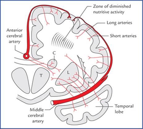
FIG. 15.9 Coronal section of cerebral hemisphere showing the arterial supply of the deep cerebral structures by middle and anterior cerebral arteries. (T = thalamus, C = caudate nucleus, L = lentiform nucleus. Also note the distribution of the cortical and central branches of the cerebral arteries, and zone of diminished nutrition.)
• The thalamus is supplied mainly by central branches of posterior communicating, posterior cerebral and basilar arteries.
• The midbrain is supplied by posterior cerebral, superior cerebellar and basilar arteries.
• The pons is supplied by basilar, superior cerebellar and anterior inferior cerebellar arteries.
• The cerebellum is supplied by superior, anterior inferior, and posterior inferior cerebellar arteries.
For details see the individual parts of the brain.
Clinical Correlation
• The cerebral angiography is a radiological technique to visualize the vessels of the brain. A radiopaque solution is injected into one of the major arteries supplying the brain, and serial radiographs of skull are taken at approximately 1 second intervals. The injection into the common carotid artery or the internal carotid artery (carotid angiogram) shows the distribution of middle and anterior cerebral arteries; whereas the injection into the vertebral artery permits the visualization of vertebral, basilar and posterior cerebral arteries together with their branches. The cerebral angiography is valuable in identifying vascular malformations and aneu-rysms. It is also provides useful information about occlusive vascular disease and space-occupying lesions.
• The cortical and central branches of cerebral arteries do not anastomose with each other. The short cortical arteries do not penetrate beyond the cerebral cortex while the long cortical arteries do traverse the cortex to supply the white matter up to a depth of 4 or 5 cm only. The deep portion of the white matter is supplied by the central branches. There is however, a zone of diminished nutrition between the territories of cortical and central branches. The formation of cavities due to senile softening of brain mostly seen in this zone.
Venous Drainage of The Brain
The veins of the brain drain into the intracranial dural venous sinuses, which eventually open into the internal jugular veins of the neck. The veins emerge from the brain, traverse the subarachnoid space, pierce the arachnoid mater and meningeal layer of dura mater to drain into the venous sinuses.
The characteristic features of venous drainage of the brain are:
• The venous return in the brain does not follow the arterial pattern.
• The veins of the brain are extremely thin-walled due to absence of muscular tissue in their walls.
• The veins of the brain possess no valves.
• The veins of the brain run mainly in the subarachnoid space.
• The cerebral veins, generally enter obliquely into the dural venous sinuses against the flow of blood in the sinuses to avoid their possible collapse following an increased intracranial pressure as they are thin walled.
The veins of the brain comprise, cerebral veins, cerebellar veins, and veins of the brainstem.
Cerebral Veins
The cerebral veins are divided into external and internal cerebral veins which drain the external surfaces and the internal regions of the cerebral hemisphere respectively.
External (superficial) cerebral veins
The external cerebral veins drain the surface (cortex) of the hemisphere and are divided into three groups: (a) superior, (b) middle, and (c) inferior.
Superior cerebral veins (Fig. 15.10)
Superior cerebral veins are about 8 to 12 in number and drain the upper parts of the superolateral and medial surfaces of the cerebral hemisphere. They ascend upwards, pierce the arachnoid mater and traverse the subdural space to enter the superior sagittal sinus.

FIG. 15.10 Veins on the superolateral surface of the cerebral hemisphere.
The anterior veins open at right angle while the posterior open obliquely against the flow of blood stream in the superior sagittal sinus (embryologically it is due to backward growth of the rapidly increasing cerebrum), thereby preventing their collapse by increased CSF pressure.
Middle cerebral veins
Middle cerebral veins are four in number, two on each side: (a) superficial middle cerebral vein, and (b) deep middle cerebral vein.
• The superficial middle cerebral vein (Fig. 15.10) lies superficially in the lateral sulcus.
Anteriorly it runs forwards to drain into the cavernous sinus while posteriorly it communicates with the superior sagittal sinus via superior anastomotic vein (of Trolard) and with the transverse sinus via inferior anastomotic vein (of Labbe).
• The deep middle cerebral vein lies deep in the lateral sulcus on the insula along with middle cerebral artery. It runs downwards and forwards and joins the anterior cerebral vein to form the basal vein.
Inferior cerebral veins
Inferior cerebral veins are many in number but smaller in size. They drain the inferior surface and lower parts of medial and superolateral surfaces of the cerebral hemisphere into nearby intracranial dural venous sinuses, viz. transverse sinus, etc.
Other veins
Anterior cerebral vein (Fig. 15.11)
It accompanies the anterior cerebral artery around the corpus callosum and drains the parts of medial surface which cannot be drained into the superior and inferior sagittal sinuses.

FIG. 15.11 Veins on the medial surface of the cerebral hemisphere.
N.B. The anterior cerebral vein is the only large vein of the brain which has a similar name and course as its companion artery (anterior cerebral artery).
Basal vein (of Rosenthal)
It is formed at the base of brain in the region of anterior perforated substance by the union of three veins: anterior cerebral, deep middle cerebral and striate veins (Fig. 15.12). The striate veins emerge from the anterior perforated substance.
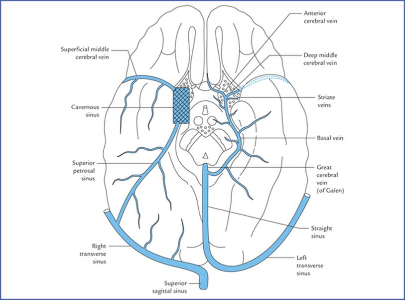
FIG. 15.12 Veins at the base of the brain. Note the formation, course and tributaries of the basal vein.
The basal vein runs posteriorly around the midbrain, medial to the uncus and parahippocampus and terminate into the great cerebral vein (of Galen) below the splenium of corpus callosum.
Tributaries
Besides the formative three veins, the basal vein receives the tributaries from:
• Cerebral peduncle
• Uncus and parahippocampus
• Structures of interpeduncular fossa
• Optic tract and olfactory trigone
• Inferior horn of the lateral ventricle.
Internal cerebral veins (Fig. 15.13)
There are two internal cerebral veins located one on either side of midline in the tela choroidea of the third ventricle.
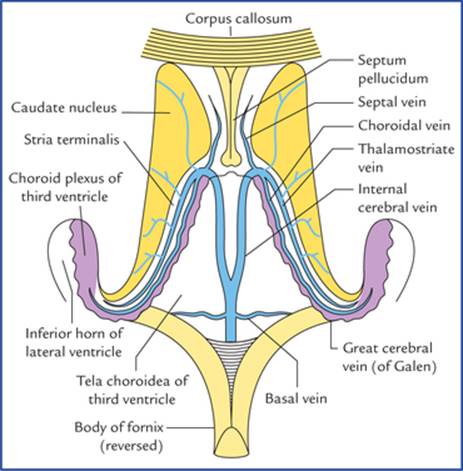
FIG. 15.13 Formation, course, and tributaries of the internal cerebral veins and the great cerebral vein of Galen.
Each internal cerebral vein is formed at the interven-tricular foramen (of Monro) by the union of three veins: thalamostriate, septal and choroidal. The two internal cerebral veins run posteriorly one on either side of mid-line, between the two layers of tela choroidea of third ventricle and unite together beneath the splenium of corpus callosum to form the great cerebral vein (of Galen) which empties into the straight sinus.
The thalamostriate, septal and choroidal veins are the most important deep veins of the cerebrum. As their names imply, the thalamostriate (striothalamic) vein drains the thalamus and basal ganglia; the septal vein drains the septum pellu-cidum, and the choroidal vein drains the choroid plexus.
Great cerebral vein (of Galen)
Great cerebral vein is a single vein (about 2 cm in length). It is formed by the union of two internal cerebral veins below and behind the splenium of corpus callosum. It immediately receives the two basal veins and after a short backward course it joins the inferior sagittal sinus to form the straight sinus.
Tributaries
• Internal cerebral veins
• Basal veins
• Veins from colliculi (tectum of midbrain)
• Veins from cerebellum and adjoining parts of the occipital lobes of the cerebrum.
Venous Drainage of the Different Surfaces of the Cerebral Hemisphere
Venous drainage of the superolateral surface
Superolateral surface is drained by the following veins:
• Superior cerebral veins drain the upper part into the superior sagittal sinus.
• Inferior cerebral veins drain the lower part into the superficial middle cerebral vein, however some from the pos-teroinferior part drain into the transverse sinus.
Venous drainage of the inferior surface
Inferior surface is drained by the inferior cerebral veins:
• Inferior cerebral veins, from the orbital part drains into the superficial, middle cerebral and anterior cerebral veins
• Inferior cerebral veins, from the tentorial part drains into:
1. Venous sinuses at the base of skull, viz. cavernous, superior petrosal, straight and transverse sinuses;
2. Superficial middle cerebral vein which drains into cavernous sinus and basal vein which drains into the straight sinus.
Venous drainage of the medial surface
Medial surface is drained by the following veins:
• Superior cerebral veins drain the upper part into superior sagittal sinus.
• Inferior cerebral veins drain the lower part into the inferior sagittal sinus.
• Some of the veins from the posterior part drain into the great cerebral vein.
• Anterior cerebral vein drains the anterior part.
N.B. From the above description, it is clear that the superficial veins drain mainly into the superior sagittal sinus, which ultimately drain into the right internal jugular vein. On the other hand the deep veins drain mainly into the great cerebral vein, which ultimately drain into left internal jugular vein.
Clinical Correlation
Subdural haemorrhage
It occurs due to rupture of cerebral veins in the sub-dural space. The cerebral veins while traversing the subdural space en route to drain into the dural venous sinuses have little support and are torn following moderate trauma on head. The superior cerebral veins are most commonly torn, where they enter the superior sagittal sinus. The cause is usually a blow on the front or back of the head, resulting in excessive anteroposterior displacement of the brain within the skull. Consequently the cerebral veins in the sub-dural space (called bridging veins) are unduly stretched and torn. The subdural haemorrhage is generally extensive because of the loose attachment between the dura and arachnoid.
the types of intracranial haemorrhage and vessels most commonly involved are summarized in Table 15.2.
Table 15.2
Types of intracranial haemorrhage
|
Type of haemorrhage |
Vessels/vessels most commonly involved |
|
Extradural haemorrhage |
Rupture of anterior division of middle meningeal artery |
|
Subdural haemorrhage |
Tearing of superior cerebral (bridging) veins |
|
Subarachnoid haemorrhage |
Leakage or rupture of congenital berry aneurysms on the arterial circle of Willis |
|
Intracerebral (cerebral) haemorrhage |
Rupture of thin-walled lenticulostriate artery (Charcot's artery of cerebral haemorrhage), a branch of the middle cerebral artery |
Various types of intracranial haemorrhages can be easily detected by CT scan of brain Figures 15.14 to 15.17, shows extradural, subdural, subarachnoid and intracerebral haemorrhage respectively.

FIG. 15.14 CT scan of brain showing extradural haemorrhage/ haematoma. Source: Drake, Richard L, Vogl, Mitchell, Adam WM. Grays Anatomy for Students. Philadelphia: Elsevier, Inc., 2005

FIG. 15.15 CT scan of brain showing subdural haemorrhage/ haematoma. Source: Drake, Richard L, Vogl, Mitchell, Adam WM. Grays Anatomy for Students. Philadelphia: Elsevier, Inc., 2005
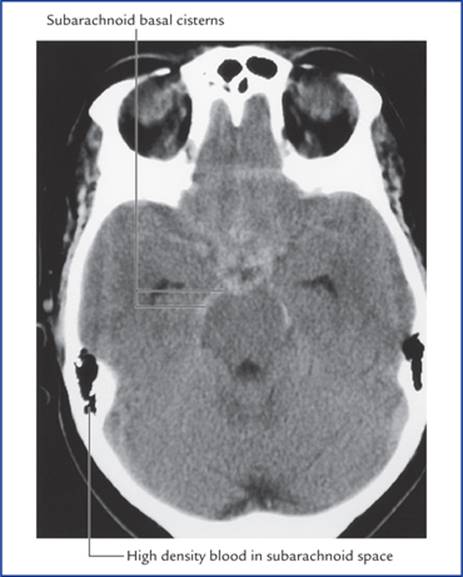
FIG. 15.16 CT scan of brain showing subarachnoid haemorrhage (arrows). Source: Drake, Richard L, Vogl, Mitchell, Adam WM. Grays Anatomy for Students. Philadelphia: Elsevier, Inc., 2005

FIG. 15.17 CT scan of brain showing intracerebral haemorrhage. (arrows) Source: Haslet, Christopher, Chilvers, Edwin R, Boon, Nicholas A, et al., editors. Davidson's Principles and Practice of Medicine. 19th edn. Oxford: Elsevier Science Ltd., 2002
Blood-Brain Barrier (BBB)
The brain and spinal cord need a stable environment to function normally. This is provided by the presence of a semipermeable barrier, called blood-brain barrier (the term blood-brain-spinal cord barrier would be more accurate name). This barrier protects the brain and spinal cord from potentially harmful substances (toxic drugs and other exogenous materials) while allowing the gases and nutrients to enter the nervous tissue.
Structure of Blood-Brain Barrier
The BBB consists of following structures (Fig. 15.18) which intervene between the blood in the capillaries and the extracellular spaces surrounding the neurons and neuroglia in the brain:

FIG. 15.18 Structure of blood-brain barrier (BBB).
• capillary endothelial cells and tight junctions between them.
• a basement membrane on which the capillary endothelial cells are arranged, and
• the foot processes of the astrocytes that adhere to the outer surface of the capillary wall.
N.B. The tight junctions between the endothelial cells of the blood capillaries form the most important component of blood-brain barrier.
Areas of the Brain Devoid of Blood-Brain Barrier
The following areas of the brain are devoid of blood-brain barrier:
• Pineal gland
• Posterior lobe of the pituitary gland ^ Tuber cinereum
• Wall of the supraoptic recess of the third ventricle
• Area postrema at the lower end of the floor of fourth ventricle.
In fact in these areas, the capillary endothelium has fenes-trations, across which protein and small organic molecules may pass from blood to the nervous tissue. These are the sites where the neuronal receptors may sample the chemical contents of the plasma directly. The hypothalamus which is involved in the regulation of the metabolic activities of the body might react suitably and modify the activities thereby protecting the nervous tissue.
Clinical Correlation
• Any injury to the brain by trauma, chemical toxins or inflammation causes a breakdown of the blood-brain barrier allowing free diffusion of larger molecules into the nervous tissue. For example, normally when penicillin is administered systemically, only a small amount of it enters the CNS because penicillin in high concentration is toxic to the nervous tissue. In the presence of meningitisthe menin-ges become more permeable at the site of inflammation thus permitting sufficient antibiotic to reach the site to check the infection.
• The drugs like, chloramphenicol, tetracyclines, sul-phonamides, thiopental (lipid soluble), atropine (lipid soluble) easily pass through the blood-brain barrier. The drugs like, phenylbutazone, and neuro-transmitters like exogenous epinephrine and dopa-mine cannot cross the blood-brain barrier.
• In Parkinson's disease, there is deficiency of neu-rotransmitter dopamine in the corpus striatum.
Unfortunately dopamine cannot be used for the treatment, as it will not cross the blood-brain barrier. Instead levodopa (L-dopa) a precursor of dopamine is used which readily crosses the blood-brain barrier. The L-dopa is converted into dopamine by the neurons within CNS.
In infants, the blood-brain barrier is not fully developed, therefore if the serum bilirubin level is high, the bilirubin readily enters the brain tissue producing bilirubin encephalopathy (syn. kernicterus) a severe form of jaundice seen only in newborn babies.
Blood-Csf Barrier
Similar to BBB, there is barrier between the blood in the capillaries of choroid plexus and the cerebrospinal fluid (CSF) within the ventricles of the brain. It allows the free passage of water, gases and lipid soluble substances from the blood to the CSF, but prevents the entry of macromolecules such as proteins and most hexoses other than glucose.
Structure of Blood-CSF Barrier
The blood in the lumen of blood capillary (within the vil-lus of choroid plexus) is separated from the CSF in the cavity of the ventricle by the following structures which constitute the blood-CSF barrier:
• The endothelial cells, which are fenestrated (the fenes-trations are not true perforations but are filled with thin diaphragms).
• The basement membrane of the capillary endothelial cells.
• The basement membrane of the choroidal epithelial cells.
• The tight junctions between the choroidal epithelial cells.
• The scattered pale cells with their flattened processes between the two basement membranes.
Clinical Problems
1. The resuscitation (a device to restore circulation and respiration) should be done within 4 minutes of cardiorespiratory arrest. Why?
2. On examining a carotid angiogram it is commonly noted that the contrast medium fills the anterior and middle cerebral arteries but fails to fill the posterior communicating artery beyond a certain point. Mention its anatomical basis.
3. The subarachnoid haemorrhage usually occurs at the base of the brain.
4. A 50-year-old hypertensive patient suddenly complained of severe headache. A few movements later he lost his consciousness and developed the following neurological defects: (a) loss of speech (aphasia), (b) right sided hemiplegia, and hemianaesthesia. Make the clinical diagnosis and mention the anatomical basis.
5. Why do the patients suffering from chronic subdural haematoma complain of headache, drowsiness and mental confusion and they are relieved of these symptoms once the blood clot is removed?
Clinical Problem Solving
1. This is because the irreversible brain damage starts to occur at about 4 minutes.
2. This is because, in the posterior communicating artery the streams of blood from internal carotid and vertebral arteries do not mix (also seepage 172).
3. The subarachnoid haemorrhage commonly occurs due to rupture of congenital berry aneurysms which develop in the arteries of circle of Willis. Since the circle of Willis is located at the base of brain, the suba-rachnoid haemorrhage commonly occurs at the base of brain.
4. It is a typical case of brain stroke/cerebral stroke. The brain stroke is clinically defined as sudden development of neurological deficits due to cerebrovascular accident. A sudden severe headache followed by a loss of consciousness is a common clinical finding in patient with a blockage or rupture of cerebral artery. The neurological deficits noticed in this patient suggest the involvement of left middle cerebral artery.
N.B. The cerebral haemorrhage usually occurs due to rupture of an atheromatous* artery and is most common in middle-aged patients suffering from hypertension.
5. The subdural haemorrhage occurs in the subdural space, usually due to tearing of superior cerebral veins. (Also seepage 183) The headache, drowsiness and mental confusion occur due to raised intracranial pressure caused by the presence of subdural haematoma, consequently when subdural haematoma (blood clot) is removed the symptoms disappear.
*Atheroma = A disease of arterial wall due to degenerative changes characterised by the formation of a tumour containing porridge-like matter (matter like a gruel). The affected artery is called atheromatous artery.