
White Matter Of The Cerebrum
The cerebral white matter is a compact mass of a vast number of nerve fibres and associated neuroglia. It lies deep to the cerebral cortex and forms the large volume of each cerebral hemisphere.
Types of Fibres in the White Matter
The fibres of white matter connect the various parts of cerebral cortex with each other and to the other parts of the central nervous system. They are classified into following three types, on the basis of the types of connections they provide (Fig. 14.1):

FIG. 14.1 Frontal view of coronal section of the brain showing association, commissural, and projection fibres. (C = caudate nucleus, T = thalamus, L = lentiform nucleus.)
1. Association fibres.
2. Commissural fibres.
3. Projection fibres.
Association fibres
The association fibres interconnect the different regions of the cerebral cortex in the same hemisphere (intrahemi-spheric fibres). These are of two types (Fig. 14.2):

FIG. 14.2 Bundles of short and long association fibres within the cerebral hemisphere.
• Short association fibres (arcuate or ‘U’ fibres) which interconnect the adjacent gyri by hooking around the sulcus, hence they are also called arcuate fibres.
• Long association fibres travel for long distances and interconnect the widely separated gyri, viz. gyri of different lobes. The long association fibres are grouped into bundles. The examples of bundles of long association fibres are as follows:
1. Uncinate fasciculus which connects the motor speech area and orbital cortex of frontal lobe with the cortex of temporal pole by hooking around the stem of lateral sulcus. It is narrow in the middle and fanned out at both ends.
2. Cingulum (also called limbic association bundle): It is thick bundle of fibres occupying the cingulate and parahippocampal gyri. It extends from the parater-minal gyrus to the uncus forming almost a circle like a girdle (cingulum = girdle) hence its name.
3. Superior longitudinal bundle: It is the longest association bundle which connects the frontal lobe to the occipital and temporal lobes.
4. Inferior longitudinal bundle: It connects the visual association area of occipital lobe to the temporal lobe.
5. Fronto-occipital bundle: It commences in the frontal pole, runs backwards to radiate into the occipital and temporal lobes. The fronto-occipital bundle pursues a similar course to that of superior longitudinal fasciculus. However, it lies deep to the superior longitudinal bundle and is separated from it by the fibres of the corona radiata.
Commissural fibres
The commissural fibres interconnect the identical/corresponding areas of the two cerebral hemispheres (interhemispheric fibres). The bundles of such fibres are termed commissures.
The important commissures of the brain are as follows:
1. Corpus callosum.
2. Anterior commissure.
3. Posterior commissure.
4. Hippocampal commissure (commissure of fornix).
5. Habenular commissure.
N.B. The commissural fibres are essential for interhemi-spheric transfer of information for bilateral responses and learning processes.
The primary visual area and hand region of primary somato-sensory area of cortex have no known commissural fibres.
Corpus callosum (Figs 14.3-14.5)
The corpus callosum is the largest commissure of the brain connecting the cerebral cortex of the two cerebral hemispheres. Since it connects the neocortex (neopallium) of the two sides, it attains enormous size in man. It is 10 cm long, nearly half of the anteroposterior length of the hemispheres, and consists of about 300 million fibres.
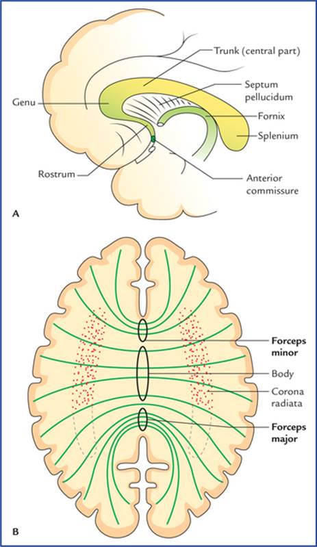
FIG. 14.3 Median sagittal section of the cerebrum showing shape and different parts of corpus callosum.
The corpus callosum connects all the parts of neocortex of two hemispheres except for the lower and anterior parts of temporal lobes which are connected by the anterior commissure.
External features and relations of corpus callosum
• Corpus callosum forms a massive arched interhemispheric bridge in the floor of the median longitudinal cerebral fissure connecting the medial surfaces of the two cerebral hemispheres.
• It forms massive, arched, interhemispheric bridge flooring the midline longitudinal fissure and roofing both the lateral ventricles.
• It lies nearer the anterior end (4 cm behind the frontal pole) of the hemisphere than the posterior end (6 cm in front of the occipital pole).
• In sagittal section of cerebrum it is seen as C-shaped mass of white fibres on the medial surface of the hemisphere forming the roof of the lateral ventricle (Fig. 14.4).
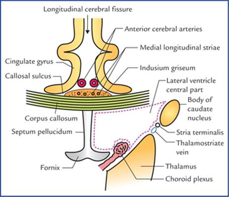
FIG. 14.4 Some of the relations of corpus callosum in the coronal section of the brain passing through the central parts of lateral ventricles parts.
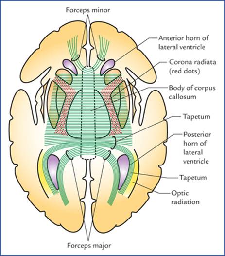
FIG. 14.5 Horizontal section of the cerebrum (schematic) to show the course and components of the fibres of corpus callo-sum. Note that the fibres of corona radiata are cut at right angle to their course.
• The concave inferior aspect of corpus callosum is attached with the convex superior aspect of the fornix by the septum pellucidum and its convex superior aspect is covered by a thin layer of grey matter, the indusium griseum, embedded in which are the fibre bundles of bilateral medial and lateral longitudinal striae.
• The anterior cerebral vessels often lie on the pia mater covering its superior aspect of corpus callosum.
• The superior aspect of corpus callosum is covered on each side by cingulate gyrus from which it is separated by a callosal sulcus.
Parts of the corpus callosum
The corpus callosum is divided from before backwards into four parts: (a) rostrum, (b) genu, (c) trunk/body, and (d) splenium (Fig. 14.3).
Genu: It is thick curved anterior extremity of corpus callosum which lies 4 cm behind the frontal pole. Genu forms the anterior boundary of the anterior horn of the lateral ventricle. The fibres of genu sweep (curve) forwards on either side into the anterior parts of the frontal lobes, forming a fork-like structure, the forceps minor.
Rostrum: The genu extends downwards and backwards as a thin prolongation to join the lamina terminalis forming, rostrum of corpus callosum. The rostrum forms the floor of the anterior horn of lateral ventricle and its fibres extends inferiorly to connect the orbital surfaces of the two frontal lobes.
Trunk: The trunk is main (middle) part of the corpus callosum between its thick anterior (genu) and posterior (splenium) extremities. Its fibres connect most of the frontal and anterior parts of the parietal lobes of the two cerebral hemispheres. The central part of the corpus callosum forms the roof of the central part of the lateral ventricle.
Splenium: The splenium is the massive posterior extremity of the corpus callosum, lying 6 cm in front of the occipital pole. It overhangs the thalamic pulvinars, pineal gland and tectum of the midbrain. The transverse fissure containing tela choroidea of third ventricle, posterior choroidal arteries and great cerebral vein of Galen is located inferior to the splenium.
The fibres of the splenium connect the posterior parts of the parietal lobes, and temporal and occipital lobes of the two hemispheres. The fibres connecting the occipital lobes sweep backwards on either side above the calcarine sulcus forming a large fork-like structure, the forceps major (Fig. 14.5). The forceps major forms a swelling in the upper part of the medial wall of the posterior horn of lateral ventricle, the bulb of the posterior horn.
N.B. The tapetum is the thin lamina of white fibres (2 cm thick) which forms the roof and lateral wall of the posterior horn; and lateral wall of the inferior horn of the lateral ventricle.
The tapetum is formed by those fibres of the trunk and splenium of corpus callosum which are not intersected by the fibres of corona radiata. In the coronal section, the tapetum looks whiter than the surrounding white matter because section passes parallel to the fibres of the tapetum.
Functions of the corpus callosum
The corpus callosum is largely responsible for interhemi-spheric transfer of information which is essential for bilateral responses and in learning processes. However, its congenital absence or surgical division does not produce any change in personality or intelligence and patients remain completely unnoticed of any definite neurological disorder in day-to-day life. Only special tests of tactile and visual systems will reveal any abnormality.
Clinical Correlation
Split-brain (Split-brain syndrome)
If the corpus callosum is congenitally absent or sectioned surgically each cerebral hemisphere becomes isolated and patient responds as if he/she has two separate brains, a condition called split-brain syndrome.
The severance of corpus callosum in young monkeys produces split-brain syndrome, viz. if they are trained to perform a task with one hand, they are unable to repeat the same act with the other hand.
Lesions of anterior corpus callosum result in akinetic mutism and tactile anomia whereas lesions of posterior corpus callosum result in alexia without agraphia.
N.B. The section of corpus callosum in past has been attempted surgically to prevent the spread of severe epileptic seizures from one hemisphere to the other.
Anterior commissure
The anterior commissure is a small round bundle of white fibres which crosses the midline in the upper part of the lamina terminalis, immediately in front of the anterior column of the fornix and the interventricular foramen.
Anterior commissure consists of two components (Fig. 14.6):
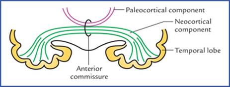
FIG. 14.6 Anterior commissure. The large neocortical component is shown in green and the small paleocortical component in maroon.
• A large posterior neocortical component, which interconnects the lower and anterior parts of the temporal lobes.
• A smaller anterior paleocortical component, which interconnects the olfactory regions (olfactory bulbs, olfactory tubercles, etc.) of the two hemispheres.
Seen from below the full extent of anterior commissure has the shape of a cupid's bow.
Posterior commissure
The posterior commissure is a slender bundle of white fibres which crosses the midline through the inferior lamina of the stalk of pineal gland.
It interconnects the superior colliculi, pretectal and interstitial nuclei of two sides.
Habenular commissure
The habenular commissure is a slender bundle of white fibres which crosses the midline through the superior lamina of the stalk of pineal gland. It interconnects the habe-nular nuclei of the two sides.
Hippocampal commissure (commissure of fornix)
Hippocampal commissure interconnects the crura of fornix and thus the hippocampal formations of the two sides (Fig. 19.10).
Projection fibres
The projection fibres connect the cerebral cortex to the subcortical centres (such as the corpus striatum, thalamus, brainstem) and spinal cord. These fibres are of two types:
• Corticofugal fibres go away from the cortex (cortical efferents) to centres in the other parts of the CNS.
• Corticopetal fibres come to the cerebral cortex from the other centres in the CNS.
The projection fibres of neocortex constitute the corona radiata and internal capsule while those of allocortex (i.e. archicortex and paleocortex) constitute the fimbria and fornix.
The most important bundles of projection fibres are: internal capsule and fornix.
Internal capsule
The internal capsule is a compact bundle of projection fibres between the thalamus and caudate nucleus medially and the lentiform nucleus laterally.
These fibres fan out rostrally to form the corona radiata and condense caudally to continue as the crus cerebri of the midbrain. The ascending (corticopetal/sensory) and descending (corticofugal/motor) fibres of internal capsule chiefly interconnect the cerebral cortex with the brainstem and spinal cord.
These fibres are mainly responsible for the sensory and motor innervation of the opposite half of the body.
N.B. Because of high concentration of motor and sensory nerve fibres within the internal capsule, even a small lesion may produce a widespread paralytic effects and sensory loss in the opposite half of the body.
Shape and boundaries of the internal capsule
In a horizontal section of the cerebral hemisphere, the internal capsule appears as a V-shaped compact bundle of white fibres with its concavity directed laterally (Fig. 14.7):

FIG. 14.7 Location, shape, boundaries and parts of the internal capsule.
It is bounded medially by the caudate nucleus and thal-amus, and laterally by the lentiform nucleus.
Parts of the internal capsule
The internal capsule is divided into following five parts (Fig. 14.7)
• Anterior limb, lies between the head of caudate nucleus medially and the anterior part of the lentiform nucleus laterally.
• Posterior limb, lies between the thalamus medially and the posterior part of the lentiform nucleus laterally.
• Genu, is the bend between the anterior and posterior limbs with concavity of the bend facing laterally.
• Retrolentiform part, lies behind the lentiform nucleus.
• Sublentiform part, lies below the lentiform nucleus.
Constituent fibres of the internal capsule (Figs 14.8-14.10)
Motor fibres (Fig. 14.8)
• Corticopontine fibres originate from the cerebral cortex of all the lobes of the cerebral hemisphere and form the largest single group of projection fibres in the internal capsule (about 2/3rd of the total fibre component). They are named according to the lobe from which they arise, e.g. frontopontine, parietopontine, occipitopontine and temporopontine, arising from frontal, parietal, occipital and temporal lobes respectively.

FIG. 14.8 The disposition of motor fibres passing through the internal capsule.
The frontopontine fibres are most numerous and pass through the anterior limb, genu, and posterior limb. The parietopontine and occipitopontine fibres pass through the retrolentiform part. The temporopontine fibrespass through the sublentiform part (Fig. 14.10).
The corticopontine fibres relay (synapse) in the ipsilat-eral pontine nuclei. The fibres arising from pontine nuclei cross the midline to relay in the cortex of the opposite cerebellar hemisphere, thus forming the cortico-ponto-cerebellar pathway. The corticoponto-cerebellar pathway is most recent in development and best developed in man.
• Pyramidal fibres arise in the cerebral cortex and relay in the lower motor neurons within the brainstem and spinal cord.
The pyramidal fibres are of two types:
– Corticonuclear fibres synapse with the contralateral motor nuclei of the cranial nerves which innervate the head and neck muscles. The corticonuclear fibres occupy the genu of the internal capsule.
– Corticospinal fibres synapse with the anterior horn cells of the opposite half of the spinal cord, which innervate the muscles of the upper limb, trunk and lower limb. The corticospinal fibres form several discrete bundles in the anterior two-third of the posterior limb. The fibres for the upper limb are most anterior, followed in that order, by the fibres for the trunk and the lower limb.
• Extrapyramidal fibres arise in the cerebral cortex and relay into the subcortical grey matter belonging to the extrapyramidal system, viz. red nucleus, corpus stria-tum, substantia nigra, etc. They are named according to their destinations, viz. corticorubral, corticostriate, cor-ticonigral, etc. respectively. Most of the extrapyramidal fibres occupy the position near the corticospinal fibres in the internal capsule, and are therefore affected in the lesions of the posterior limb.
Sensory fibres (Fig. 14.9A,B)
Sensory fibres are mostly thalamocortical fibres, which radiate from thalamus in different directions to reach the widespread areas of the cerebral cortex and constitute most of thalamic radiation. Most of the thalamocortical fibres, are the tertiary (3rd order) sensory neurons of various sensory pathways conveying somesthetic sensations to the cerebral cortex. The other fibres contributing the formation of thalamic radiation are corticothalamic fibres.

FIG. 14.9 (A) Direction of fibres of different groups of thalamic radiation. (B) Location of different groups of thalamic radiation in different parts of the internal capsule.

FIG. 14.10 Fibres passing through the sublentiform part of the internal capsule. (MGB = medial geniculate body.)
According to the direction of these fibres the thalamic radiation is divided into following subgroups:
• Anterior thalamic radiation: the fibres of anterior thal-amic radiation are directed anteriorly and connects the anterior and dorsomedial nuclei of thalamus to frontal lobe cortex.
• Superior thalamic radiation is directed superiorly. Its fibres pass through anterior limb of internal capsule and connect the ventral tier of thalamic nuclei with the sensory cortex of the frontal and parietal lobes.
• Posterior thalamic radiation is directed posteriorly. The fibres of posterior thalamic radiation pass through the retrolentiform part of internal capsule and connect the lateral geniculate body to the primary visual cortex of the occipital lobe forming optic radiation (geniculocal-carine tract) (seeFig. 14.11).

FIG. 14.11 Main parts of the internal capsule and fibres/tracts passing through them.
• Inferior thalamic radiation is directed inferiorly. Its fibres pass through sublentiform part of internal capsule and most of them connect the medial geniculate body with the primary auditory area of the temporal lobe forming auditory radiation (Fig. 14.11).
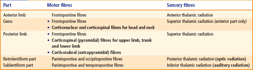
The constituent motor and sensory fibres in different parts of the internal capsule are summarized in Table 14.1 and shown in Figure 14.11.
Table 14.1
Constituent motor and sensory fibres in different parts of the internal capsule
Arterial supply of the internal capsule (Fig. 14.12)
The arterial supply of the internal capsule is of great clinical significance, due to high incidence of vascular lesions of internal capsule (called capsular lesions).
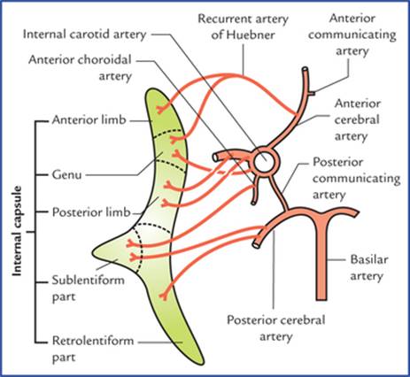
FIG. 14.12 Arteries supplying the internal capsule.
Various arteries supplying the internal capsule are:
• Medial and lateral striate branches of the middle cerebral artery. One of the lateral striate branches is larger and more frequently ruptured. It is often termed Charcot's artery of cerebral haemorrhage. It enters through the anterior perforated substance and supplies the posterior limb of the internal capsule.
• Striate branches of anterior cerebral artery. One of these branches is larger and takes a recurrent course. It is termed recurrent artery of Huebner. It arises just proximal to the anterior communicating artery, runs superior to the optic chiasma and penetrates the anterior perforated substance to supply the genu and anterior limb of the internal capsule.
• Central branches of the anterior choroidal artery supply the sublentiform part.
• Some direct branches from the internal carotid artery supply the genu.
• Central branches of the posterior communicating artery.
• Posterolateral central branches of the posterior cerebral artery supply the retrolentiform and sublentiform parts of the internal capsule.
Clinical Correlation
• Damage to the internal capsule, due to haemorrhage or infarction leads to loss of sensations and spastic paralysis of the opposite half of the body (contralateral hemiplegia).
The haemorrhage commonly occurs due to rupture of artery of cerebral haemorrhage (also called Charcot's artery of cerebral haemorrhage), which supplies the posterior limb of the internal capsule. The spastic paralysis of the opposite half of the body occurs due to the involvement of the pyramidal and extrapyramidal fibres for the upper limb, trunk and lower limb.
N.B. Rupture of Charcot's artery of cerebral haemorrhage is the most common cause of the hemiplegia.
• Involvement of recurrent artery of Huebner (due to thrombosis/rupture) results in paralysis of the face and upper limb on the opposite side (because of the involvement of corticonuclear fibres in genu and adjacent pyramidal fibres in the posterior limb for the upper limb).
• Lesions of the posterior one-third of the posterior limb, and sublentiform and retrolentiform parts of the internal capsule lead to visual (hemianopia) and auditory (loss of hearing) defects. These lesions usually occur due to thrombosis of the anterior choroidal artery, a branch of internal carotid artery.
Fornix
The fornix is described in detail on page 231.
Lateral Ventricles
There are two lateral ventricles one in each cerebral hemisphere. Each lateral ventricle is a roughly C-shaped cavity situated within each cerebral hemisphere. The lateral ventricle wraps itself around the thalamus, the lentiform nucleus, and the caudate nucleus. It is lined with ependyma and filled with cerebrospinal fluid. It has a capacity of about 7–10 ml. The main parts of two ventricles are separated from each other by a septum extending between corpus callosum and fornix called septum pellucidum. The septum pellucidum is a thin vertical sheet of nervous tissue consisting of grey and white matter, and covered on either side by the ependyma.
Each lateral ventricle communicates with the third ventricle through the interventricular foramen (of Monro). Most of the CSF in the CNS is produced by the choroid plexuses of two lateral ventricles.
The projection of ventricles on the surface of the brain is shown in Figure 14.13.
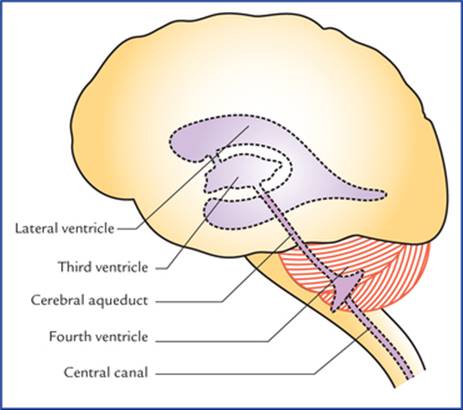
FIG. 14.13 Surface projection of ventricles on the left lateral aspect of the brain.
Parts of Lateral Ventricle (Fig. 14.14)
For descriptive purposes, each lateral ventricle is divided into four parts:
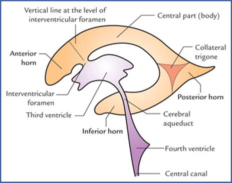
FIG. 14.14 Ventricular system of the brain; lateral view. Note the different parts of the lateral ventricle.
• Central part or body lies mostly within the parietal lobe and extends from interventricular foramen in front to the splenium of the corpus callosum behind.
• Anterior horn is the anterior extension from the central part into the frontal lobe and, lies in front of interven-tricular foramen and behind the posterior surface of the genu of corpus callosum.
• Posterior horn is the backward extension from the central part into the occipital lobe towards the occipital pole.
• Inferior horn is considered as the direct continuation of the main ventricular cavity into the temporal lobe. The inferior horn is the largest of the three horns. It begins where the central part and posterior horn meet and curves round the pulvinar of thalamus into the temporal lobe to end about 2.5 cm behind the temporal pole.
N.B. The posterior horn is the most recent one in evolution scale amongst the three horns.
Boundaries of the Different Parts of Lateral Ventricle
Central part or body
Central part or body is triangular in shape in coronal section with a medial wall, a roof, and a floor (Fig. 14.15).

FIG. 14.15 Boundaries of the central part of the lateral ventricle.
The roof is formed by the under surface of the body (trunk) of the corpus callosum.
The floor slopes downwards from lateral to the medial side and is formed in that order by:
• Body of caudate nucleus.
• Stria terminalis and thalamostriate vein.
• Lateral part of the upper surface of the thalamus.
• Choroid plexus covering the medial part of the upper surface of the thalamus.
• Upper surface of the body of fornix.
The medial wall is formed by the septum pellucidum.
Anterior (frontal) horn
Anterior horn is roughly triangular in coronal section, and presents roof, floor, anterior, medial and lateral walls (Fig. 14.16).
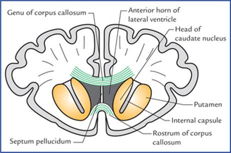
FIG. 14.16 Coronal section of brain at the level of rostrum of corpus callosum, showing boundaries of anterior horn of the lateral ventricle.
The roof is formed by the undersurface of the anterior part of the body of corpus callosum.
The narrow floor is formed by the upper surface of the rostrum of corpus callosum.
The anterior wall is formed by the genu of the corpus callosum.
The medial wall is formed by the septum pellucidum.
The lateral wall is formed by the bulging head of the caudate nucleus.
Posterior (occipital) horn
Posterior horn is quadrangular or diamond-shaped in coronal section and presents roof, lateral wall, floor, and medial wall (Fig. 14.17).

FIG. 14.17 Boundaries of posterior horn of the lateral ventricle.
The roof, lateral wall, and floor are formed by a sheet of fibres (tapetum) from the splenium of the corpus callosum. The posteriorly sweeping fibres of the optic radiation remain separated from the cavity of the posterior horn by the tapetum.
The medial wall is invaginated by two ridges; the upper of these, the bulb of posterior horn is formed by the fibres of forceps major and the lower one, the calcar avis is produced by the anterior part of the calcarine sulcus.
Inferior (temporal) horn
The inferior horn is the largest and longest of the three horns. It begins where the central part and posterior horn meet. From here it curves ventrally downwards and forwards into the temporal lobe. The area where inferior horn and posterior horn diverge is called collateral trigone (Fig. 14.14). The inferior horn lies more or less parallel to the superior temporal sulcus and in coronal section appears as a transverse-slit presenting roof and floor (Fig. 14.18).

FIG. 14.18 Boundaries of inferior horn of lateral ventricle as seen in coronal section of brain.
The lateral part of the roof is formed by tapetum of corpus callosum, and the medial part by the tail of caudate nucleus and stria terminalis.
The floor presents following features from lateral to medial side:
• Collateral eminence, an elongated swelling in the lateral part of the floor, produced by the collateral sulcus, which is deep enough to produce this elevation.
• Hippocampus, an another longitudinal elevation lying parallel and medial to the collateral eminence.
The fibres of hippocampus form a thin sheet of white matter called alveus that covers its ventricular surface. The fibres of alveus converge medially to form a ridge called fimbria (for detail seepage 232).
• Choroid plexus: Most medially the floor is occupied by the choroid plexus that passes into the inferior horn through the choroid fissure lying between the fimbria below, and the stria terminalis and the tail of the caudate nucleus above.
Choroid plexus and choroid fissure
On the medial aspect of the cerebral hemisphere, along the C-shaped line between the diencephalon and hemisphere, the medial wall of the central part and inferior horn of the lateral ventricle is made up of only ependyma. The pia mater covering the ependyma along this C-shaped line is invagi-nated by the fringe-like tuft of blood vessels into the central part and inferior horn to form the choroid plexus of the lateral ventricle. The line of invagination of the choroid plexus into the lateral ventricle is called cho roid fissure.
At the interventricular foramina, the choroid plexus of one lateral ventricle is continuous with its counterpart of the other side across the third ventricle.
The choroid plexus of lateral ventricle is derived from anterior choroidal artery, a branch of internal carotid artery and the posterior choroidal artery a branch of posterior cerebral artery.
Clinical Correlation
• The general form of the ventricular system of the brain can be visualised by:
(a) computerized axial tomography (CT scanning), and magnetic resonance imaging (MRI), and
(b) ventriculography, a radiological technique in which a small quantity of air or oxygen is introduced via a needle into the lateral ventricle through a burr hole in the skull (in children below two years of age, the needle is inserted through the lateral angle of the anterior fontanelle).
• The outline of ventricles and cerebral gyri can be visualized by air encephalography (also called pneu-moencephalography) in which the air or oxygen is introduced into the subarachnoid space through lumbar puncture. The air readily replaces the CSF within the ventricles and subarachnoid space. The air or gas being less dense than the fluid or neural tissue, the ventricles and cerebral gyri are easily visualized.
Clinical Problems
1. Explain, why the congenital absence of corpus callosum goes unnoticed by an individual or even by the physician?
2. Explain, why a small lesion in the internal capsule has a widespread sensory and motor effect as compared to that in the corona radiata?
3. A ‘CT scan’ of skull of older individuals (transverse cuts) through the level of the anterior horns of the lateral ventricles often shows three small calcified shadows: one in the midline, and one on each side in the region of posterior horn of lateral ventricle. Name the structures of the brain which give rise to these shadows and why?
Clinical Problem Solving
1. The cortex of the right cerebral hemisphere controls the muscular activity of, and receives sensory input from the left half of the body; on the other hand, the left cerebral hemisphere controls the muscular activity of and receives the sensory input from the right half of the body. The two hemispheres thus can perform most of the day-to-day motor and sensory functions independently. Therefore, in day-to-day life the patient does not notice any definite neurological disorder and carry on his day-to-day activities perfectly well. For this reason, it goes unnoticed by an individual or even by the physician (also seepage 164).
2. A small lesion in the internal capsule produces extensive motor and sensory effects because the internal capsule consists of the huge number of motor and sensory fibres which are packed densely in a narrow space between the lentiform nucleuses laterally, and the caudate nucleus and thalamus medially. A similar lesion in corona radiata may have little or no effects as here, the fibres are widely separated from each other.
3. The midline shadow indicates the calcified pineal gland while the lateral shadows are due to calcified deposits in the choroid plexuses of the lateral ventricles.
It is important to note that with age:
(a) the calcium phosphates and carbonates get deposited in the pineal gland (seepage 133), and
(b) the choroid plexuses of lateral ventricles especially in their posterior horns show calcified deposits.