
Reticular Formation
The reticular formation is defined as diffuse ill-defined mass of intermingled neurons and nerve fibres occupying the entire core of brainstem (Fig. 19.1). The reticular formation has derived its name from its light microscopic appearance of a vague network of nerve cells and nerve fibres. It has been defined to include all areas within the brainstem (except the named nuclei and tracts) which when stimulated will produce arousal.
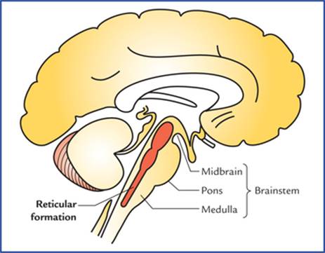
FIG. 19.1 Location of reticular formation in the brainstem.
Phylogenetically it represents the old reticular core of brain and contains within it the vital cardiac and respiratory centres which control respiration, heart rate and blood pressure. In primitive vertebrates, the diffuse arrangement of neurons was named ‘reticular’.
The reticular formation receives data from most of the sensory systems and has efferent (direct or indirect) connections with all the levels of neuraxis.
The knowledge of reticular system is important, because:
(a) it regulates levels of consciousness, and alertness,
(b) i t regulates respiration, blood pressure, heart rate and other vegetative functions,
(c) it regulates tone of skeletal muscles, and
(d) it modulates the impulses in the pain pathways.
Clinical Correlation
Damage of the reticular activating system in the core of the brainstem leads to progressive loss of consciousness, followed by stupor, coma and death.
Anatomical Extension
The reticular formation extends cranially to the dienceph-alon (subthalamus, hypothalamus and thalamus) and caudally to the spinal cord in the cervical region. These extensions are either actual or projectional. According to some authorities some centres of cerebrum and cerebellum are also closely related functionally to the reticular formation of brainstem.
Although reticular formation is described to be consisting of network of nerve fibres and scattered neurons, among them a number of regions with fairly localized cell groups called reticular nuclei, have been recognised.
Reticular nuclei in the brainstem
The reticular nuclei in brainstem are arranged into three longitudinal columns (Fig. 19.2).

FIG. 19.2 Schematic diagram to show the reticular nuclei in the brainstem.
• Median column lies in the midline and consists of intermediate size neurons. The nuclei of this column are termed raphe nuclei.
• Medial column consists of nuclei which are made up large-size neurons, hence this column is also termed magnocellular column.
• Lateral column consists of nuclei which are made up of small neurons, hence this column is also termed parvo-cellular column (parvus = little, small).
The nuclei belonging to these columns are shown in Figure 19.2. Since it is not advisable for the student to burden his memory with the names of all the nuclei, only those which have a functional or descriptive value are labelled.
The reticular nuclei as seen in transverse sections of the midbrain, pons and medulla are shown in Figure 19.3.
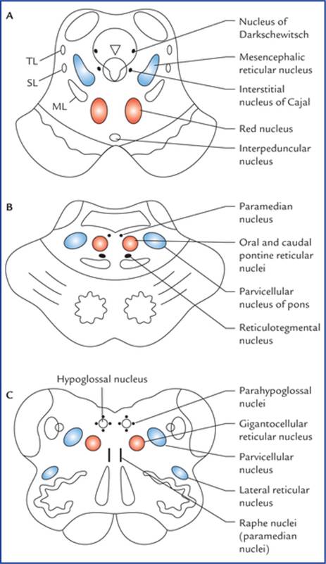
FIG. 19.3 The transverse sections of: midbrain (A), pons (B), and medulla (C) showing the location of reticular nuclei. (ML = medial lemniscus, SL = spinal lemniscus, TL = trigeminal lemniscus.)
Raphe nuclei (median group of nuclei)
The raphe nuclei form a contiguous column in the mid-line. The neurons of raphe nuclei produce serotonin, a substance that they use as a neurotransmitter. The dorsal raphe nucleus located in the midbrain projects to the spinal cord and forms the pain controlling pathway.
The nucleus raphe magnus in medulla projects to the caudal part of the spinal nucleus of the trigeminal nerve and influences perception of pain through spinal nucleus.
In fact once the raphe nuclei (vide supra) are activated, the serotogenic neurons inhibit the transmission of pain impulses from dorsal grey horns and spinal nucleus of trigeminal nerve respectively.
Clinical Correlation
The electrical stimulation of either the dorsal raphe nucleus (the periaqueductal grey matter) or the nucleus raphe magnus results in loss of the ability to experience pain from sites of injury or disease. The former procedure has been used clinically in the management of otherwise intractable pain.
Medial group of nuclei
The medial group of nuclei includes ventral reticular nucleus (in medulla), gigantocellular nucleus (in medulla and pons) and oral and caudal pontine nuclei (in pons). Nuclei of this group receive afferents from nuclei of lateral group and efferents from these nuclei ascend or descend longitudinally in the brainstem and give collaterals to the other reticular cells, thus forming a polysynaptic pathway—a characteristic of impulse transmission through the reticular formation.
Lateral group of nuclei
The lateral group of nuclei includes parvicellular nuclei of medulla and pons, nucleus locus ceruleus of pons and pedunculopontine nucleus of the midbrain.
These nuclei receive collaterals from several ascending pathways and project to the medial group of nuclei of the reticular formation. They are regarded as an association region of the reticular formation.
Connections of Reticular Formation
The reticular formation receives information from almost all the principal parts of the nervous system and in turn, projects (directly or indirectly) to all these parts (Fig. 19.4).

FIG. 19.4 Connections of reticular formation. (P = putamen, and GP = globus pallidus.)
The reticular pathways are polysynaptic, both ascending, descending, and crossed and uncrossed. As a result a unilateral stimulation produces bilateral responses.
Afferent connections
The afferents are classified into three types:
Afferents from various sensory pathways or systems
– Optic system—through tectoreticular fibres,
– Olfactory and limbic systems—through variety of descending pathways,
– Auditory system—through tectoreticular fibres,
– Gustatory system,
– Spinal pathways—through spinoreticular fibres. A considerable number of fibres of spinothalamic tract terminate in the lateral reticular nucleus of medulla, which in turn project to the cerebellum. Spinoreticulo-cerebellar pathway is an important pathway for carrying exteroceptive sensations to the cerebellum,
– Trigeminal pathways.
Afferent fibres from other parts of central nervous system
– Cerebellum from both but mainly from contralateral fastigial nucleus.
– Basal ganglia, mainly from corpus striatum.
– Thalamus, hypothalamus and subthalamus.
– Limbic system, mainly from septal areas, amygdaloid nuclei, and hippocampus.
– Cerebral cortex mainly from motor and sensory areas of the cerebral cortex.
– Red nucleus, substantia nigra and habenular nuclei.
Other factors influencing the activity of reticular formation
– Hormones and chemical substances, viz. adrenaline, ace-tylcholine and carbon dioxide.
– Drugs, viz. barbiturates, anaesthetics and tranquillizers.
Efferent connections
The efferent connections of reticular formation are to all the parts of CNS from which it receives afferents but mainly to:
• Autonomic and locomotor control centres of brainstem and spinal cord.
• Cranial nerve nuclei, e.g. dorsal nucleus of vagus.
• Cerebral cortex—indirectly through diencephalic nuclei.
• Red nucleus, substantia nigra and tectum of midbrain.
Functional Divisions of Reticular Formation
Functionally the reticular formation is divided into two systems: (a) the ascending reticular activating system (ARAS), and (b) the descending reticular system (DRS). The ascending reticular activating system is commonly termed by the clinicians simply as reticular activating system (RAS).
Ascending reticular activating system (Fig. 19.4)
Most of the ascending tracts, viz. spinothalamic tract, trigem-inal lemniscus, lateral lemniscus and central vestibular pathway, while passing through the brainstem give collaterals to the lateral part of the reticular formation which projects to the intralaminar and reticular nuclei of the thalamus. These nuclei in turn project to the widespread areas of the cerebral cortex.
When this part of reticular formation is stimulated, the individual becomes alert hence it is termed ascending reticular activating system.
The ascending reticular activating system is believed to be responsible for maintaining a state of alertness and consciousness.
Clinical Correlation
• The visual and acoustic stimuli can stimulate the reticular activating system to maintain alertness and attention, therefore the stimuli such as sounds of ringing alarm clock or sudden bright light, can arouse consciousness. Conversely, removal of visual and auditory stimuli may lead to drowsiness and sleep.
• The functions of reticular activating system (RAS) can be affected by certain drugs. For example, general anaesthetics and tranquillizers cause its suppression. On the other hand, ammonia and other irritants stimulate it.
• A coma is a state of unconsciousness (due to inactivity of RAS). In coma, even the most powerful external stimuli cannot cause arousal.
The sleep is thought to occur because of a decrease in activity within the RAS.
N.B. The nuclei within the reticular formation, generate a continuous flow of impulses unless they are inhibited by other parts of the brain or drugs.
Descending reticular system
Descending reticular system consists of descending pathways from reticular formation to the autonomic centres in the brainstem and, the lateral and anterior horn cells in the spinal cord (see reticulospinal tracts in Chapter 7).
Clinical Correlation
The descending fibres from reticular formation constitute one of the most important motor pathways. The fibres from reticular formation to autonomic centres in the brainstem are critical in controlling respiratory and cardiac rhythms and other vital functions.
Functions of Reticular Formation
• Maintains the normal state of consciousness or wakefulness through its connections with cerebral cortex by way of ascending reticular activating system.
• Regulates respiration, heart rate blood pressure and other vegetative functions through autonomic reflex centres present within it in the brainstem.
• Controls muscular activity, directly through reticulospi-nal projections to lower motor neurons and indirectly by influencing the activities of cerebellum, red nucleus, substantia nigra, corpus striatum, and cerebral cortex.
• Controls receptivity of sensory end organs.
• Controls threshold of central sensory pathways.
• Regulates endocrine, visceral and emotional functions, through its connections with hypothalamus and limbic lobe.
Therefore, reticular formation constitutes the one, if not the most important regulatory mechanisms within the CNS.
Limbic System
The word limbus means ring, the term limbic system is applied to the parts of the cortical and subcortical structures that form a ring around the upper end of the brainstem.
The limbic system was formerly called rhinencephalon because of its association to olfaction, but in human beings only a small part of it is actually concerned with smell.
The limbic cortex is phylogenetically oldest part of the cerebral cortex and made up of primitive type of cortical tissue called allocortex which consists of only three layers and surrounds the hilum of the cerebral hemisphere. There is second ring of transitional cortex called juxta-allocortex between the allocortex and the neocortex. It consists of three to six layers. The cortical tissue of remaining non-limbic portion of the hemisphere is called neocortex which is made up of six layers and most highly developed in man.
The limbic system plays a vital role in elaboration of emotional behaviour, drive, and memory.
Functions of the Limbic System
The limbic system is functionally associated with following neural activities:
• Emotional aspects of behaviour together with visceral responses accompanying these emotions, particularly the reactions of fear and anger and emotions associated with sexual behaviour which are necessary for:
– survival of an individual including procuring of food and eating behaviour, and
– survival of the species including the sex behaviour.
• Brain mechanisms responsible for recent memory.
• Integration of olfactory, visceral and somatic impulses reaching the brain.
N.B. The visceral responses following activities in limbic system are expressed through hypothalamus by way of autonomic nervous system. Because of visceral responses to activities in the limbic system, it is also known as visceral brain.
Components of the Limbic System
The structures forming the limbic system are interposed between the superolateral surfaces of the diencephalon and the inferomedial surfaces of the two cerebral hemispheres. Many of these structures have highly arched forms.
A large number of structures of the brain are included in the limbic system. However, a fairly accepted list of these structures is presented here.
Regions of grey matter in limbic system
Cortical structures
• Limbic lobe, consisting of cingulate gyrus, isthmus, parahippocampal gyrus and uncus (anterior part of the parahippocampal gyrus) (Fig. 19.5).
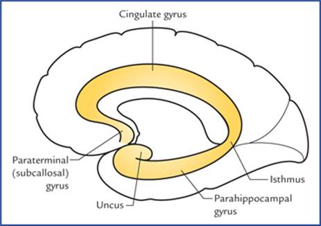
FIG. 19.5 Limbic lobe consisting of cingulate gyrus, isthmus, parahippocampal gyrus, and uncus.
• Hippocampal formation (Fig. 19.6) which includes hippocampus (cornu ammonis), dentate gyrus, gyrus fasciolaris and indusium griseum.
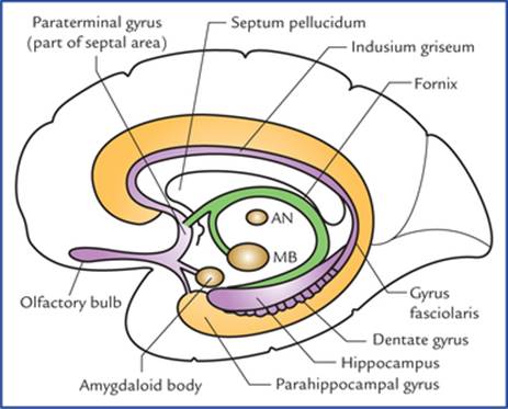
FIG. 19.6 Structures forming hippocampal formation (viz. hippocampus, dentate gyrus, gyrus fasciolaris and indusium griseum) and associated structures. (MB = mammillary body of hypothalamus, AN = anterior nucleus of thalamus.)
N.B. The cingulate gyrus is a ‘satisfaction centre’ of brain and associated with the feeling of satisfaction after a meal or after sexual intercourse.
Subcortical nuclei
• Amygdaloid nuclear complex (also called amygdaloid body).
• Septal region and nuclei.
• Olfactory areas (see Chapter 18).
• Hypothalamus especially the mammillary bodies.
• Anterior nucleus of thalamus.
Amygdaloid Nuclear Complex (Also Called Amygdaloid Body or Amygdala)
Amygdaloid nuclear complex is an almond-shaped mass of grey matter underlying the rostral part of the parahippocampal gyrus on the anteriormost part of the roof of the inferior horn of lateral ventricle.
Posteriorly the amygdaloid body becomes continuous with tail of caudate nucleus and stria terminalis (Fig. 13.4).
Connections (Fig. 19.7)
Afferents: Main afferents to amygdaloid body are from primary olfactory regions.
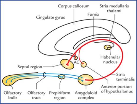
FIG. 19.7 Main afferent and efferent connections of the amygdaloid complex. Note the course of the stria terminalis. (AC = anterior commissure).
Efferents: Stria terminalis forms the main efferent tract of the amygdaloid body. It takes a circuitous route along with (but not functionally related to) the tail of caudate nucleus in close relation to the lateral ventricle until the level of anterior commissure, where majority of its fibres terminate in the septal area and anterior portion of the hypo-thalamus. The others join the anterior commissure and are distributed to the contralateral amygdaloid body. Some fibres run caudally to reach the habenular nucleus through stria medullaris thalami.
In general the amygdaloid body plays an important role in controlling the somatic responses to internal needs, drives or instincts. Since part of it receives olfactory input, it is believed that the amygdaloid body plays an important role in smell-mediated sexual behaviour.
Stimulation of amygdaloid body produces excitability, fear and rage. Bilateral damage of amygdaloid body reduces fear and increases sexual activity.
N.B. People in late sixties become pervasive in their sexual behaviour, probably due to atrophy of amygdaloid bodies.
Septal Region
The septal region is on the medial aspect of the frontal lobe beneath the genu and rostrum of corpus callosum and in front of the lamina terminalis. The septal region includes paraterminal and parolfactory gyri. The cerebral cortex in this region is called septal area.
The septal area has been shown to be a pleasure zone of brain in rats.
Hippocampal Formation
The hippocampal formation consists of: (a) hippocampus, (b) dentate gyrus, (c) subiculum, (d) indusium griseum, and (e) medial and lateral longitudinal striae.
Hippocampus (also called ram's horn or Ammon's horn)
Hippocampus is an area of cerebral cortex which has rolled into the floor of the inferior horn of the lateral ventricle during fetal life. In adult brain it forms a longitudinal elevation in the floor of inferior horn of the lateral ventricle and is continuous medially with the subiculum and para-hippocampal gyrus.
The name ‘hippocampus’ meaning ‘sea horse’, is derived from its appearance in coronal section (Fig. 19.8).
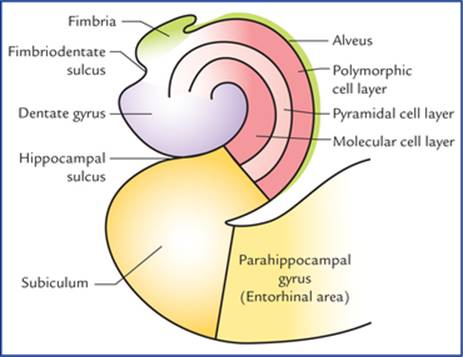
FIG. 19.8 Coronal section of the hippocampus and related structures.
In the frontal section the hippocampus is ‘C-shaped’ and its outline bears a resemblance to a ram's horn, hence the name ram's horn. It is also called Ammon's horn after an Egyptian deity with ram's head. Its anterior extremity is expanded and bears few grooves and intervening ridges. Because of its resemblance to an animal's paw it is termed pes hippocampi (pes = foot). Traced posteriorly the hippocampus gradually narrows and ultimately ends beneath the splenium of corpus callosum.
The ventricular surface of hippocampus is covered by a thin layer of white fibres called alveus. The fibres of alveus originate in the hippocampal cortex, course towards the medial border of hippocampus where they converge to form a narrow strip of white matter, the fimbria of hippocampus.
Phylogenetically, hippocampus represents the archicortex and consists of three layers. These are:
• Superficial molecular layer.
• Middle pyramidal cell layer.
• Deep polymorphic cell layer.
N.B. The parahippocampal cortex (neocortex) is made up of six layers. In the region known as subiculum, there is gradual transition from six-layered neocortex to the three-layered archicortex.
Connections (Fig. 19.9)
Afferents: Hippocampus receives fibres mainly from ento-rhinal area (area 28).
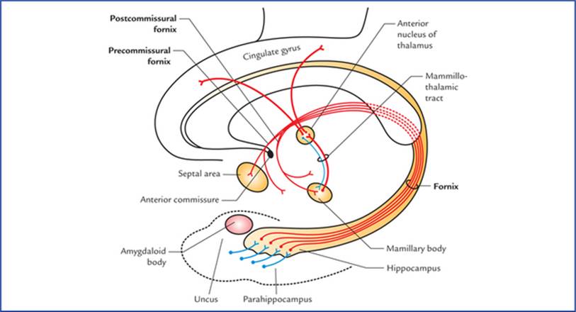
FIG. 19.9 Connections of hippocampus.
Efferents: The fornix is the main efferent tract of the hippocampus.
The fibres leaving the hippocampus pass:
• To the opposite hippocampus through the commissure of fornix/hippocampal commissure.
• To the septal and anterior hypothalamic regions.
• To the mammillary body which sends impulses to cin-gulate gyrus through anterior nucleus of thalamus.
Functions of hippocampus
• Formerly it was regarded as the part of olfactory system but it has no direct connections with the sense of smell in man.
• In man it is an integrative centre which influences endocrine and visceral functions and emotional states through its connections with hypothalamus, septal nuclei, and the cingulate gyrus. It was once considered as the seat of soul.
• It plays an important role in recent memory.
Clinical Correlation
• The hippocampus categorizes the afferent information related to recent memory and forms the new concepts. Then it correlates the new concepts learned for the first time with the pre-existing memory. The conceived facts then become stored in the cerebral cortex as memory. Thus if the hippocampus is damaged or is in a state of shock, the new memory is not formed. As a result the patient cannot tell about, the happenings at the time of accident (loss of recent memory/amnesia) but remembers the past happenings for the old memory is already stored in different parts of the cerebral cortex.
Emotionally charged memories are affected more than the non-emotional ones.
• The hippocampus is the most epileptogenic part of the cerebral hemisphere. Its lesions may cause psychomotor epilepsy.
Dentate gyrus, indusium griseum, and medial and lateral longitudinal striae
In the fetal brain, the dentate gyrus develops as a further extension of the hippocampus and occupies the interval between the hippocampus and the parahippocampal gyri, lying deep to fimbria. It has a three-layered archicortex. Its surface is toothed hence the name dentate gyrus. When traced anteriorly the dentate gyrus runs medially across the inferior surface of uncus. This part is called tail of dentate gyrus. The posterior end of dentate gyrus is continuous with the splenial gyrus or gyrus fasciolaris, which continues as thin layer of grey matter over the corpus cal-losum called indusium griseum.
The indusium griseum is the vestigial grey matter and contains two delicate longitudinal bands of fibres buried in it, the medial and lateral longitudinal striae.
Subiculum
It is a transition zone between three-layered archicortex and six-layered neocortex. It receives input from hippocampus and projects through the fornix to the mammillary nuclei and anterior nucleus of the thalamus.
Fibre bundles of limbic system
• Fornix.
• Mammillothalamic tract.
• Stria medullaris thalami.
• Stria terminalis.
• Medial forebrain bundle.
• Anterior commissure.
• Cingulum.
• Diagonal band (of Broca).
Several of these components of limbic system are already discussed with olfactory system and diencephalon. The remaining ones are described here.
Fornix (Fig. 19.10)
The fornix is a large bundle of mainly projection fibres which connects the hippocampus with the mammillary body. It constitutes the sole efferent system of the hippocampus.
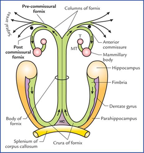
FIG. 19.10 Schematic diagram showing parts of fornix and its connections. (T = anterior nucleus of thalamus, MT = mammillothalamic tract, HC = hippocampal commissure).
On the medial surface of cerebral hemisphere, it is seen as an arched prominent bundle of white fibres below the corpus callosum, along the lower border of septum pellucidum.
There is one fornix in each cerebral hemisphere but two are so closely related/fused beneath the middle of the body of corpus callosum that they are usually described as a single structure.
Origin, course and distribution of its fibres
The fibres of fornix arise mainly from the pyramidal cells of the hippocampus and form a thin layer of white fibres on its ventricular surface called alveus.
The fibres of ‘alveus’ collect on the medial margin of hippocampus to form a narrow strip of white matter, the fim-bria, lying flat over the dentate gyrus. The fimbria becomes a rounded band, the crus of fornix as it arches upwards, medially and forwards underneath the splenium of corpus callosum. The two crura, one of each hemisphere, curving over the thalamus, converge and unite in the midline beneath the trunk of corpus callosum to form the body of fornix.
Anteriorly, the body of fornix divides into two columns, the columns of fornix. Each column of fornix arches downwards towards the anterior commissure, and forms the anterior boundary of interventricular foramen. Then it curves posteriorly through the hypothalamus to end in the mammillary body. These fibres being located posterior to anterior commissure are referred to as postcommissural fornix. For some fibres of column pass in front of anterior commissure to end in the septal area and anterior hypo-thalamic region, etc. to constitute the precommissural fornix.
N.B.
• Hippocampal commissure of fornix. The two crura are interconnected by fibres passing from one to another. These crossing fibres interconnect the two hippocampi and form the commissure of fornix/hippocampa! commissure.
• Some fibres of fornix pass above the splenium of corpus callosum to end in the cingulate gyrus of the same side and constitute the dorsal fornix made up of association fibres.
• Most of the fibres of fornix is made up of projection fibres connecting hippocampus with the mamillary body
Thus, fornix consists of three types of white fibres: (a) projection fibres, (b) commissural fibres, and association fibres.
Clinical Correlation
Bilateral transection of the fornix may cause a clinical condition called ‘acute amnestic syndrome’ in which an individual is unable to consolidate his short-term memory into long-term memory
Mammillothalamic tract (also called bundle of Vicq d'Azyr)
Mammillothalamic tract is a prominent bundle of fibres which carry impulses from mammillary body to the anterior nucleus of thalamus. It also includes some thalamomammary fibres. Mammillothalamic tract is readily demonstrable by gross dissection. The efferents from anterior nucleus of thala-mus are projected mainly to areas 23 and 24 of the cingulate gyrus but some fibres are also shunted to the tegmental nuclei of the midbrain through mammillo-tegmental tract.
Papez Circuit
The Papez circuit includes the following limbic structure (Flowchart 19.1):
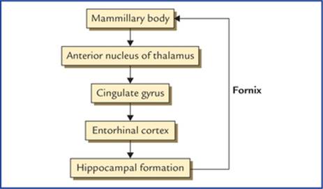
FLOWCHART 19.1 Papez circuit.
1. Hippocampal formation.
2. Mammillary body/nucleus.
3. Anterior nucleus of thalamus.
4. Cingulate gyrus.
5. Entorhinal cortex.
The route followed by the circuit of Papez is as under: Hippocampal formation to mammillary nucleus to cingu-lated gyrus to entorhinal cortex to hippocampal formation.
The structures included in the Papez circuit and their connections constitute a harmonious mechanism which elaborate central emotion and emotional expressions.
Clinical Problems
1. A light slapping or tickling of person's face or splashing water on it, is a common and effective technique for arousing him from sleep. Similarly cotton-wisp soaked in liquid ammonia is put near the nostrils to arouse an unconscious patient. Why?
2. What is the neuroanatomical basis of the state of consciousness?
3. A 55-year-old individual met a road-traffic accident and became unconscious thereafter. He was taken to the hospital where he regained consciousness. His CT scan of head did not reveal any brain lesion. When he was enquired—where and how accident took place he could not answer but he could tell the address of his residence and place of work. Provide the anatomical basis.
4. What is the anatomical basis of schizophrenia? Discuss its presenting features.
5. What is Kiuver-Bucy syndrome?
Clinical Problem Solving
1. This is because these activities stimulate the trigeminal nerve on the face and in the nasal mucosa respectively, which in turn stimulates the reticular activating system (RAS).
2. The state of consciousness means that the patient should be oriented to time, place, and person. Further, he should be able to appropriately respond to questions and environmental stimuli.
The consciousness has two facets, namely arousal and awareness, which depend on two brain structures: (a) the brainstem reticular activating system (RAS), and (b) the cerebral cortex.
The arousal is the phenomenon of being awake, and it is primary function of the RAS, a nonspecific transmission system for sensory inputs which activate the cerebral cortex. The awareness is more sophisticated function requiring intact cortical activity in order to interpret the sensory input and respond accordingly.
3. The reticular activating system (RAS) is very sensitive part of the brain, as a result, even a blow on head can stop its functioning for sometime leading to unconsciousness. The loss of recent memory occurred due to involvement of hippocampus.
4. The schizophrenia is a mental disorder which occurs due to involvement of limbic system. It is characterized by: (a) chronically disordered thinking, (b) blunting of emotional responses, (c) depression and anxiety, and (d) amnesias and phobias.
5. The Kiuver-Bucy syndrome consists of number of signs and symptoms in monkeys following removal of both temporal lobes, viz. (a) docility, (b) loss of fear and anger, (c) increased appetite, and (d) increased sexual activity which is often perverse.
N.B. This syndrome has also been described in humans following removal of large areas of temporal lobe on both the sides.