
The autonomic (self-controlling) nervous system is that part of the nervous system which regulates most of the involuntary activities of the body, such as the activities of smooth muscles of bronchial tree, gut, genitourinary system, pupil, arrector pili muscles of the hair, cardiac muscle and secretion of the glands. Thus it represents the visceral component of the nervous system, hence sometimes also called ‘visceral nervous system’.
The autonomic nervous system, like the somatic nervous system is made up of afferent and efferent pathways.
The visceral afferent pathways resemble somatic afferent pathways. The cell bodies of first order sensory neurons (pseudo-unipolar) are located in the cranial and dorsal root ganglia. There peripheral processes are distributed through autonomic ganglia or plexuses or through somatic nerves. The central processes accompany somatic afferent fibres through cranial nerves or dorsal spinal roots into the central nervous system when they establish connections to mediate autonomic reflexes and visceral sensation.
Thus the afferent fibres of the autonomic nervous system are identical to the afferent fibres of the somatic nervous system and form part of general afferent component of the entire nervous system. Visceral efferent pathways, however, differ from those of somatic efferent pathways (i.e. the efferent fibres, differ in the somatic and autonomic nervous systems).
Somatic efferent fibres pass directly from the CNS to the skeletal muscle, whereas autonomic (visceral) efferent fibres do not pass directly to the visceral effector organs from the CNS, instead they first relay in the autonomic ganglia outside the CNS, and then the postganglionic fibres supply the effector organs. The only exception is the innervation of the adrenal medulla.
Thus the efferent pathway of autonomic nervous system is made up of two neurons, preganglionic and postganglionic.
For all practical purposes, the autonomic nervous system (ANS) is a general visceral efferent motor system which controls and regulates smooth muscle, cardiac muscle and glands. These three divisions of autonomic nervous system differ in their organization and structure but they are closely related functionally.
N.B. All the efferent peripheral nerve fibres belong to the autonomic nervous system except those to the skeletal muscles.
Divisions of the Autonomic Nervous System
The autonomic nervous system is divided into three divisions, the sympathetic nervous system and parasympathetic nervous system, and the enteric nervous system.
1. Sympathetic nervous system: The preganglionic sympathetic fibres arise from lateral horn cells of the thoracic and upper two lumbar segments (T1 to L2) of the spinal cord, hence sympathetic nervous system constitutes the thoracolumbar outflow (Fig. 20.1). The axons of preganglionic neurons project to the autonomic ganglia (the sympathetic ganglia). The ganglia are connected to each other and form a beaded chain.

FIG. 20.1 Schematic diagram to show the craniosacral outflow (parasympathetic) and thoracolumbar outflow (sympathetic). Note that postganglionic parasympathetic fibres are shorter in length as compared to postganglionic sympathetic fibres.
2. Parasympathetic nervous system: The preganglionic parasympathetic fibres arise from general visceral efferent (GVE) nuclei of brainstem and lateral horn cells of the second, third and fourth sacral segments of the spinal cord, hence parasympathetic nervous system constitutes the craniosacral outflow (Fig. 20.1). The axons of preganglionic fibres project to the autonomic ganglia (the parasympathetic ganglia) which are located near or embedded in the wall of viscera/glands.
The sympathetic ganglia are located near the CNS along the sympathetic chain in the paravertebral region or in front of vertebral column, hence the postganglionic sympathetic fibres are longer in length, on the other hand the parasympathetic ganglia are located near effector organ, hence the postganglionic fibres are shorter in length (Fig. 20.1).
The effects of sympathetic and parasympathetic control are mainly stimulation or inhibition of glandular secretion and contraction or relaxation of smooth/cardiac muscle.
3. Enteric nervous system: It is a network of neurons in the wall of gastrointestinal tract.
N.B. In general the sympathetic and parasympathetic nervous systems produce opposite effects, viz. sympathetic stimulation causes dilation of pupil, whereas parasympathetic stimulation causes constriction of the pupil. In tissues or organs innervated by both parasympathetic and sympathetic systems, the two systems function in an integrated reciprocal manner to produce a balanced action.
Sympathetic Nervous System
The activities (functions) of the sympathetic nervous system are such that as if it prepares the body to deal with the emergency (exciting and stressful) situations. The heart rate is increased, arterioles of the skin and intestine are constricted, those of skeletal muscle are dilated, and the blood pressure is raised. There is redistribution of the blood so that it leaves the skin and GIT and pass to the brain, heart and skeletal muscles. In addition there is dilatation of pupils, inhibition of the smooth muscle of the bronchi, intestine, and urinary bladder, and closure of sphincters.
It is generally said that the sympathetic stimulation mobilizes the body energy for flight or fight.
Functions of the sympathetic nervous system
• Dilatation of pupil
• Widening of the palpebral fissure
• Secretion of sweat glands
• Motor for arrector pili muscles
• Vasoconstriction of blood vessels all over the body, except those in skeletal muscles
• Stimulation of heart
• Bronchodilatation and inhibition of secretion from bronchial glands
• Inhibition of gastrointestinal motility and secretions
• Sensory for pain from most of the viscera
• Contraction of sphincters of the bladder and bowel
• Motor for ejaculation.
Efferent nerve fibres (thoracolumbar outflow)
The preganglionic sympathetic fibres arise in the lateral grey column of the spinal cord from segments T1 to L2. From each of these segments they emerge as small myelinated axons into the corresponding anterior primary ramus and pass via white ramus communicans into the ganglion of a sympathetic trunk.
Once preganglionic fibres reach the paravertebral ganglia (ganglia in the sympathetic chain) they are distributed as follows (Figs 20.2, 20.3):

FIG. 20.2 Sympathetic efferent and afferent fibres. The preganglionic sympathetic fibres are shown by solid red lines and postganglionic sympathetic fibres by interrupted red lines. The sympathetic afferent fibres are shown by green lines. Note the fate of preganglionic sympathetic fibres. These may (a) relay in their corresponding ganglion and pass to their corresponding spinal nerve for distribution, (b) ascend or descend in the sympathetic chain and relay in higher or lower ganglia, or (c) pass without synapse to a peripheral (prevertebral) ganglion for relay.

FIG. 20.3 Sympathetic nervous system (thoracolumbar outflow). The postganglionic sympathetic fibres are shown by interrupted lines.
• Synapse with the cells in the ganglion. The postganglionic fibres (non-myelinated axons) arising from ganglion cells rejoin the spinal nerves (grey rami communicantes) and distributed through its branches to the blood vessels, sweat glands and arrector pili muscles.
• Ascend in the sympathetic trunk to synapse in the cervical sympathetic ganglia.
• Descend in the sympathetic trunk to synapse in lumbar sympathetic ganglia.
• Leave the ganglia without synapsing as splanchnic nerves and relay in the prevertebral ganglia. Some of these fibres pass to the suprarenal gland where they synapse with the cells within the medulla. These medullary cells which may be regarded as modified postganglionic sympathetic neurons, secrete epinephrine and norepinephrine.
Sympathetic trunks
The sympathetic trunks are two ganglionated chains of nerve fibres lying one on either side of the vertebral column, extending along its whole length, i.e. from atlas vertebra above to the coccyx below. Each chain (trunk) bears a number of knot-like enlargements, the sympathetic ganglia. The number of ganglia is variable, but generally there are three in the cervical region, 11 in the thoracic region, four in the lumbar region, and four in the sacral region. Thus each sympathetic chain possesses 22 sympathetic ganglia. The ganglia associated with sympathetic chain are termed paravertebral ganglia.
N.B. The terminal ganglion, the ganglion impar is formed by fusion of two sympathetic trunks as they converge in front of the coccyx.
Cervical sympathetic ganglia
The cervical part of sympathetic trunk extends from base of skull to the neck of first rib. It consists of three large ganglia, superior, middle and inferior. They are believed to be formed during development by the fusion of smaller segmental ganglia.
• The superior cervical ganglion is largest and formed by the fusion of upper 4 cervical ganglia. It is spindle-shaped and lies in front of transverse processes of C2 and C3 vertebra.
Branches
– Grey rami communicantes to ventral rami of upper four cervical spinal nerves.
– Arterial branches to, both external and internal carotid arteries. These branches form plexuses around these arteries and are distributed along their branches. The fibres carried by external carotid artery are vasomotor, sudomotor, and pilomotor and supply areas of skin mainly supplied by trigeminal nerve.
The fibres carried by internal carotid artery (internal carotid nerve) are (motor) to the dilator pupillae and smooth muscle of levator palpebrae superioris (Muller's muscle).
– Cardiac branch (superior cervical cardiac branch) to the heart. It is motor (visceral) to cardiac muscle.
• The middle cervical ganglion is small and formed by the fusion of 5th and 6th cervical ganglia. It lies in close relationship to inferior thyroid artery at the level of cricoid cartilage (C6).
Branches
– Grey rami communicantes to 5th and 6th cervical spinal nerves.
– Arterial branch to inferior thyroid artery for distribution to pharynx, larynx, and upper parts of the trachea and oesophagus, thyroid and parathyroid.
– Cardiac branch (middle cervical cardiac branch) to the heart.
• The inferior cervical ganglion is formed by the fusion of 7th and 8th cervical ganglia. It fuses with the first thoracic ganglion to form the cervicothoracic ganglion (also called stellate ganglion because its numerous branches give it a star-shaped appearance).
Branches
– Grey rami communicantes to C7 and C8 (in most cases T1 also) spinal nerves.
– Arterial branches to both subclavian and vertebral arteries.
– Cardiac branch (inferior cardiac branch) to the heart.
N.B. The cervical ganglia receive their preganglionic fibres from first and second thoracic (T1 and T2) spinal segments by the white rami communicantes of first and second thoracic spinal nerves.
Thoracic ganglia
The thoracic part of sympathetic trunk usually consists of 11 ganglia (because first thoracic ganglion gets fused with the inferior cervical ganglion).
• They are connected by grey and white rami communicantes to the thoracic spinal nerves.
• The first five thoracic ganglia (T1-T5) give postganglionic fibres to the heart, lung and oesophagus (through cardiac, pulmonary and oesophageal plexuses).
• The lower eight ganglia give preganglionic fibres, which are grouped to form the three splanchnic nerves:
• Greater splanchnic nerve arises from 5th to 9th thoracic ganglia.
• Lesser splanchnic nerve arises from 10th and 11th thoracic ganglia.
• Least splanchnic nerve arises from 12th (or last thoracic) ganglion.
The three splanchnic nerves pierce the crus of diaphragm to end in coeliac and superior mesenteric plexuses and ganglia on abdominal aorta. From there postganglionic fibres arise and supply the abdominal viscera.
Lumbar ganglia
The abdominal part of sympathetic trunk consists of four ganglia:
• Only upper two ganglia receive white rami communicantes from first and second lumbar spinal nerves.
• All ganglia give grey rami communicantes to corresponding lumbar spinal nerves.
• Visceral branches and lumbar splanchnic nerves from ganglia pass downwards, to form a plexus in front of the 5th lumbar and upper sacral vertebrae, called superior hypogastric plexus. It lies between the common iliac vessels. It divides into two inferior hypogastric plexuses, one on each side of the rectum.
Sacral ganglia
The pelvic part of sympathetic trunk consists of four ganglia. They give grey rami communicantes to sacral and coccy-geal nerves to supply lower limbs, buttocks and perineum; and visceral branches to the pelvic plexuses.
Prevertebral ganglia
There are three prevertebral ganglia, which are situated in front of vertebral column in the abdominal cavity. They are located close to the origins of superior, middle and inferior mesenteric arteries and are named after them, viz.
• Coeliac ganglion, at the origin of coeliac artery
• Superior mesenteric ganglion, at the origin of superior mesenteric artery
• Inferior mesenteric ganglion, at the origin of inferior mesenteric artery.
Parasympathetic Nervous System
The activities of parasympathetic nervous system are directed towards conserving and restoring energy. The heart rate is slowed, the pupils are constricted, the peristalsis and glandular activity is increased, the sphincters are relaxed (opened), and the bladder wall is contracted.
In general the parasympathetic stimulation slows down the body processes except that of GIT and genitourinary system, and allows restoration processes to occur quietly and peacefully.
Functions of the parasympathetic nervous system
• Constriction of pupil.
• Increase in lens curvature (antero-posterior) for accommodation.
• Secretion of respiratory and digestive glands.
• Inhibition of cardiac excitation, conduction and contraction.
• Bronchoconstriction and increased secretion from bronchial glands.
• Increased gastrointestinal motility and inhibition of pyloric and anal sphincters.
• Sensory differentiation of faeces and flatus.
• Contraction of muscular wall of urinary bladder (detrusor muscle) and inhibition of the internal urethral sphincter (sphincter vesicae).
• Motor for initiation and maintenance of erection of penis.
The functions of sympathetic and parasympathetic nervous system are compared in Table 20.1.
Table 20.1
Comparison of sympathetic and parasympathetic functions
|
Structure |
Sympathetic function |
Parasympathetic function |
|
Eye |
Dilatation of pupil |
Constriction of pupil |
|
Lacrimal gland and Salivary glands |
Viscous secretion |
Watery secretion |
|
Bronchial smooth muscle |
Relaxation |
Contraction |
|
Heart |
Increases heart rate |
Decreases heart rate |
|
GIT |
Decreases peristalsis and constricts the sphincters |
Increases peristalsis and relaxes sphincters |
|
Genitourinary tract |
||
|
• Bladder wall and sphincter |
Relaxes bladder wall and constricts the sphincter |
Contracts the bladder wall and relaxes the sphincter |
|
• Penis |
Ejaculation |
Erection |
|
Skin |
||
|
• Sweat glands |
Produce sweating |
No effect |
|
• Arrector pili |
Contraction of arrector pili leading erection of hair |
No effect |
|
Blood vessels |
||
|
• Skin, splanchnic vessels |
Contraction |
No effect |
|
• Skeletal muscles |
Relaxation |
No effect |
Efferent nerve fibres (Fig. 20.4)
The preganglionic parasympathetic fibres arise from brain-stem and spinal cord.
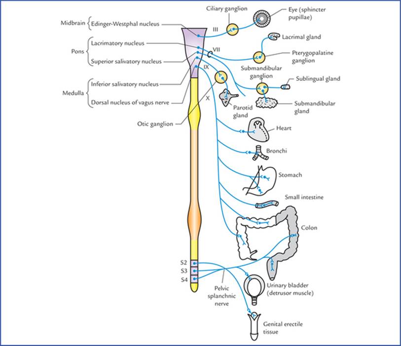
FIG. 20.4 Parasympathetic nervous system (craniosacral outflow).
From the brainstem, they arise from general visceral efferent nuclei (viz. Edinger-Westphal nucleus, lacrimatory nucleus and superior salivatory nucleus, inferior salivatory nucleus, and dorsal nucleus of vagus) and run through IIIrd (oculomotor), Vllth (facial), IXth (glossopharyngeal) and Xth (vagus) cranial nerves, respectively to terminate in the parasympathetic ganglia in the region of head and neck (viz. ciliary, pterygopalatine, submandibular, and otic) and ganglion cells in the autonomic nerve plexuses in the thorax and abdomen. These fibres form what is termed cranial outflow. Preganglionic fibres from Edinger-Westphal nucleus relay in the ciliary ganglion, from lacrimatory nucleus in the pterygopalatine ganglion, from superior salivatory nucleus in the submandibular ganglion, from inferior salivatory nucleus in the otic ganglion, and from dorsal nucleus of vagus in the ganglion cells of autonomic nerve plexuses in the thorax and abdomen.
From the spinal cord, the preganglionic parasympathetic fibres arise from the cells in the grey matter of the 2nd, 3rd, and 4th sacral spinal segments, pass through anterior nerve roots of the corresponding sacral spinal nerves (sacral outflow), leave the sacral nerves and form the pelvic splanchnic nerves, through which they terminate in parasympathetic ganglia located near or within the walls of pelvic organs.
The postganglionic parasympathetic fibres arise from parasympathetic ganglia or autonomic plexuses and supply the viscera.
Autonomic plexuses
The large collections of afferent and efferent autonomic fibres and their associated ganglia form autonomic plexuses in the thorax, abdomen, and pelvis. The branches from these plexuses innervate the viscera.
The autonomic plexuses in different regions of body are listed in Table 20.2.
Table 20.2
Autonomic plexuses and their locations
|
Region |
Autonomic plexuses |
|
Thorax |
Cardiac, pulmonary, and oesophageal |
|
Abdomen* |
Coeliac, superior mesenteric, inferior mesenteric and aortic |
|
Pelvis |
Superior and inferior hypogastric |
*In the abdomen the plexuses are associated with aorta and its branches, and named after them.
Autonomic ganglia
Autonomic ganglia are the sites where preganglionic fibres synapse on postganglionic neurons and thus serve as a relay station. The autonomic ganglia are situated along the course of peripheral efferent autonomic pathways.
The sympathetic ganglia form part of sympathetic chain/trunk and are also located in front of vertebral column, away from the viscera.
The parasympathetic ganglia, on the other hand, are situated close to or within the walls of the viscera.
Neurotransmitters involved in autonomic nerve impulse transmission
The autonomic nerve fibres release one of the two transmitter substances, acetylcholine (cholinergic endings) or noradrenaline (adrenergic endings).
All the preganglionic autonomic fibres (i.e. both sympathetic as well as parasympathetic) and postganglionic parasympathetic fibres are cholinergic, i.e. their terminals liberate acetylcholine as their transmitter substance. All the postganglionic sympathetic fibres are adrenergic, i.e. their terminals liberate noradrenaline (norepinephrine) except for those supplying the sweat glands (i.e. sudomotor fibres), hair muscles (arrector pili) which are cholinergic (Fig. 20.5).

FIG. 20.5 The autonomic efferent fibres and chemical transmitter substances released by their terminals.
Points to Note
• The postganglionic sympathetic transmitter substance, norepinephrine produces both excitatory and inhibitory actions at the postsynaptic sites. This is, due to presence of two types of adrenergic receptors, α and β in the target organs. The response depends upon the type of receptors being activated. In general the activation of α receptors causes excitation and activation of (β receptors causes inhibition.
• The cholinergic receptors are also of two types, muscarinic and nicotinic. The muscarinic receptors are activated by muscarine which elicits the same effects as acetylcholine in the target organs. The nicotinic receptors are excited by low doses, while blocked by high doses of nicotine.
Clinical Correlation
The classification of autonomic receptors has led to the development of many drugs which can modify autonomic activities. These drugs are commonly used to treat shock, hypertension and cardiac problems.
The anatomical, physiological and pharmacological differences between sympathetic and parasympathetic parts of autonomic nervous system are provided in Table 20.3.
Table 20.3
Anatomical, physiological, and pharmacological differences between sympathetic and parasympathetic parts of the autonomic nervous system
|
Sympathetic system |
Parasympathetic system |
|
|
Anatomical differences |
||
|
• Cells of origin of preganglionic fibres |
Are located in the thoracic and upper 2 or 3 lumbar segments (T1-L2/3) of the spinal cord (thoracolumbar outflow) |
Are located in the brainstem and second, third, and fourth sacral segments (S2, S3 & S4) of the spinal cord (craniosacral outflow) |
|
• Ganglia |
Are located along the sympathetic chain and in front of prevertebral region, hence preganglionic fibres are shorter in length and postganglionic sympathetic fibres are longer in length |
Are located near or in the effector organs, hence preganglionic fibres are longer in length and postganglionic are shorter in length |
|
• Outflow from CNS occurs through: |
Spinal nerves |
Cranial nerves |
|
• Distribution of postganglionic fibres |
Wide distribution |
Limited distribution |
|
Physiological differences |
Prepares the body for emergency |
Conserves and restores the energy |
|
Pharmacological differences |
(Also see page 239) |
(Also see page 239) |
|
Neurotransmitter liberated by the postganglionic fibres |
All postganglionic sympathetic fibres liberate norepinephrine except for those supplying the sweat glands or blood vessels in the skeletal muscles which liberate acetylcholine |
All postganglionic parasympathetic fibres liberate acetylcholine |
Afferent nerve fibres of autonomic nervous system (visceral afferents)
The cell bodies of the afferent nerve fibres (first order sensory neurons) of the autonomic nervous system are located either in the dorsal root ganglia of spinal nerves or in the sensory ganglia of the cranial nerves. These neurons are general visceral afferent neurons as they carry general sensations from the viscera. The central processes of these neurons enter the central nervous system and either take part in the formation of local reflex arcs, or pass to higher centres of the autonomic nervous system, such as the hypothalamus.
The impulses carried by these fibres are associated with:
• Visceral reflexes, usually at the unconscious level.
• Sensations of hunger, thirst, nausea, sex desire and bladder and bowel distension.
• Visceral pain.
Once the autonomic afferents enter the spinal cord or brain, they travel alongside, or mixed with, the somatic afferent fibres.
N.B. The nerve endings of autonomic afferents (visceral afferents) are not activated by heat, cut or touch but rather by stretch or lack of oxygen.
Visceral Pain
The visceral pain is of great clinical significance, hence needs an elaborate description.
Usually the viscera are insensitive to cutting and such sensations as touch and heat. However, visceral pain is experienced when it is stretched or suffers from ischaemia (lack of O2) or if there is accumulation of metabolites.
The visceral pain is dull, boring and poorly localized due to stimulation of large number of fibres, on the other hand the somatic pain is sharp, precise and well localized. The pain of appendicitis is a classical example to put forward these facts. Initially when there is an inflammation of appendix and its covering layer of visceral peritoneum (both innervated by autonomic nerve fibres), the pain is dull and vaguely located in the midline in the periumbilical region. The midgut from which the appendix is derived is a median structure during the development. As the inflammation progresses and involves parietal peritoneum (innervated by somatic nerve fibres), the pain is sharp and clearly located in the right iliac fossa at McBurney's point.
Clinical Correlation
Referred pain
The visceral pain is frequently referred to the skin areas (dermatomes) that are innervated by the same segments of the spinal cord as the diseased viscus. Hence referred pain may be defined as pain felt at a site remote from its source of origin.
Anatomical basis of referred pain
The exact basis is not known. However, there are two theories to explain it:
• One theory is that when nerve fibres from the viscus and the skin (dermatome) enter the same segment of spinal cord, they synapse with and stimulate the same (common) second order neurons in the sensory pathway to the cerebral cortex (Fig. 20.6). As a result stimuli from viscus and skin ascend to the central nervous system along a common pathway, consequently the cerebral cortex fails to distinguish between the sites of origin of stimuli.

FIG. 20.6 The first order sensory neurons carrying sensations from viscera and dermatome are entering in the same segment of the spinal cord and synapsing with the common second order sensory neuron.
• Another theory is that under normal conditions the viscera do not send painful stimuli to the brain, whereas the skin repeatedly sends noxious stimuli to the brain. Since afferent fibres from both enter the same segment of the spinal cord, the brain interprets that the information is coming from the skin rather than from the viscus.
Some important examples of referred pain (Fig. 20.7) are listed in Table 20.4.
Table 20.4
The organs and sites of referred pain
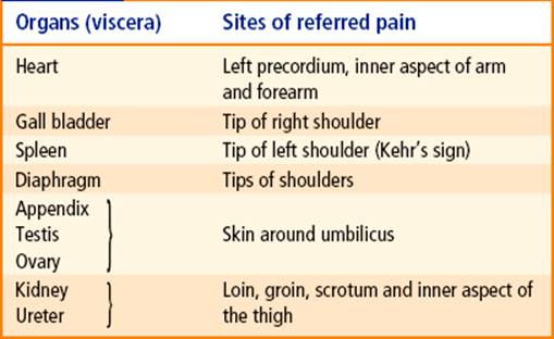

FIG. 20.7 Sites of referred pain from some viscera.
Higher control of the autonomic nervous system
The normal activities of autonomic nervous system are governed by regulatory centres in the brainstem, hypothalamus and cerebral cortex.
The hypothalamus is regarded as the most important higher centre for the control of autonomic nervous system. Its anterior part controls the parasympathetic responses, whereas its posterior part controls the sympathetic responses.
Almost all the autonomic responses can be modified by the activity of same area of the cerebral cortex, presumably through hypothalamus.
Clinical Correlation
Miller et al. (1970) suggested that the autonomic nervous system can be brought under voluntary control to some extent and that the patients with hypertension, for example, possibly can be trained to reduce their blood pressure.
The higher centres of the brain can abnormally influence the functions of autonomic nervous system and induce clinical conditions like cardiac palpitations and myocardial infarction.
Enteric nervous system
The enteric nervous system is a network of intrinsic neurons and ganglia located in the wall of the gastrointestinal tract. These intrinsic neurons are derived from neural crest cells and are independent of sympathetic and parasympathetic nerves. There exists reflex pathway (local reflex mechanisms) in the enteric nervous system through which it controls contraction of muscle coats of the alimentary tract, the secretion of gastric acid, intestinal transport of water and electrolytes, mucosal blood flow, etc.
N.B. Though complex interactions occur between enteric and parasympathetic and sympathetic nervous systems, the enteric nervous system is capable of performing its reflex activities independent of CNS.
Clinical Correlation
Since intrinsic neurons survive following section of extrinsic sympathetic and parasympathetic nerves, the organs/parts of GIT that are transplanted are not truly denervated. Consequently there is no cessation of their activity, i.e. contraction remains unaffected and no structural changes occur.
Important autonomic innervations
The Eyeball
The main structures supplied by autonomic motor fibres in the eyeball are: sphincter pupillae, ciliary muscle and dilator pupillae.
• Sphincter pupillae and ciliary muscle, are innervated by parasympathetic fibres (seeChapter 9).
• Dilator pupillae, it is innervated by sympathetic fibres. The preganglionic sympathetic fibres arise from lateral horn cells of first thoracic spinal segment (T1), pass through anterior root of the first thoracic nerve, to enter the inferior cervical sympathetic ganglion and ascend in the cervical sympathetic chain to relay in its superior cervical sympathetic ganglion. The postganglionic fibres from ganglion, pass successively through internal carotid nerve (or plexus), ophthalmic nerve, nasociliary nerve and long ciliary nerves (occasionally short ciliary nerves) to supply the dilator pupillae (Fig. 20.8).
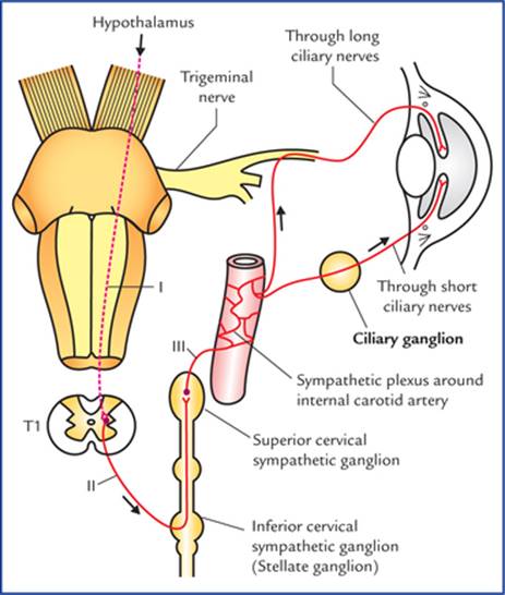
FIG. 20.8 Sympathetic pathway to the eye. I. First order neuron (hypothalamospinal fibres project) without interruption from hypothalamus to the ciliospinal centre in the intermediolateral cell column of T1 spinal segment, II. Second order neuron (preganglionic fibres), arise from T1 spinal segment and relay in the superior cervical sympathetic ganglion, III. Third order neuron (postganglionic fibres) arise from superior cervical sympathetic ganglion. The second and third order neurons form the peripheral part of the sympathetic motor pathway.
N.B. Sympathetic fibres also supply the orbitalis muscle in the floor of orbit, smooth muscle fibres of levator palpe-brae superioris (Muller's muscle) and sudomotor and vaso-motor fibres to orbit, eyelid and face.
Clinical Correlation
Horner's syndrome
It occurs due to interruption of sympathetic pathway to the head and neck (Fig. 20.8). The common sites of lesion are: brainstem, cervical part of spinal cord, or stellate ganglion.
Characteristic features
|
• Miosis (i.e. constriction of pupil) |
Due to paralysis of dilator pupillae and unopposed action of sphincter pupillae. |
|
• Partial ptosis (i.e. slight drooping of the upper eyelid) |
Due to paralysis of smooth muscle fibres of levator palpebrae superioris called Muller's muscle. |
|
• Anhidrosis (i.e. loss of sweating) |
Due to involvement of sudomotor fibres which are concerned with sweating in the region of head and neck. |
|
• Enophthalmos (sunken eyeball) |
It is often apparent but not real and is caused by ptosis. The paralysis of orbitalis muscle may also be responsible. |
|
• Flushing of the face |
Due to involvement of vasoconstrictor fibres leading to vasodilation of skin arterioles in the region of head and neck. |
The Salivary Glands
Parotid gland
Parasympathetic innervation (secretomotor supply): The preganglionic fibres arise from inferior salivatory nucleus in the pons, pass successively through glossopharyngeal nerve and its tympanic branch, the tympanic plexus and the lesser petrosal nerve to terminate in the otic ganglion. The postganglionic fibres from the otic ganglion, pass through the auriculotemporal nerve to supply the parotid gland (Fig. 20.9).

FIG. 20.9 Secretomotor pathway for parotid gland.
Submandibular and sublingual glands
Parasympathetic innervation (secretomotor nerve supply): The preganglionic fibres arise from superior salivatory nucleus in the pons, pass successively through facial, chorda tympani and lingual nerves to terminate in the submandibular ganglion. The postganglionic fibres from the submandibular ganglion supply the submandibular gland directly, and sublingual gland through the lingual nerve (Fig. 20.10).
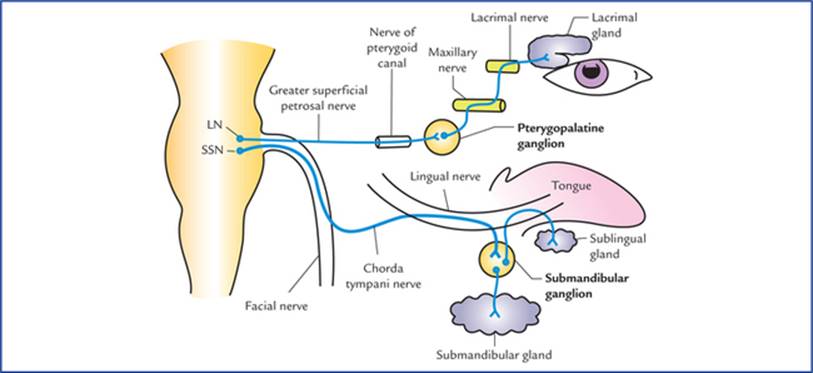
FIG. 20.10 Secretomotor pathways for lacrimal gland, and submandibular and sublingual salivary glands. (LN = lacrimatory nucleus, and SSN = superior salivatory nucleus.)
Lacrimal gland
Parasympathetic innervation (secretomotor nerve supply): The preganglionic parasympathetic fibres arise from lacrimatory nucleus in the pons, and pass successively through nerves intermedius, geniculate ganglion of facial nerve, greater petrosal nerve and nerve of pterygoid canal to terminate in the pterygopalatine ganglion. The postganglionic fibres arise from pterygopalatine ganglion, and pass successively through maxillary nerve, zygomatic nerve and its zygomatico-temporal branch, and lacrimal nerve to supply the lacrimal gland (Fig. 20.10).
Gastrointestinal Tract
Motor supply
Parasympathetic innervation
• The most of GIT, i.e. from oesophagus to the junction of right two-third and left one-third of transverse colon, is supplied by vagus nerve. Preganglionic parasympathetic fibres arise from dorsal nucleus of vagus, and pass successively through vagus nerve coeliac, superior and inferior mesenteric plexuses, and then run along the coeliac, superior mesenteric and inferior mesenteric arteries and their branches to enter the GIT, where they relay in the ganglia within myenteric (Auerbach's) and submucosal (Meissner's) plexuses located in the wall of the gut.
• From junction of right two-third and left one-third of transverse colon to the junction of upper and lower halves of the anal canal the GIT is supplied by sacral parasympathetic outflow. The preganglionic parasympathetic fibres arise from anterolateral grey columns of S2, S3 and S4 spinal segments, pass successively through the ventral roots of spinal nerves, pelvic splanchnic nerves and superior and inferior hypogastric plexuses to terminate in the ganglia within myenteric (Auerbach's) and submucosal (Meissner's) plexuses located in the gut wall.
The postganglionic fibres arise from parasympathetic ganglia located in the gut wall and supply the muscle and glands of GIT.
Sympathetic innervation: The preganglionic sympathetic fibres arise from T5 to L2 spinal segments, pass through the sympathetic trunks without relay, travel through the splanchnic nerves to terminate in the ganglia related to the coeliac (coeliac ganglion), superior mesenteric (superior mesenteric ganglion) and inferior mesenteric (inferior mes-enteric ganglion) arteries. The postganglionic fibres arise from these ganglia and travel along these arteries to reach the gut which they supply.
Functional significance
The parasympathetic nerves stimulate peristalsis (gastrointestinal motility) and relax the sphincters. They also stimulate secretion from mucosal glands.
The sympathetic fibres inhibit peristalsis and cause contraction of the sphincters; they also inhibit secretion from mucosal glands.
Clinical Correlation
Congenital megacolon (Hirschsprung disease)
Sometimes, during embryonic period the neural crest cells fail to migrate into the wall of descending colon at its junction with the sigmoid colon, leading to lack of formation of parasympathetic ganglia in the myenteric (Auerbach's) plexus. As a result the part of colon lacking ganglia shows marked decrease in its peristalsis leading to functional obstruction. Consequently the proximal part of descending colon becomes greatly distended due to accumulation of faeces, causing congenital megacolon (Hirschsprung disease).
Sensory supply
The afferent fibres travel along both sympathetic and parasympathetic pathways.
Pain from most of the GIT travel along the sympathetic nerves except from upper part (i.e. pharynx and oesophagus) and lower part (i.e. lower part of pelvic colon, rectum and upper part of anal canal) from where it is carried by parasympathetic fibres through vagus and pelvic splanchnic nerves respectively.
Urinary Bladder
Motor supply (Fig. 20.11)
Parasympathetic innervation: The preganglionic parasympathetic fibres arise from S2, S3 and S4 spinal segments (spinal micturition centre), pass through the pelvic splanchnic nerves and inferior hypogastric plexus to terminate in the nerve cells located in the vesical plexus. The postganglionic fibres arise from vesical plexus and supply the urinary bladder.
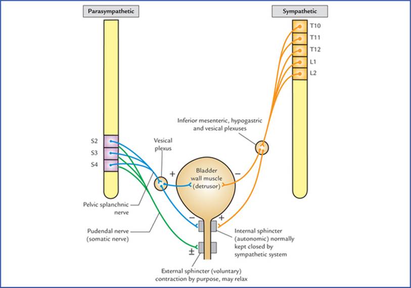
FIG. 20.11 Motor innervation of the urinary bladder, showing physiological and neurological principles of bladder function. (+) = contraction, (−) = relaxation, (±) = contraction and relaxation.
Sympathetic innervation: The preganglionic sympathetic fibres arise from T10 to L2 spinal segments, and terminate in the cells of inferior mesenteric, superior hypogastric, inferior hypogastric and vesical plexuses. The postganglionic fibres arise from the cells within these plexuses and supply the urinary bladder.
The parasympathetic fibres stimulate the contraction of the smooth muscle of the bladder wall (detrusor muscle) and relax the internal urethra! sphincter (sphincter vesicae).
The sympathetic fibres have little or no action on the detrusor muscle and play only a minor role in maintaining urinary continence by causing contraction of sphincter vesicae. However, in males during ejaculation (brought out by sympathetic action) the sympathetic stimulation causes active contraction of sphincter vesicae and prevents the seminal fluid from entering the bladder.
Sensory supply
Sensory fibres carry impulses of distension and pain from the bladder. They travel through both sympathetic and parasympathetic fibres to the CNS.
Clinical Correlation
• The fibres carrying sensations of distension (filling of the bladder) travel through the posterior white column of the spinal cord, while the fibres carrying pain travel through the anterior and lateral white columns of the spinal cord.
The important point to note here is that sensory pathways carrying sensation of pain and distension take different routes in the spinal cord. Therefore, intractable bladder pain due to carcinoma bladder may be relieved by cutting anterior and lateral white columns of spinal cord on both the sides (bilateral anterolateral cordotomy).
• Effects of spinal cord injuries on the bladder function
– The atonic bladder occurs during the phase of spinal shock (temporary loss/halt of all spinal functions) immediately after the injury. The muscle of bladder wall (detrusor muscle) is relaxed, the internal sphincter (sphincter vesicae) is tightly contracted (due to loss of inhibition from higher centres) and the external sphincter is relaxed. As a result the bladder becomes greatly distended with urine which finally overflows.
– The automatic reflex bladder occurs after the patient has recovered from the spinal shock, and if the cord is injured above the level of segments responsible for the parasympathetic outflow, i.e. spinal micturition centre (S2, S3 and S4). Due to involvement of descending fibres from paracentral lobule, the cortical micturition centre, the voluntary control on bladder is lost. As a result, the bladder fills and empties reflexly. The stretch receptors in the bladder wall are stimulated as the bladder fills and the afferent impulses pass to the spinal cord (S2, S3 and S4). From spinal cord the efferent impulses pass to the bladder, causing detrusor muscle to contract and internal and external sphincter urethrae to relax leading to emptying of the bladder. This spinal reflex for emptying the bladder occurs every 1 to 4 hrs.
– The autonomous bladder occurs if the sacral segments (S2, S3 and S4) of the spinal cord are destroyed. In this condition, the bladder has neither reflex control nor voluntary control because both, reflex centre for bladder emptying (S2, S3 and S4) and descending fibres from voluntary cortical centre have become non-functional.
As a result the bladder wall becomes flaccid, and the capacity of the bladder is greatly increased. It fills to capacity arid overflows, causing continuous dribbling of urine.
Bladder function
The process of micturition requires proper integration of autonomic and voluntary nervous systems. The contraction of smooth muscle of bladder wall (the detrusor muscle) is initiated by parasympathetic activity, while the internal sphincter mechanism (which is normally closed by sympathetic activity) is allowed to relax by decreased sympathetic activity. The external sphincter mechanism, which can be voluntarily relaxed to allow micturition is controlled by pudendal nerves (S2, S3 and S4).
Heart
Motor supply
Parasympathetic innervation: The preganglionic parasympathetic fibres arise from dorsal nucleus of vagus in the medulla oblongata, run through vagus nerve and its cardiac branches to synapse on the cells or ganglia in superficial and deep cardiac plexuses. The postganglionic fibres arise from these cells or ganglia and supply the sinuatrial and atrioventricular nodes and coronary arteries.
Sympathetic innervation: The preganglionic sympathetic fibres arise from T1 to T5 spinal segments and terminate in the cells of upper 3 or 4 thoracic ganglia and cervical sympathetic ganglia.
The postganglionic sympathetic fibres arise from these ganglia, pass first through the cardiac branches of these ganglia and then through cardiac plexuses to supply sinuatrial and atrioventricular nodes, and coronary arteries of the heart.
Functions
The sympathetic stimulation increases whereas the parasympathetic stimulation decreases the heart rate. During normal circumstances, the heart rate is normal because a balance is maintained between the inhibitory and the stimulatory effects of the parasympathetic and sympathetic innervation. The exertion and emotional emergencies tilt the balance towards the sympathetic tone, hence responsible for increased heart rate. The excessive vagal stimulation can bring the activity of the heart standstill.
Further, the sympathetic stimulation causes dilatation, and parasympathetic stimulation causes constriction of the coronary arteries, thus increasing or decreasing blood supply to the heart respectively.
Sensory supply
The afferent fibres from heart travel through both sympathetic and parasympathetic pathways. The sensations of pain from heart (due to anoxia, viz. angina) are carried by sympathetic fibres. They travel through cardiac branches of middle and inferior cervical sympathetic ganglia, pass successively through these ganglia, sympathetic trunks and spinal nerves to enter the spinal segments from T1 to T4/5.
Afferent fibres running along the cardiac branches of vagus nerves are concerned with reflexes controlling the activity of the heart.
Arteries of the Upper Limb
The arteries of the upper limb are innervated by sympathetic fibres only.
The preganglionic fibres arise from T2 to T8 thoracic spinal segments, and pass to the sympathetic trunk through white rami communicantes. After entering the trunk they ascend to synapse with the cells within the stellate and middle cervical sympathetic ganglia. The postganglionic fibres arise from these ganglia, join the nerves forming brachial plexus and are distributed to the arteries through the branches of brachial plexus. The stimulation of sympathetic nerves causes vasoconstriction of cutaneous arteries and vasodilatation of arteries supplying the skeletal muscles.
Clinical Correlation
The Raynaud's disease occurs due to vasospasm of the digital arteries of the upper limb following exposure to cold. It is common in women. There is pallor or cyanosis of the fingers associated with severe pain. The gangrene of the tips of the fingers may occur. It can be treated by preganglionic sympathectomy.
The symptoms are relieved: (a) by avoiding exposure to cold, and stopping smoking (smoking causes vasoconstriction), and (b) by giving drugs that inhibit sympathetic activity to bring about arterial dilatation.
The removal of stellate (cervicothoracic) ganglion (cervicothoracic preganglionic sympathectomy) has also been used in the past.
Arteries of the Lower Limb
The arteries of the lower limb are supplied by sympathetic nerves. The preganglionic fibres arise from lower three thoracic and upper two or three lumbar segments of the spinal cord (T10-L2), and pass to the lower thoracic and upper lumbar ganglia of the sympathetic chain. These fibres synapse in the lumbar and sacral ganglia. The postganglionic fibres arise from these ganglia and reach the arteries through the branches of lumbar and sacral plexuses.
Clinical Correlation
Buerger's disease is an arterial occlusive disease of the legs causing ischaemia of muscles. It is common in men, particularly those who smoke a lot. These individuals complain of intermittent claudication, i.e. appearance of pain on exercise which disappears after rest and reappears on exercise.
They may suffer from ischaemia and dry gangrene of the toes.
The preganglionic lumbar sympathectomy (removal of upper three lumbar ganglia and intervening parts of the sympathetic chain) is advocated in order to bring about vasodilatation and increase in the blood flow through the collateral circulation.
Clinical problems
1. What is the embryological basis of congenital megacolon (Hirschsprung disease)?
2. What are the effects of spinal cord injury on the bladder function?
3. Explain, why there is enuresis (bed-wetting) during infancy.
4. Pain arising from gall bladder is often referred to the tip of right shoulder. Mention the anatomical basis.
5. Explain, why the pain of renal and ureteric colic is referred to loin, groin scrotum/labium majus and inner aspect of the upper thigh.
6. Pain from appendix, testis and ovary is often referred to the skin around umbilicus. Mention the anatomical basis.
7. Explain, why the cardiac pain is felt over the mid-sternum in the precordium and often spreads to the medial side of arm and forearm.
Clinical problem solving
1. Seepage 244.
2. Seepage 245.
3. Because there is no cortical (voluntary) control on the spinal micturition centre in infants (as the descending corticospinal fibres have yet not myelinated).
N.B. The corticospinal fibres get myelinated and begin to function about the age of 3 to 6 years. Therefore bed-wetting after the age of 6 years attracts the parent's concerns.
4. Irritation of parietal peritoneum lining the diaphragm by diseased gall bladder (viz. cholecystitis) accounts for referred pain to the tip of right shoulder, because parietal diaphragmatic peritoneum and skin over tip of right shoulder are supplied by phrenic and supraclavicular nerves respectively. Both of which arise from same spinal segments, i.e. C3 and C4.
5. The kidney and ureter are supplied by sympathetic fibres (responsible for carrying pain sensations) derived from T10 to L2 spinal segments. Therefore, the pain of renal and ureteric colic is referred to the regions of skin innervated by T10 to L2 spinal segments. It therefore commences in the back over the lower ribs (loin) and shoots downwards and forwards to the inguinal region (groin), scrotum/labium majus and upper inner aspect of the thigh (Fig. 20.7).
6. Pain from appendix, testis and ovary is referred to the skin around the umbilicus because all these organs and skin around the umbilicus are supplied by T10 spinal segment.
7. The heart is supplied by upper four thoracic spinal segments (T1–T4). The skin over sternum and precordium is supplied by T4, T3 and T2 spinal segments, the skin on the medial aspect of arm by T2 spinal segment and that on the medial aspect of forearm by T1 spinal segment.
The cardiac pain therefore being visceral in nature, is not felt in the heart, rather it is referred to the skin areas supplied by the same spinal segments which supply the heart.