
An accurate assessment of the degree and type of cutaneous sensory impairment and muscular activity play a key role in making the correct diagnosis of a neurological disease.
The precise knowledge of cutaneous sensory impairment enables the physician to identify the peripheral nerve lesion, or the segmental nerve loss or the level of spinal cord involvement.
The observation of specific muscles at rest and during action indicates whether the muscle itself is diseased or it is affected due to impairment of its innervation.
The students, therefore, should have clear concept of segmental innervation of skin and muscles.
Segmental Innervation of Skin
The area of skin supplied by a single spinal nerve, and therefore, a single spinal segment, is called dermatome. With the exception of the first cervical nerve (C1), all of the spinal nerves are associated with specific dermatomes.
In the trunk the arrangement of dermatomes is simple because the thoracic and upper lumbar spinal nerve supplying it are arranged segmentally. On the trunk the dermatome extends around the body from the posterior median line to the anterior median line. Figures 5.1 and 5.2 depict the dermatomal maps for the anterior and posterior surfaces of the body. Adjacent dermatomes overlap considerably (Fig. 5.3); therefore to produce a region of complete anaesthesia at least three contiguous nerves have to be severed. Further, it is important to note that the area of tactile loss is always larger than the area of loss of thermal and painful sensations. This is due to the fact that the degree of overlap of fibres carrying pain and temperature sensations is much more extensive than the overlap of fibres carrying tactile sensations.

FIG. 5.1 Anterior aspect of the body showing the pattern of dermatomes on the left side, and distribution of cutaneous nerves on the right side.
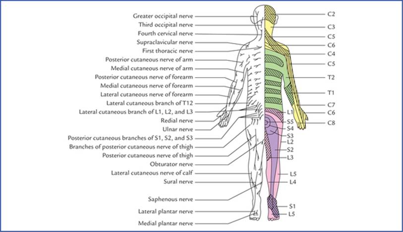
FIG. 5.2 Posterior aspect of the body showing pattern of dermatomes on the right side and distribution of cutaneous nerves on the left side.
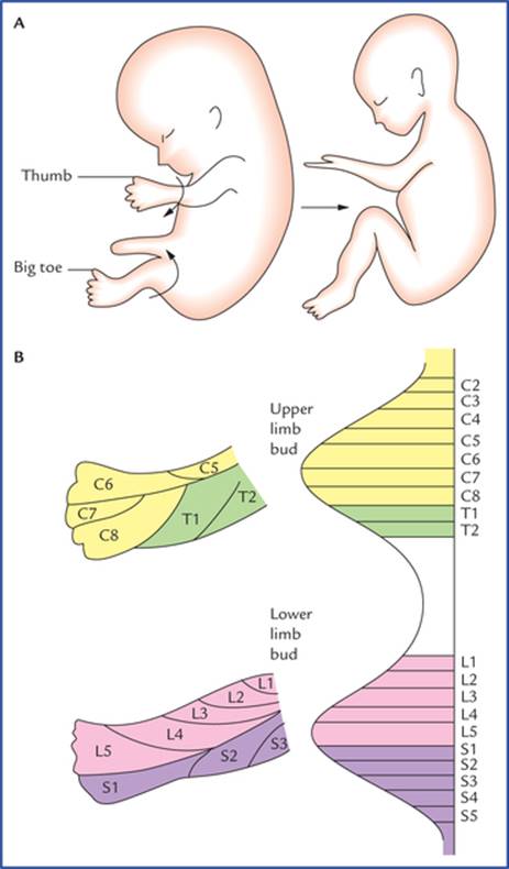
FIG. 5.3 (A) The rotation of the limbs during embryonic development. (B) The arrangement of dermatomes in the upper and lower limbs of the embryo.
In the limbs the arrangement of dermatomes is complicated and this is because of two reasons: (a) the embryo-logical rotation of the limbs as they grow out from the trunk, and (b) the spinal nerves that supply them form plexuses.
In early embryonic development, the upper and lower limbs appear as paired paddle-shaped limb buds. Each bud has preaxial and postaxial borders, with former being towards the head. Later in the development the ends become expanded and flattened to form hand and foot plates, in which the digits develop. The digits along the preaxial border are the thumb and big toe. The limbs then rotate. The upper limb rotates laterally so that its preaxial border and thumb come to lie on the lateral side. On the other hand, the lower limb rotates medially and its preaxial border and big toe come to lie on the medial side. It is now easier to understand why the dermatomes innervated by spinal nerves of brachial plexus are arranged consecutively down on the lateral side and up on the medial side of the upper limb, and dermatomes innervated by spinal nerves of lumbosacral plexus are arranged down the medial side and up the lateral side and back of the lower limb (Fig. 5.3 A, B).
The spinal nerves that supply the upper limb form plexuses from which individual named nerves arise. Each of these named nerves may contain fibres derived from a number of spinal nerves so that the area of skin supplied by one of the named nerves will be quite different from the area supplied by the individual spinal nerves.
In the head and face region, each of the three divisions of the trigeminal nerve supply a precise area of skin and there is little or no overlap to the cutaneous area of the another division (Fig. 9.8).
Points to Note
• Because of the development of the upper limbs the anterior primary rami of lower cervical and first thoracic spinal nerves have lost their cutaneous innervation of the trunk anteriorly, and at the level of the sternal angle, the fourth cervical der-matome is contiguous with the second thoracic dermatome (Fig. 5.1).
• During the development of head and neck, due to upward migration of upper cervical dermatomes the back of the head is supplied by upper cervical spinal nerves, and at the interauricular line, the second cervical dermatome is continuous with the cutaneous area innervated by the trigeminal nerve (Figs 5.1 and 9.9).
Clinical Correlation
• Abnormalities of muscle tone: In disease conditions, the muscle tone is either increased (hypertonia) or decreased (hypotonia). In hypertonia there is increased resistance to the passive movement. If this increased resistance is greatest at the initial phase of the movement, and then suddenly it gives way during the later phase, it is termed as clasp-knife rigidity. When resistance to passive movement is uniformly increased throughout the range of the movement, it is termed as plastic-type of rigidity or lead-pipe rigidity.
If the increased resistance is felt intermittently throughout the range of movement, it is termed cogwheel-type of rigidity.
• Abnormalities of Sensory Perception
In disease conditions, the patient may have areas of reduced sensitivity to pain (hypoalgesia) or areas of reduced sensitivity to touch (hypoaesthesia) or areas of exaggerated sensitivity (hyperaesthesia) or areas of abnormal sensations (paraesthesia). The paraesthesia includes sensations like feeling of pins and needles, burning, prickling, etc.
Segmental Innervation of Muscles
The skeletal muscles are also segmentally innervated. Most of these muscles are innervated by more than one spinal nerve and therefore by the same number of spinal segments. Therefore, to paralyze a muscle completely it would be necessary to section several spinal nerves or damage several segments of the spinal cord.
It is important to note the following facts carefully:
• Most of the skeletal muscles are supplied by two or more spinal segments (the intrinsic muscles of the hand are exception as they are unisegmental).
• Muscles sharing a common primary action are supplied by the same spinal segments and the opposing muscles by the lower segments in sequence with the former. For example, the flexors of the elbow joint are supplied by C5 and C6 spinal segments and the extensors by the C7 and C8 spinal segments.
• There is correlation among the innervation of muscles, joints and the skin, i.e. the branches of those spinal nerves which supply the muscles moving a joint also supply the skin over the insertions of the same muscles and the interior of the joint.
It is neither necessary nor possible to remember the seg-mental innervation of all the muscles of the body but one should remember the segmental innervation of the muscles given in Table 5.1 because they can be tested by eliciting the simple muscle reflexes/stretch reflexes (also called tendon jerks) in the patient during physical examination.
Table 5.1
Segmental innervation of muscles used for eliciting tendon reflexes
|
Muscle |
Segmental innervation |
Reflex |
|
Biceps brachii |
C5, 6 (Musculocutaneous nerve) |
Biceps jerk (flexion of elbow by tapping the biceps tendon) |
|
Triceps brachii |
C7, 8 (Radial nerve) |
Triceps jerk (extension of elbow by tapping the triceps tendon) |
|
Brachioradialis |
C5, 6 (Radial nerve) |
Brachioradialis/supinator jerk* (flexion of forearm in midprone position by tapping the insertion of brachioradialis) |
|
Quadriceps femoris |
L2, 3, 4 (Femoral nerve) |
Quadriceps jerk/knee jerk/patellar jerk (extension of knee by tapping the ligamentum patellae/patellar tendon) |
|
Triceps surae (gastrocnemius and soleus) |
S1, 2 (Tibial nerve) |
Achilles tendon reflex/ankle jerk (plantar flexion of ankle by tapping the tendocalcaneus, also called Achilles tendon) |
*The brachioradialis jerk is commonly termed supinator jerk by clinicians after the old name of the brachioradialis muscle, the supinator longus.
Clinical Correlation
The knee jerk (patellar jerk) is a classical example of the stretch reflex and used by clinicians to determine if higher centres that normally influence the reflex are functional. When ligamentum patellae are tapped with a reflex hammer the quadriceps muscle is stretched. As a result the intrafusal fibres within the muscle spindles are also stretched. Consequently sensory fibres innervating the intrafusal fibres of muscle spindle are stimulated and send afferent action potentials to the spinal cord segments (L2, L3 and L4) where they stimulate alpha motor neurons directly. The efferent fibres arising from these neurons transmit action potentials to the extrafusal fibres of the quadriceps muscle, causing a rapid contraction of the stretched muscle leading to extension of leg—positive knee jerk response (Fig. 5.4).
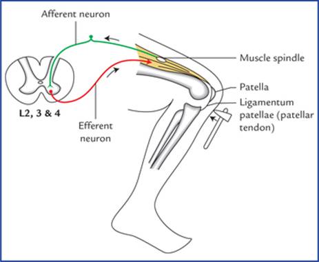
FIG. 5.4 Knee jerk (patellar jerk). Note that the reflex arc passes through L2-4 spinal segments of the spinal cord. The stretch reflex is monosynaptic.
N.B. The stretch reflex is unique in that it does not require an association neuron between the afferent and efferent neurons (i.e. monosynaptic).
Muscle Tone
Every muscle while at rest is in a state of partial contraction, which provides a tone to the muscle. The muscle tone is tested by assessing the resistance offered by the muscles to passive stretch. Since muscle fibres remain either in the state of full contraction or full relaxation (they are never in state of partial contraction or relaxation). This means that a few muscle fibres within a muscle are fully contracted all the time. Therefore, to keep the tone maintained continuously and simultaneously not allowing the muscle to fatigue, the different groups of muscle fibres belonging to different motor units are brought into action at different times.
Basically the muscle tone is dependent on the integrity of a simple monosynaptic reflex arcs composed of two neurons. The nerve impulses travel along the afferent fibres to the spinal cord, where they synapse with the alpha (a) motor neurons in the anterior horn, which in turn send impulses along their axons (efferent fibres) to the muscle fibres (Fig. 5.4).
Two types of sensory receptors are present in the muscle including its tendon: (a) muscle spindles, and (b) tendon spindles. The former detects the lengthening and shortening in the muscle while the latter detects the tension in the tendon.
The muscle spindles themselves are innervated by small gamma (γ) efferent fibres which regulate the response of the muscle spindles. In this way the muscle tone is maintained reflexly according to the needs of posture and movement.
Clinical Correlation
If the afferent or efferent limb of the reflex arc is cut, the muscle will loose its tone immediately and become flaccid. On palpation, the flaccid muscle feels like a mass of dough (dough = mass of flour) and shows no resilience at all. It quickly atrophies and becomes reduced in volume.
Movement and Posture
Movement
The movement is accomplished by the action of muscles. The muscle acts by contraction and with rare exceptions single muscle does not contract alone, because the whole movement rather than individual muscles are represented in the cerebral cortex. Therefore, each movement generally requires the contraction and relaxation of a whole group of muscles. For example, the lifting of the arm from the side not only requires the contraction of abductors of the shoulder joint but also simultaneous relaxation of the adductors of the shoulder joint. It follows that muscle movement is accomplished by bringing into action increasing number of motor units of synergist muscles which contract and at the same time, reducing the activity of the motor units of the antagonist muscles that oppose the movement.
The muscle fatigue occurs due to progressive loss of strength of muscle with prolonged contraction. It occurs due to reduction in the amount of adenosine triphosphate (ATP) within the muscle fibres.
Muscle power
The muscle power is the force or strength of contraction of muscle or muscle groups. In normal individuals the power in different groups of muscles varies according to their physiological requirements. In general, the larger the muscle, the greater the muscle power.
Grading of muscle power
The power of muscles is objectively expressed in grades. The Medical Research Council (MRC) grading is generally adopted. It is given in Table 5.2.
Table 5.2
The grades of muscle power
|
Grade |
Movement |
|
Grade 0 |
Complete paralysis with not even a flicker of movement |
|
Grade 1 |
A flicker of movement is possible |
|
Grade 2 |
The part can be moved, if the gravity is eliminated by suitable positioning |
|
Grade 3 |
The muscle can contract against gravity but not against the resistance |
|
Grade 4 |
The muscle can contract against mild or moderate resistance |
|
Grade 5 |
Normal power is present. Movement against full resistance is possible |
Clinical Correlation
• The lesions of the peripheral nerve that supplies the muscle or involvement of anterior horn cells in diseases (e.g. poliomyelitis) reduce the power of or paralyze the muscles involved.
– The muscular wasting occurs within 2-3 weeks after peripheral nerve lesion.
– The muscular fasciculations, i.e. twitching of group of muscle fibres is very commonly seen in patients with chronic disease that affects the anterior horn cells.
– The muscular contracture occurs most commonly in the muscles that normally oppose the paralyzed muscles. The contracture leads to permanent shortening of the muscle.
• The localized wasting of muscles is associated with lower motor neuron (LMN) type of paralysis and disuse atrophy.
• Generalized wasting of muscles occur in debilitating diseases such as cancer, pulmonary tuberculosis, etc.
Posture (= physical disposition of the body)
The posture may be defined as the position adopted by an individual in his/her environment. The posture of an individual depends on the degree and distribution of muscle tone and, therefore, on the activity of motor nerves which supply the muscles. The normal postural tone of skeletal muscle is dependent not only on the integrity of the ‘simple muscle reflex’ (stretch reflex) but also on the summation of nerve impulses or information received by anterior horn cells of spinal cord from other sources, viz.
(a) from membranous labyrinths of internal ears,
(b) from cerebellum, midbrain and cerebral cortex, and
(c) from the eyes.
While standing erect, the line of gravity passes through the dens of the second cervical vertebra, behind the centres of hip joints, and in front of the centres of knee and ankle joints.
Since humans have acquired an erect posture in animal kingdom, it is not surprising that in humans in order to stabilize and prevent the body from falling, the antigravity muscles (extensors of leg and flexors of the arm) are well developed and exhibit maximal degree of tone.
Clinical Problems
1. Explain, why a physician while testing exteroceptive sensations (pain, touch and temperature) applies stimulus in a longitudinal direction in limbs and in a horizontal direction in the trunk.
2. What are the various types of muscular rigidities that are found in patients suffering from paralysis and how their knowledge helps to know whether the paralysis is due to upper motor neuron lesion or extrapy-ramidal lesion?
3. A 55-year-old man developed skin rashes (vesicles containing clear fluid) in his right 5th intercostal space and experienced shooting/burning pain. Name the disease and the spinal nerve involved.
Clinical Problem Solving
1. The sensations of pain, touch and temperature are tested by using sterile pin, wisp of cotton wool, and holding hot and cold test tubes respectively.
The stimulus is applied by moving the above-mentioned objects longitudinally along the long axis of the limb because dermatomes run longitudinally along the long axis of the limb. In the trunk the dermatomes run almost horizontally, therefore the stimulus is applied in a horizontal direction.
2. The muscular rigidity (increased muscle tone) can be classified into three types: (a) clasp-knife type, (b) lead-pipe type, and (c) cogwheel type.
The following tabular presentation of facts is of great diagnostic help in determining the site of lesion.
|
Rigidity |
Feature |
Lesion |
|
Clasp-knife |
The initial resistance to the movement is suddenly overcome |
Upper motor neuron lesion |
|
Lead-pipe |
A steady increase in resistance throughout the movement |
Extrapyramidal lesion |
|
Cogwheel |
Ratchet-like (intermittent) increase in resistance to movement |
Extrapyramidal lesion |
3. The herpes zoster is a viral infection that affects the posterior root (sensory) ganglia of the spinal nerves and sensory ganglia of cranial nerves. The posterior root ganglia of thoracic spinal nerves and sensory ganglion of trigeminal nerve (geniculate ganglion) are most commonly affected. Since the patient is experiencing severe pain and has skin vesicles in the areas of right fifth intercostal space. It is for sure that dorsal root ganglion of right 5th intercostal nerve is infected by herpes zoster virus.
N.B. The infection of trigeminal ganglion by herpes zoster virus mostly involves the ophthalmic division of trigeminal nerve and vesicles appear above the eye along the distribution of supratrochlear and supraorbital nerves. This condition is termed herpes zoster ophthalmicus.