
The central nervous system (CNS) consists of the brain and spinal cord, which are located in the cranial cavity and vertebral canal respectively (Fig. 6.1).
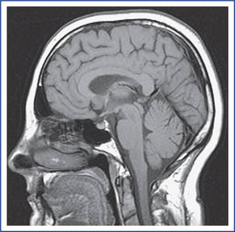
FIG. 6.1 A sagittal view of MRI of the skull, brain and cranial portion of the spinal cord.
The delicate CNS is well protected because it is enclosed by the skull and vertebral canal and meninges, and is bathed in cerebrospinal fluid (CSF).
Structurally, the brain and spinal cord consist of grey and white matter. Therefore, sections through the CNS present greyish and whitish regions.
The grey matter consists of nerve cell bodies and dendrites, and the white matter is made up predominantly of myelinated nerve fibres.
N.B. Both grey matter and white matter contain neuroglia and blood vessels.
In the brain (except the brainstem), the grey matter is present at the periphery and the white matter in the centre. Contrary to it, in the spinal cord the grey matter is present in the centre and white matter at the periphery.
Brain
The brain is that part of the CNS which lies within the cranial cavity.
The functions of the brain are as follows:
• It receives information from, and controls the activities of trunk and limbs mainly through its connections with the spinal cord.
• It receives the information from, and controls the activities of head and neck structures through cranial nerves.
• It assimilates experiences, a requisite to higher mental processes such as memory, learning and intelligence.
• It is also responsible for one's personality, thoughts and aspirations.
N.B. Plato (427 BC) referred to the brain as the “divinest part of the body”.
The adult brain constitutes about one-fiftieth of body weight and weighs about 1400 g in males and 1200 g in females. It consists of six major parts: (a) the cerebrum, (b) the diencephalon, (c) the midbrain, (d) the pons, (e) the medulla oblongata, and (f) the cerebellum (Fig. 6.2).
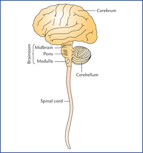
FIG. 6.2 The diagram showing parts of central nervous system. The diencephalon is not seen. The superior, middle and inferior cerebellar peduncles, which connect the cerebellum with the mid-brain, pons and medulla oblongata are shown schematically.
The midbrain, pons and medulla oblongata collectively form the brainstem.
The Figure 6.3 shows the superolateral aspect of the brain, as seen in dissection.

FIG. 6.3 Superolateral surface of the brain (left lateral view) as seen in the dissection.
Cerebrum
The cerebrum is the largest part of the brain. It consists of two large, hemispheres (the left and the right cerebral hemispheres), which occupy the anterior and middle cranial fossae, and the supratentorial region of posterior cranial fossa.
Each cerebral hemisphere consists of a surface layer of grey matter, called cerebral cortex and a central core of white matter. In the basal part of the latter are located large masses of grey matter, known as basal nuclei/ganglia (Fig. 6.4).
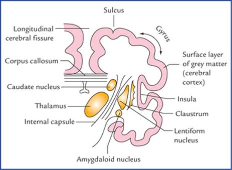
FIG. 6.4 A coronal section of the cerebral hemisphere showing surface layer of grey matter (cerebral cortex), central core of white matter and basal nuclei.
The two hemispheres are partly separated from each other by a deep median longitudinal fissure, called longitudinal cerebral fissure. A massive commissure, the corpus callosum, whose fibres interconnect the corresponding cortical areas of the two cerebral hemispheres, lies in the floor of the fissure.
The surface of cerebral hemisphere is convoluted, i.e. it has a series of elevations, the gyri, separated by shallow depressions, the sulci or deep grooves called fissures (Fig. 6.3).
A central sulcus which runs downwards and forwards from superomedial border to the lateral sulcus is located about midway along the length of the hemisphere.
There are individual differences in the appearance of the sulci and gyri, but some sulci are constant in their position and appearance, and serve as an important landmark.
The superolateral surface of each cerebral hemisphere is divided into four lobes, which are named after the overlying skull bones (Fig. 6.5):
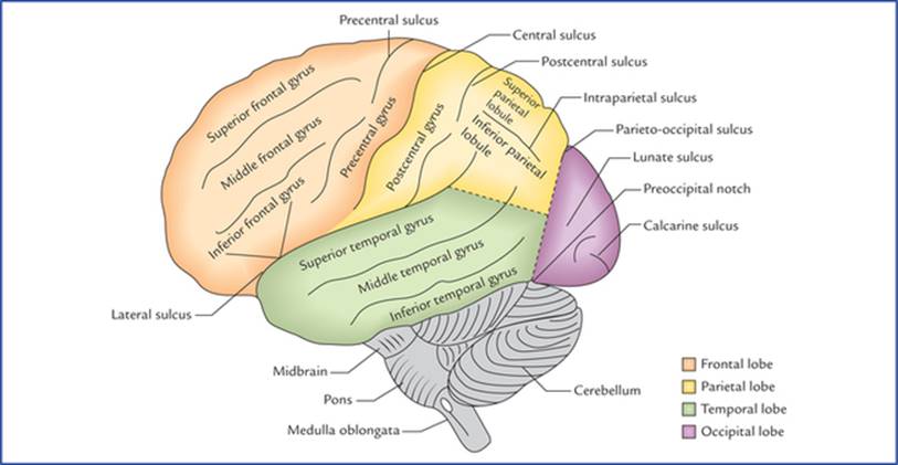
FIG. 6.5 Superolateral surface of the brain. Note the demarcation of lobes on the superolateral surface of the cerebral hemisphere.
• Frontal lobe is anterior to the central sulcus and above the lateral sulcus.
• Parietal lobe is posterior to the central sulcus and above the lateral sulcus.
• Occipital lobe is behind a line extending from parieto-occipital sulcus to the pre-occipital notch.
• Temporal lobe is below the lateral sulcus and in front of parieto-occipital notch.
The frontal lobe is important for voluntary motor functions, motivation, aggression, emotions, effect, drive and awareness of self.
The parietal lobe is the major centre for reception and evaluation of all sensory informations except for smell, hearing, and vision.
The occipital lobe is responsible for reception and integration of visual input.
The temporal lobe receives and evaluates input for smell and hearing and plays an important role in memory.
N.B. Deep within the lateral sulcus lies a submerged portion of cerebral cortex, the insula which is often referred to as fifth lobe or central lobe.
The so-called limbic lobe is a composite bordering zone (limbus = border) between the telencephalon and diencephalon. It is somewhat ring-shaped. It is associated with basic survival instincts, viz. the acquisition of food and water and reproduction. It provides ability to store and retrieve information and is particularly important for short-term memory.
The medial surface of the cerebral hemisphere is visualized in the sagittal section of brain and presents a number of features as shown in Figure 6.6.
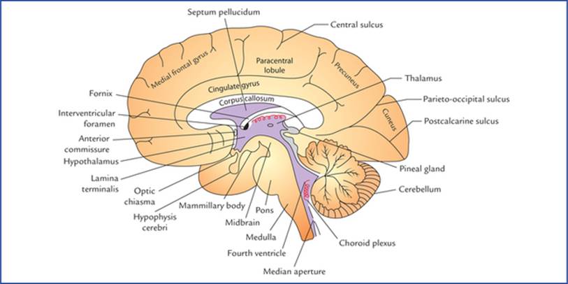
FIG. 6.6 Median sagittal section of the brain.
The inferior surface of the cerebral hemisphere is uneven and presents orbital and tentorial surfaces.
Basal ganglia/nuclei
The basal ganglia are subcortical masses of grey matter which are situated in the white core of each cerebral hemisphere.
The basal ganglia include the lentiform nucleus, caudate nucleus, claustrum, and amygdaloid body (Fig. 6.4).
During development of connections between the cerebral cortex and the brainstem, the bundles of fibres, converging as the internal capsule, partly divide the corpus striatum into a medial caudate nucleusand a lateral lentiform nucleus. Between the internal capsule and the cerebral cortex, the nerve fibres diverge as the corona radiata.
Functionally, the basal ganglia also include the subthalamic nucleus of diencephalon, and the substantia nigra and red nucleus of midbrain.
The basal ganglia influence the quality of motor functions and are sometimes termed extrapyramidal nuclei.
The major effect of basal ganglia is to decrease the muscle tone and inhibit the unwanted muscular activity.
Diencephalon
The diencephalon is the part of brain between the cerebrum and the brainstem. Its main components are: (a) two thalami, (b) hypothalamus, (c) metathalamus, (d) epithalamus, and (e) subthalamus.
Thalamus
The two thalami form by far the largest mass of the diencephalon and are separated from each other by a cavity of third ventricle, and joined with each other by an interthalamic adhesion. Each thalamus is a large ovoid mass of nuclei, lying obliquely across the path of cerebral peduncle as it ascends into the cerebral hemisphere. It forms a great relay station for all sensory impulses except smell, and appreciate crude sensations like pain and touch. The thalamic neurons send projections to the sensory area of the cerebral cortex.
The thalamus also integrates motor functions, relaying impulses from the cerebellum and corpus striatum to the motor area of the cerebral cortex. The connections of thalamus with limbic system influence mood, behaviour and memory.
Hypothalamus
The hypothalamus is the most inferior portion of the diencephalon and contains several small nuclei and nerve tracts. The most conspicuous nuclei called mammillary bodies appear as rounded elevations on the base of the brain in the region of interpeduncular fossa.
The hypothalamus regulates visceral activity through the autonomic nervous system and hormonal activity through the hypophysis cerebri.
Metathalamus
The metathalamus consists of two rounded medial and lateral geniculate bodies which protrude from the postero-inferior surface of the thalamus.
The medial and lateral geniculate bodies form the relay stations for the special senses of hearing and vision respectively.
Epithalamus
The epithalamus is a small area of diencephalon, postero-superior to the thalamus. It consists of pineal gland and habenular nuclei. The pineal gland plays an important role in controlling the onset of puberty through its secretion, the melatonin. The reduction of pineal secretion precipitates puberty. Pineal gland is also involved in sleep-wake cycle.
The habenular nuclei have olfactory and limbic connections. They are influenced by smell and are involved in emotional and visceral responses to odours.
Subthalamus
The subthalamus is a small area of diencephalon, that lies between the thalamus and the midbrain. It contains several nerve tracts, and the subthalamic nuclei which are associated with basal ganglia and are involved in controlling motor functions.
Midbrain
The midbrain is the smallest segment of the brainstem (Fig. 6.7). It is just superior to the pons, and contains the nuclei of IIIrd (oculomotor), IVth (trochlear), and Vth (trigeminal) cranial nerves.
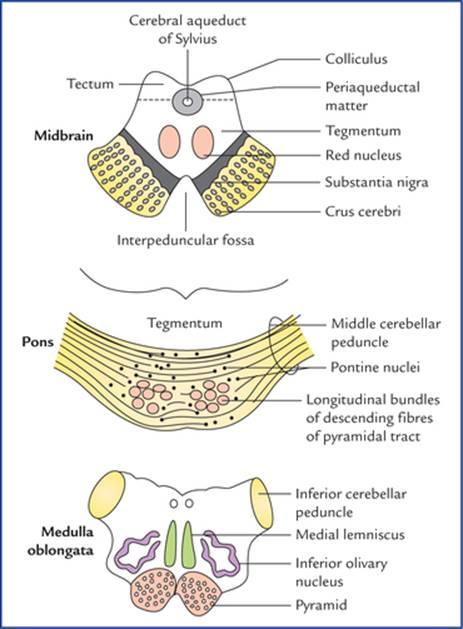
FIG. 6.7 Transverse sections through the three components of the brainstem.
The midbrain is traversed by the cerebral aqueduct. The part dorsal to the aqueduct, is called tectum, and consists of four surface elevations, the corpora quadrigemina (comprising two superior and two inferior colliculi). The superior and inferior colliculi receive visual and auditory impulses respectively, and are concerned with reflexes involving these senses. The part ventral to the aqueduct is divided into right and left halves, the cerebral peduncles. Each cerebral peduncle consists of a central part, the teg-mentum which is separated ventrally from the crus cerebri by a mass of pigmented grey matter, the substantia nigra.
The crus cerebri is continuing ipsilaterally with the internal capsule above and contains descending fibres from cerebral cortex to the brainstem and spinal cord. The space between the two crura is termed interpeduncular fossa.
The tegmentum largely consists of ascending tracts from the spinal cord to the thalamus and contains two large cigar-shaped nuclei, called red nuclei. In cross-section the red nuclei appear as oval masses of pinkish grey matter.
The red nuclei help in unconscious regulation and coordination of motor activities.
Pons (= Bridge)
The pons is the large middle part of the brainstem. It is continuous above with the midbrain and below with the medulla oblongata. It is so named because it forms a broad bridge between the two cerebellar hemispheres by its transverse fibres constituting the middle cerebellar peduncles. The vertical median sulcus on its ventral aspect lodges the basilar artery and therefore, termed basilar sulcus.
In transverse section (Fig. 6.7) the pons is seen to consist of a large ventral and a smaller dorsal region.
The ventral portion contains large number of nuclei, the pontine nuclei and longitudinal bundles of descending fibres of pyramidal tract. The pontine nuclei relay information from cerebrum to the cerebellum (cortico-ponto-cerebellar pathway).
The dorsal portion or tegmentum of pons contains important pontine sleep and respiratory centres. The pontine respiratory centre functions with the respiratory centre in the medulla to help control the respiratory movements.
Medulla Oblongata
The medulla oblongata is the lower part of the brainstem, and is continuous inferiorly with the spinal cord at the foramen magnum.
On the ventral aspect of medulla, there are two pyramid-shaped elevations, one on either side of median plane called pyramids. These elevations are produced by the descending fibres of the corticospinal tracts involved in the conscious control of the skeletal muscles. Lateral to the pyramids there are two rounded oval elevations, called olives containing inferior olivary nuclei. Dorsal to each olive is an inferior cerebellar peduncle (Fig. 6.7).
On the dorsal aspect, the medulla has cuneate and gracile tubercles produced by similarly named nuclei on either side of the median plane.
Medulla contains various vital autonomic centres responsible for several reflexes, such as those involved in the regulation of the heart rate, blood vessel diameter, breathing, swallowing, vomiting, coughing and sneezing.
Cerebellum (= Little Brain)
The cerebellum lies dorsal to the pons and medulla, and consists of two lateral hemispheres, the cerebellar hemispheres and a median worm-like part called vermis. It is connected to the midbrain, pons and medulla by superior, middle and inferior cerebellar peduncles respectively. The surface of cerebellum has narrow transverse ridges called folia (leaf-like in sections) separated by deep fissures.
Functionally, the cerebellum is mainly concerned with involuntary control of somatic motor activities, essential for maintenance of equilibrium, muscle tone, and posture.
Base of the Brain
The base of the brain as seen in dissection is shown in Figure 6.8. The base of the brain presents: (a) orbital and tentorial surfaces of the frontal and temporal lobes of both the cerebral hemispheres, (b) interpeduncular fossa, (c) ventral aspects of midbrain, pons, medulla oblongata and cerebellum, and (d) superficial attachment of the cranial nerves (Fig. 6.9).
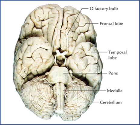
FIG. 6.8 The base of the brain; the midbrain lying between the pons and hypothalamus is not seen.
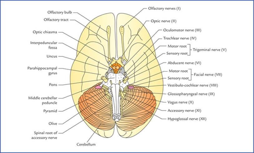
FIG. 6.9 Base (inferior aspect) of the brain. (Source: Singh Vishram. Anatomy of Head, Neck and Brain. New Delhi: Elsevier, 2009.)
The interpeduncular fossa and superficial attachments of cranial nerves are described in brief.
Interpeduncular fossa (Fig. 6.10)
The interpeduncular fossa is a rhomboidal space bounded on either side by crus cerebri of cerebral peduncles, anteriorly by optic chiasma and optic tracts; and posteriorly by the pons. The interpeduncular fossa contains: (a) a raised area of grey matter lying anterior to the mammillary bodies called tuber cinereum, (b) two small spherical bodies called mammillary bodies, (c) a narrow stalk which connects the hypophysis cerebri with the tuber cinereum called infundibulum, (d) posterior perforated substance, which is a layer of grey matter in the angle between the crus cerebri, and is pierced by central branches of the posterior cerebral arteries, and (e) oculomotor nerve which emerges immediately dorsomedial to the corresponding crus.
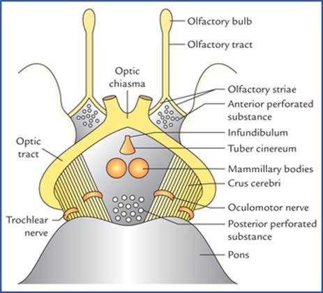
FIG. 6.10 Boundaries and contents of interpeduncular fossa.
The divisions and functions of the brain are summarized in Table 6.1.
Table 6.1
Divisions and functions of the brain
|
Brain region |
Function |
|
Cerebrum |
Conscious perception of sensory modalities, conscious motor activity, cognition,* etc. |
|
• Basal ganglia |
Control of muscular activity and posture. Largely inhibit unintentional movement |
|
• Limbic system |
Autonomic response to smell, emotion, mood, recent memory, etc. |
|
Diencephalon |
|
|
• Thalamus |
Major sensory relay centre; influences mood and movement |
|
• Hypothalamus |
Major control centre for maintaining homeostasis and regulating endocrine function |
|
• Subthalamus |
Associated with basal ganglia and help in coordinating motor functions |
|
• Epithalamus |
Sleep-wake cycle, smell |
|
Brainstem |
Contains ascending and descending nerve tracts, houses nuclei of all cranial nerves except 1st and Ilnd |
|
• Midbrain |
Visual and auditory reflex centres |
|
• Pons |
Relay centre between cerebrum and cerebellum, houses sleep centre |
|
• Medulla |
Centre for several vital autonomic reflexes, viz. heart rate, breathing, swallowing and vomiting |
|
Cerebellum |
Control of muscle movement and tone; regulates extent of intentional movement |
*Cognition – The intellectual functions or ways of knowing and thinking including the processes of perceiving, imagining, remembering, reasoning and judging.
Superficial Attachments of the Cranial Nerves
All the 12 pairs of cranial nerves are attached on the ventral aspect of the brain except the fourth pair (trochlear nerves) which are attached on the dorsal aspect.
Each cranial nerve enters or leaves the brain surface at its superficial attachment and the fibres, which it contains, either arise from (efferent or motor fibres) or terminate in (afferent or sensory fibres) nuclei within the brain.
The first two pairs are attached to the forebrain; the third and fourth to the midbrain; the fifth to the pons, and the remainder to the medulla oblongata. The eleventh cranial nerve also receives a root from the upper part of spinal cord.
Sites of Attachments
• Olfactory nerves. There are about 20 olfactory nerves on each side. They arise from olfactory epithelium of nasal cavity and pass through the cribriform plate of ethmoid and end in the olfactory bulb, which lies on the orbital surface of the frontal lobe. They are so delicate that no trace of them is seen on the olfactory bulb when the brain is removed from the cranial cavity.
• Optic nerve. This is a thick cylindrical nerve which arises from the retina and joins the anterolateral angle of the optic chiasma.
• Oculomotor nerve. It emerges from the groove on the medial aspect of the cerebral peduncle in the posterior part of the inter-peduncular fossa.
• Trochlear nerve. It is a slender nerve which emerges on the dorsal aspect of the midbrain, lateral to the median plane. It winds round the lateral aspect of the midbrain towards the interpeduncular fossa immediately superior to the pons.
• Trigeminal nerve. It is largest of the cranial nerves and attached to the junction of the pons and middle cerebellar peduncle by two roots: a large lateral sensory root, and a small medial motor root.
• Abducent nerve. It emerges at the inferior border of the pons, opposite to the upper end of the pyramid.
• Facial nerve. It emerges at the inferior border of pons lateral to the abducent nerve by two roots: a thick medial motor root and a slender lateral sensory root called nervus intermedius.
• Vestibulocochlear nerve. It consists of two nerves: vestibular and cochlear which are attached in the cerebellopontine angle. The cochlear nerve lies posterior to the inferior cerebellar peduncle and vestibular anterior to it.
• Glossopharyngeal nerve. It emerges by number of rootlets from a groove between the olive and inferior cerebellar peduncle.
• Vagus nerve. It also emerges by number of rootlets from a groove between the olive and inferior cerebellar peduncle below the rootlets of glossopharyngeal nerve.
• Accessory nerve. It has two parts: cranial and spinal. The cranial part emerges by number of rootlets from medulla below the rootlets of vagus nerve and it is joined by spinal part, the rootlets of which are attached to the upper five cervical spinal segments.
• Hypoglossal nerve. It arises by a row of rootlets from a groove between the pyramid and olive.
Spinal Cord
The spinal cord is the long cylindrical lower part of the CNS occupying the upper two thirds of the vertebral canal. It is continuous rostrally with the medulla oblongata at foramen magnum and ends caudally as tapered conus medullaris, at the level of lower border of first lumbar vertebra (L1).
It gives origin to 31 pairs of spinal nerves. The region of origin of a pair of spinal nerves is called spinal segment.
The spinal cord has central grey and peripheral white matter. In transverse section the grey matter has an irregular H-shape, and looking somewhat similar to a butterfly (Fig. 6.11) with two anterior horns, two posterior horns and a grey commissure containing a central canal. In the thoracic and upper two lumbar segments there are also lateral horns because these segments of spinal cord are associated with the autonomic nervous system.

FIG. 6.11 Transverse section of the spinal cord at the level of thoracic segments.
In three-dimensional view of grey matter of a spinal segment, the horns appear as long vertical pillars (Fig. 6.12) hence now a days, they are termed grey columns. However, the term ‘horn’ is still frequently used in texts on physiology and medicine.

FIG. 6.12 Three-dimensional view of grey matter of a spinal segment.
In each half of the spinal cord, the white matter is divided into anterior, lateral and posterior white columns or funiculi.
Each funiculus is subdivided into fasciculi or nerve tracts. The individual nerve fibres carrying action potentials to (ascending) or from (descending) the brain are usually grouped together to form fasciculi/tracts.
The spinal cord is extremely important for the overall function of the nervous system. It forms the communication link between the brain and the peripheral nervous system below the head, integrating incoming information and producing responses through reflex mechanisms.
Ventricular System and CSF in CNS
There are four ventricles of the brain (Fig. 6.13): two lateral ventricles, a third ventricle, and a fourth ventricle.

FIG. 6.13 The ventricles of the brain.
The two lateral ventricles, one in each cerebral hemisphere, form the largest component of the ventricular system.
They are occupied a considerable part of the cerebral hemisphere and are separated from each other by the septum pellucidum, extending between the corpus callosum and fornix (Fig. 6.6). The third ventricleis a narrow slitlike cavity of the diencephalon. Two lateral ventricles are connected with the third ventricle via the interventricular foramina (of Monro). The third ventricle communicates via the cerebral aqueduct of midbrain to the fourth ventricle, a cavity within the hindbrain. The fourth ventricle in turn is continuous with the central canal of the spinal cord. The central canal has a small dilatation at its inferior end, the terminal ventricle.
The cerebrospinal fluid is formed in the ventricles by the choroid plexuses and passes through apertures in the roof of fourth ventricle into the subarachnoid space around brain and spinal cord.
Meninges and CSF
The brain and spinal cord are surrounded by three protective membranes called meninges. From within outwards these are: (a) pia mater, (b) arachnoid mater, and (c) dura mater.
The subarachnoid space between the pia and arachnoid mater contains the cerebrospinal fluid (CSF) and the major arteries.
Clinical Correlation
Disorders Of The Central Nervous System
• Multiple sclerosis: It is a debilitating, degenerative disorder of unknown cause. Multiple sclerosis is characterized by multifocal demyelination throughout the white matter of the CNS.
• Seizure disorder (epilepsy): It is characterized by episodic, sudden, violent, and involuntary contractions of group of muscles, resulting from excessive discharge of cerebral neurons.
• Meningitis: It is the inflammation of meninges caused by bacteria and viruses.
• Encephalitis: The inflammation of brain and spinal cord that also involves the meninges.
• Space-occupying lesions: A space-occupying lesion is an abnormal growth of neural or non-neural tissue within the cranial cavity that may be primary or secondary (metastatic cancer).
• Herniation of the brain: The herniation of brain occurs due to an increased intracranial pressure produced by a space-occupying lesion such as tumour. The various types of brain herniation are (Fig. 6.14):
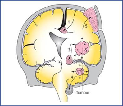
FIG. 6.14 The types of brain herniation: (1) cingulate gyrus herniation, (2) lateral tentorial herniation (temporal uncus herniation), (3) central tentorial herniation, (4) cerebellar tonsil herniation, and (5) herniation of brain-tissue through the craniotomy defect. Adapted from: Singh, Vishram. Clinical and Surgical Anatomy, 2nd edn. New Delhi: Elsevier; 2007.
– Cingulate gyrus herniation (subfalcine herniation), occurs below the falx cerebri.
– Transtentorial (uncal) herniation, is the herniation of medial edge of the temporal lobe (uncus) through the tentorial notch.
– Central tentorial herniation, is a vertical displacement of the brainstem and diencephalon through the tentorial notch.
– Tonsillar herniation, is the protrusion cerebellar tonsil through the foramen magnum. Herniation through cranial defecture may occur.
• Cerebrovascular accidents (stroke): It is a sudden, focal neurological deficit resulting from impaired circulation of the brain. It is associated with a cardiovascular disease such as thrombosis, embolism, or haemorrhage which causes impairment of circulation.
Clinical Problems
1. Explain why the older individuals often show: (a) slow response to various stimuli, (b) diminution in sensory perception, (c) decline in problem solving skills, and (d) decreased efficiency for recent memory.
2. Why do patients suffering from space-occupying lesions within skull, viz. tumour, haematoma and abscess, commonly complain of severe headache and vomiting?
3. What is contre-coup injury of brain, and how does it occur?
4. What injuries may occur to the brain in a motor-bike accident, how wearing of a crash helmet helps to prevent or minimize such injuries to the brain?
5. Explain how spinal cord injury in the cervical region above the level of 3rd spinal segment can cause sudden death.
6. What are the five most common brain tumours?
Clinical Problem Solving
1. These changes occur due to the reduction in the number of cerebral neurons at the rate of 1% a year beginning at 50 years of age. However, if the brain is kept active, the reserve cerebral neuronsmay delay the onset of such symptoms.
2. Since the skull is a rigid container of fixed volume, the space-occupying lesions add to the normal bulk of the intracranial contents producing an increased intracranial pressure. The severe headache occurs due to stretching of dura mater, and vomiting due to stimulation of ‘vomiting centre’ in brainstem because of increased intracranial pressure.
3. The contre-coup injury to brain is an injury to the pole of the brain opposite to the side of impact. The head is freely mobile on the neck and the brain is floating in the cerebrospinal fluid within the skull. Therefore, when the head is hit suddenly and with great force on the back or front, the brain is displaced in opposite direction and strikes against the cranial bones on that side and gets injured.
4. The brain is floating in the cerebrospinal fluid within the cranium, so that a severe blow to the head or sudden deceleration leads to the displacement of the brain. This may cause intracranial haemorrhage (viz. extradural, intracerebral, etc.) leading to pressure symptoms such as unconsciousness, coma, etc. A crash helmet helps to protect the brain by cushioning the blow on the head and slows down rate of deceleration of the brain.
5. Sudden death occurs due to spinal shock, and respiratory arrest following paralysis of diaphragm and intercostal muscles. As the lesion is above the origin of phrenic nerves (C3, 4 and 5) supplying diaphragm and intercostal nerves (from thoracic segments) supplying intercostal muscles.
6. The five most common brain tumours are as follows:
(a) Glioblastoma multiforme: It is the commonest and most fatal brain tumour and arises from astrocytes.
(b) Meningioma: It is the second most common tumour and arises from meninges (arachnoid granulations).
(c) Schwannoma: It arises from Schwann cells.
(d) Ependymoma: It arises from ependymal lining of the brain.
(e) Medulloblastoma: It arises from neurectodermal cells.