
INTRODUCTION
Most congenital heart diseases (CHDs) of consequence present in early childhood and are managed appropriately. Some CHDs are detected for the first time in adults either because of their natural course or because these were neglected in childhood. There is also a large pool of patients who have been operated for CHD in childhood but become symptomatic in adult life because of arrhythmias, heart failure or recurrence, or residual lesions.1-3Echocardiography is an integral tool in managing such patients. Spatial anatomy is difficult to assess in CHD and three-dimensional echocardiography has become an important part of echocardiographic assessment.
The most common congenital heart disorders affecting adults are:2,3
• Congenital valve defects (discussed in section on valvular heart disease).
• Atrial and ventricular septal defects.
• Patent foramen ovale (PFO).
• Patent ductus arteriosus.
• Anomalous pulmonary veins.
• Coarctation of the aorta discussed in section on 'Aorta'.
• Ebstein anomaly.
• Tetralogy of Fallot (TOF).
• Transposition of the great arteries.
• Pulmonary hypertension (Eisenmenger syndrome).

Fig. 16.1: Schematic diagram of the right atrium showing location of various types of atrial septal defect (ASD).
(SVC: Superior vena cava; IVC: Inferior vena cava; CS: Coronary sinus; RV: Right ventricle; TV: Tricuspid valve).
ATRIAL SEPTAL DEFECT
Three types of atrial septal defect (ASD) can occur
(Fig. 16.1): 4-6
1. Ostium secundum is the most common type (70%). It can be large, but usually does not affect the atrioventricular valves.
2. Ostium primum—the hole is situated close to the atrioventricular valves and can be associated with an atrioventricular septal defect.

Fig. 16.2: Transesophageal echocardiographic long axis view with rightward probe deflection showing secundum atrial septal defect (arrow length).
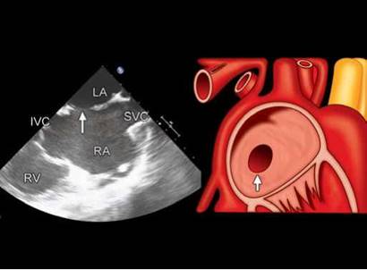
Fig. 16.3: Ostium secundum atrial septal defect shown in transesophageal echocardiographic bicaval view (left panel) and the schematic diagram (right panel).

Fig. 16.4: Transesophageal echocardiographic anatomic relationship of the secundum atrial septal defect with left-to-right shunt (interrupted line).

Fig. 16.5: Transthoracic echocardiographic apical four-chamber view in end-systole. Two rims of the atrial septal defect are shown (arrows).
3. Sinus venosus is a defect situated near the entrance of the superior vena cava (SVC) or inferior vena cava (IVC) to the right atrium.7 It is unusual and is often associated with partial anomalous pulmonary venous drainage (usually drainage of the right upper lobe into the SVC). the shunt of blood from the left atrium to the right atrium results in:
• Increased volume load and dilatation of the right atrium and right ventricle (RV).
• Increased pulmonary blood flow and enlargement of the pulmonary arteries.
• Increase in size of the pulmonary veins.
• Reduced filling of the left ventricle (LV) and aorta. the ostium secundum ASD is the most common
type of ASD, and comprises 6-10% of all CHD (Figs 16.2 to 16.6). About 70% of the ASD are ostium secundum type.
Stretched ASD dimensions are best measured in systole (Fig. 16.6).

Fig. 16.6: Apical four-chamber view with color Doppler interrogation of atrial septal defect. Supero-inferior diameter is much bigger in ventricular systole (left panel) compared to that in diastole (right panel).

Fig. 16.7: Subcostal four-chamber view showing rims of secundum atrial septal defect (arrows).
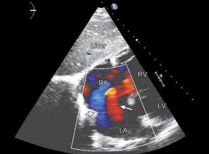
Fig. 16.8: Subcostal long-axis view showing bidirectional flow across a large secundum atrial septal defect (arrows).
The secundum ASD usually arises from an enlarged foramen ovale, inadequate growth of the septum secundum, or excessive absorption of the septum primum. Ten to twenty percent of individuals with ostium secundum ASDs also have mitral valve prolapse.
If the ostium secundum ASD is accompanied by an acquired mitral valve stenosis, that is called Lutembacher's syndrome.
Subcostal views or right low parasternal views are the best to visualise ASD because in these transthoracic views, the interatrial septum is perpendicular to the ultrasound beam (Figs 16.7 and 16.8). However, obtaining adequate views in adults may be a challenge.
Atrial septal defect is assessed by parasternal short- axis, apical and subcostal four-chamber views in transthoracic echocardiography (TTE). In transesophageal echocardiography (TEE), multiple views are used to estimate rims of the ASD and its relationship with adjoining structures (Figs 16.9A to D).
Earlier, it was sufficient to categorise ASD by transthoracic and transesophageal imaging, give its approximate size in multiple views, estimate left-to-right shunt along with pulmonary artery pressures and associated anomalies. With advent of transcatheter closure of ASD, it has become important to identify and measure rims of ASD all around and study their characteristics.8 3D THE and TEE plays a stellar role in this aspect (Figs 16.10 to 16.13). the key points to be examined are:
• Size, shape and number of ASDs.
• Panoramic view of the rims.
• Relationship to adjoining structures.
Advantages of the real-time 3DE are:8
• Unlimited viewing of the interatrial septum.
• Possibility to create an en face view.
• Examination from both right atrium and the left atrium.
• Appreciating the change in size and shape of the ASD during cardiac cycle (largest size during atrial relaxation or ventricular systole). Size can vary by about 50%.
• Feasibility for transcatheter closure and monitoring during the procedure.
• Number of defects with their relationships.

Figs 16.9A to D: Multiple transesophageal echocardiographic views to assess shape, size and margins of the secundum atrial septal defect (ASD). Comparison of upper panels with lower panels indicates that the ASD is elliptical and posterio-inferior margin (left upper panel) is deficient.
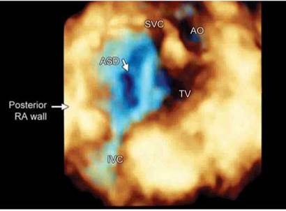
Fig. 16.10: Real-time 3DE right atrial view with atrial septal defect (ASD) in the center to show various relationships.

Fig. 16.11: 3D transthoracic echocardiographic image showing en face view of the atrial septal defect (ASD).

Fig. 16.12: Real-time 3D transesophageal echocardiographic view from the left atrium.

Fig. 16.13: Real-time 3D transesophageal echocardiographic en face view of the interatrial septum with atrial septal defect (ASD).
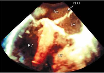
Fig. 16.14: Real-time 3D TEE examination showing patent foramen ovale (PFO) (arrow) during straining.
Atrial septal defect rims can be seen in the following manner:
• Aortic or anterosuperior rim.
• Mitral or anteroinferior rim.
• Superior or SVC rim.
• Posteroinferior or IVC rim.
• Posterior rim along posterior right atrial wall.
• Inferior or coronary sinus rim.
A PFO is a small channel that has some hemodynamic consequence; it is a remnant of the fetal foramen ovale. Clinically, it is linked to:
• Decompression sickness.
• Paradoxical embolism.
• Migraine.
On echocardiography, there may not be any shunting of blood noted except when the patient coughs. However, shunting can be easily appreciated by contrast injection.6 It is possible to visualize PFO by real-time 3D TEE (Fig. 16.14).
There is debate within the neurology and cardiology communities about the role of a PFO in cryptogenic (i.e. of unknown cause) neurologic events such as strokes and transient ischemia attacks without any other potential cause.9Some data suggested that PFOs may be involved in the pathogenesis of some migraine headaches.10 It can also be associated with fossa ovalis membranous aneurysm (Fig. 16.15).
Patent foramen ovale can be visualized by injecting agitated saline in a peripheral vein either during transthoracic examination but more often during TEE. Immediate appearance of contrast in the left atrium after it is seen in the right atrium (usually in < 3 cardiac cycles) is highly suggestive of PFO (Fig. 16.16). Sometimes, Valsalva manouvre or cough or sniff can be used to augment the effect.
Occasionally, small secundum ASD can occur in oval fossa following atrial puncture and dilatation (Fig. 16.17). These are usually restrictive with appreciable pressure gradients.
OSTIUM PRIMUM ATRIAL SEPTAL DEFECT
A defect in the ostium primum is occasionally classified as an ASD but it is more commonly classified as an atrioventricular septal defect (Figs 16.18 and 16.19). Ostium primum defects are less common than ostium secundum defects.

Fig. 16.15: Aneurysm of the patent foramen ovale membrane (arrow) seen in apical four-chamber view.

Fig. 16.16: Apical four-chamber view showing apparently intact atrial septum with contrast appearance in the left-sided chambers.

Fig. 16.17: Apical four-chamber view showing small atrial septal defect in oval fossa (arrow) following transcatheter mitral commissurotomy.

Fig. 16.18: Apical four-chamber view showing ostium primum atrial septal defect (ASD) (arrow).
SINUS VENOSUS ATRIAL SEPTAL DEFECT
A sinus venosus atrial septal defect is a type of ASD in which the defect in the septum involves the venous inflow of either the SVC or the IVC (Fig. 16.20).
A sinus venosus ASD that involves the SVC makes up 2-3% of all interatrial communication.5 It is located at the junction of the SVC and the right atrium (Fig. 16.20). It is frequently associated with anomalous drainage of the right-sided pulmonary veins into the right atrium (instead of the normal drainage of the pulmonary veins into the left atrium).
Sinus venosus defects are better appreciated by TEE and need to be differentiated from the secundum ASD (Fig. 16.21).
COMMON OR SINGLE ATRIUM
Common (or single) atrium is a failure of development of the embryologic components that contribute to the atrial septal complex. It is frequently associated with heterotaxy syndrome or tricuspid atresia (Fig. 16.22).
VENTRICULAR SEPTAL DEFECT
Ventricular septal defect (VSD) is the most common congenital heart defect (20% of all CHD) and frequently seen in adults either as a small defect or as a part of repaired but residual complex CHD. Its prevalence in adults is low, fundamentally due to spontaneous closing of some defects and to the fact that the majority
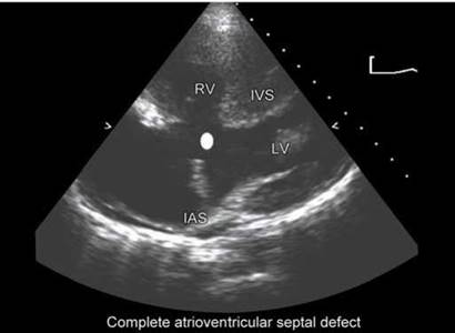
Fig. 16.19: Complete atrioventricular septal defect with primum defect (spot) seen in four-chamber view. There is no central fibrous body and inferior interatrial and superior ventricular septa are deficient.
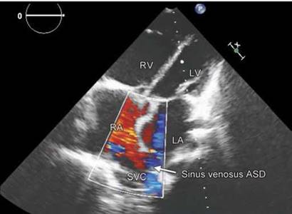
Fig. 16.20: Sinus venosus atrial septal defect (ASD) showing override of superior vena cava (SVC) and right upper pulmonary vein.

Figs 16.21A and B: 2D transesophageal echocardiographic caval view showing sinus venosus atrial septal defect. Note that the posterior wall of the superior vena cava is deficient and right pulmonary artery is jutting the defect.
of symptomatic defects are closed surgically during childhood. Symptoms depend on the size of the defect and the age of the patient. Small VSDs are usually asymptomatic and compatible with a normal life (in fact, about 40% close spontaneously in early childhood). If a large shunt does not produce symptoms during infancy, there is usually little disturbance until late adolescence or early adult life when the patient develops high pulmonary vascular resistance, breathlessness, fatigue and cyanosis. There is progression to effort syncope, recurrent hemoptysis and heart failure.
The interventricular septum has a complex and curved shape (Figs 16.23 to 16.25). The ventricular septum is directed obliquely backward to the right, and curved with the convexity toward the RV; its margins correspond
with the anterior and posterior longitudinal sulci.11 It is composed of:
• Membranous part near the central fibrous body.
• Muscular Septum.
• Outlet septum or infundibular septum or aortopulmonary septum that is spiral in orientation.
Upper and posterior part, which separates the
aortic vestibule from the lower part of the right atrium and upper part of the RV, is thin and fibrous, and is termed the membranous ventricular septum (septum membranaceum).
The muscular part of the interventricular septum derives from the bulboventricular flange that is developed due to differential growth of primitive ventricle and bulbous cordis.

Fig. 16.22: Common atrium in an adult subject with tricuspid atresia shown in apical four-chamber view.
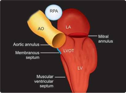
Fig. 16.23: Relationship of the membranous septum to the aortic annulus and the left ventricular outflow tract (LVOT).
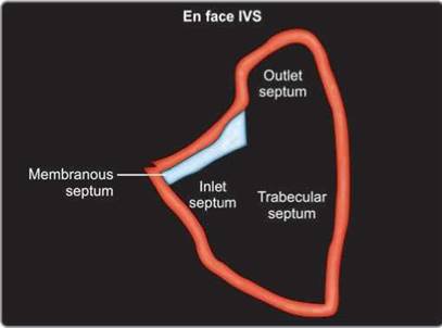
Fig. 16.24: En face view of the interventricular septum (IVS).

Fig. 16.25: Site-specific classification of the ventricular septal defect (VSD).
MORPHOLOGY OF VSD
Four types are described below:12-15
• Perimembranous—most common type in adults (80%) (Figs 16.26 and 16.27)
• Muscular—most common type in young children (Figs 16.28 to 16.30)
• Complete atrioventricular septal (endocardial cushion) defects (Figs 16.19 to16.31)
• Supracristal (subarterial) (Fig. 16.32)
PATHOPHYSIOLOGY OF VSD
• Restrictive VSD is typically small, such that a significant pressure gradient exists between the LV and RV with small shunt (Qp/Qs < 1.4: 1) (Fig. 16.33).
• Moderately restrictive VSD ^ moderate shunt (Qp/Qs 1.4 to 2.2: 1).
• Large/nonrestrictive VSD ^ large shunt (Qp/Qs > 2.2: 1). ttere is enlargement of LA and LV as well (Fig. 16.34).

Fig. 16.26: Schematic diagram of the short-axis transthoracic echocardiographic image showing location of perimembranous versus subarterial ventricular septal defect (VSD).

Fig. 16.27: Transesophageal echocardiography long-axis view showing perimembranous ventricular septal defect (arrow).

Fig. 16.28: Apical muscular ventricular septal defect (arrow) in modified short-axis view.
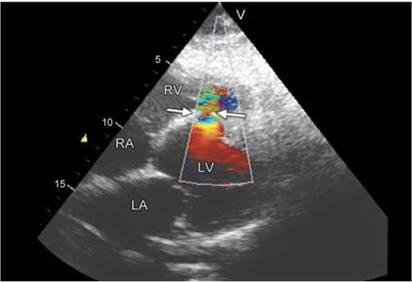
Fig. 16.29: Restrictive muscular ventricular septal defect with left-to- right shunt.

Fig. 16.30: Large muscular ventricular septal defect in posterior septum (arrow).

Fig. 16.31: 3D transthorasic echocardiographic showing atrioventricular septal defect (arrow).

Fig. 16.32: Subarterial ventricular septal defect in transthoracic echocardiography short-axis image.
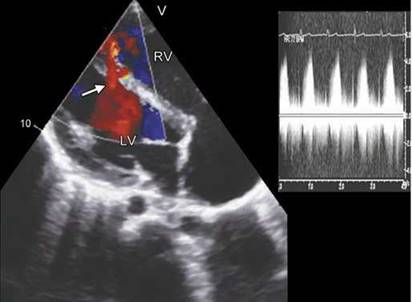
Fig. 16.33: Restrictive muscular ventricular septal defect with 100 mm Hg transventricular peak gradient.

Fig. 16.34: Large perimembranous ventricular septal defect with transventricular peak gradient of 30 mm Hg indicating presence of significant pulmonary hypertension.
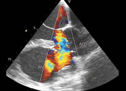
Fig. 16.35: 15 mm ventricular septal defect (VSD) size compared to 25 mm of aortic annulus (large VSD).
• Eisenmenger VSD ^ irreversible pulmonary hypertension and shunt may be reversed (i.e. R ^ L)
Defect size is often compared to aortic annulus:14 Large: > 50% of annulus size (Figs 16.35 and 16.36) Medium: 25-50% of annulus size (Fig. 16.37)
Small: < 25% of annulus size
Real-time 3DE has great utility in assessing size and location of VSD (Fig. 19.38). En face view from the left side identifies VSD more accurately.
Ebstein's anomaly is the downward displacement of a portion of the tricuspid valve with atrialization of a large
part of the RV.16-18 There is often an associated ostium secundum ASD. The atrialized portion of the ventricle hinders rather than helps the forward flow of blood and there is tricuspid regurgitation. Occasionally, Ebstein's anomaly is asymptomatic, but it generally presents in childhood or early adulthood with dyspnea, fatigue, signs of tricuspid regurgitation and right-sided cardiac failure.17
In the normal heart, the tricuspid valve has three leaflets: anterior, posterior and septal. Ebstein's anomaly is a malformation of the tricuspid valve and RV characterized by (Fig. 16.39).
• Adherence of the septal and posterior leaflets to the underlying myocardium (failure of delamination).

Fig. 16.36: Large ventricular septal defect (VSD) 25 mm in tetralogy of Fallot with secundum ASD in apical four-chamber view.

Fig. 16.37: 8 mm ventricular septal defect (VSD) size compared to 24 mm of aortic annulus (moderate VSD).

Fig. 16.38: En face view of atrioventricular septal defect from the right side (arrow).
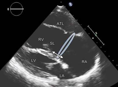
Fig. 16.39: Ebstein's anomaly in an adult in parasternal long-axis view. Note enlarged right atrium (RA), dilated annulus, small atrialised portion, septal leaflet (SL) displacement and sail-like anterior tricuspid leaflet (ATL).
Downward (apical) displacement of the functional annulus (septal>posterior>anterior).
• Dilatation of the 'atrialized' portion of the RV, with various degrees of hypertrophy and thinning of the wall.
• Redundancy, fenestrations and tethering of the anterior leaflet.
• Dilatation of the right atrioventricular junction (true tricuspid annulus).
In normal human hearts, the downward displacement of the septal and posterior leaflets in relation to the anterior mitral valve leaflet is < 8 mm/m2 body surface area. the spectrum of the malformation in Ebstein's anomaly may range from:
• Minimal displacement of the septal and posterior leaflets
• Imperforate membrane
• Muscular shelf between the inlet and trabecular zones of the RV.
the anterior leaflet is generally redundant and may contain several fenestrations. Its chordae tendineae are generally short and poorly formed. The anterior leaflet of the tricuspid valve (ATL) may be severely deformed, so that the only mobile leaflet tissue is displaced into the right ventricular outflow tract, where it may cause obstruction or form a large sail-like intracavitary curtain (Fig. 16.40).

Fig. 16.40: Ebstein's anomaly with large, redundant and deformed anterior tricuspid leaflet (ATL) attached by short aberrant chords to the right ventricle (RV) free wall.

Fig. 16.41: Type A Ebstein's anomaly.

Fig. 16.42: Type B Ebstein's anomaly.

Fig. 16.43: Type C Ebstein's anomaly. Note the right ventricular outflow tract (RVOT) obstruction caused by anterior leaflet (arrow).
In Ebstein's anomaly, the RV is divided into two regions:
1. Atrialized Ventricle: The part directly involved with the malformation (i.e. the inlet portion), which is functionally integrated with the right atrium
2. Functional RV: The part that is not involved by the anomaly, which consists of the other two components of the RV, namely the trabecular and outlet portions that constitute the functional RV.
The 'atrialized' portion of the RV (i.e. the inlet component) can become disproportionately dilated and may account for more than half of the right ventricular volume in extreme cases instead of the usual one third of the total right ventricular volume.
CARPENTIER CLASSIFICATION OF EBSTEIN'S ANOMALY19
• Type A: The volume of the true RV is adequate (Fig. 16.41).
• Type B: A large atrialized component of the RV exists, but the ATL valve moves freely (Fig. 16.42).
• Type C: The ATL is severely restricted in its movement and may cause significant obstruction of the right ventricular outflow tract (RVOT) (Fig. 16.43).
Type D: Almost complete atrialization of the ventricle except for a small infundibular component (Fig. 16.44).

Fig. 16.44: Type D Ebstein's anomaly with very small residual functional right ventricle.

Fig. 16.45: Double-chambered right ventricle (RV) in parasternal long- axis view (arrows). (DC: Distal chamber).
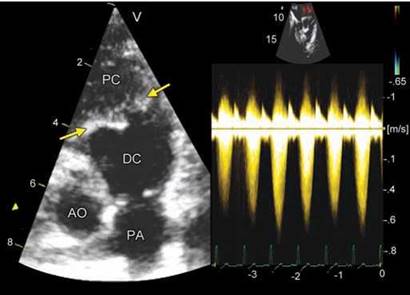
Fig. 16.46: Double-chambered right ventricle (RV; arrows) shown in modified short-axis view. A peak systolic gradient of 150 mm Hg was observed (right panel)
(PC: Proximal chamber; DC: Distal chamber).

Fig. 16.47: Shelf-like obstruction proximal to pulmonary valve (PV). (PC: Proximal chamber; DC: Distal chamber LA: Left atrium; PA: Pulmonary atresia).
double-chambered rv
In this disorder, the RV is divided into a high-pressure proximal chamber and lower-pressure distal chamber by anomalous muscle bundles, creating a double-chambered RV (Figs 16.45 and 16.46).
• the morphological features may be variable and the particular muscle bands involved vary from an anomalous septoparietal band, an anomalous apical shelf (Fig. 16.47), or an abnormal moderator band.20 The distance between the moderator band and the pulmonary artery may be abnormally short.
• the RVOT obstruction is generally believed to be an acquired one and to be progressive over time, although the basic anatomic features are congenital.
• In ~75% of cases, it is associated with a perimembranous VSD that is usually, but not always, below the level of the muscular obstruction (Fig. 16.48).
• In some instances, there is greater RV dysfunction when the VSD is restrictive or closes.
• Other associations include valvular pulmonary stenosis (PS), TOF and double-outlet RV (Fig. 16.49).
• There is subaortic obstruction in a variable number of these patients.
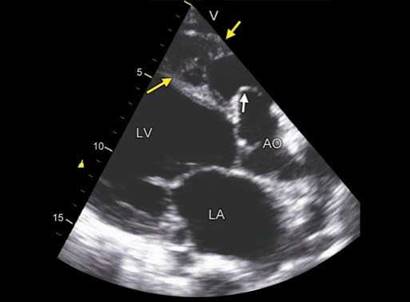
Fig. 16.48: Double-chambered right ventricle (yellow arrows) with aneurysm of the membranous ventricular septal defect (green arrow).

Fig. 16.49: Double-chambered right ventricle (yellow arrows) with pulmonary valve stenosis (right panel, white arrows).

Fig. 16.50: 3DE cross-section showing three aortic sinuses.

Fig. 16.51: Suprasternal long-axis view showing right and left aortic sinuses. Anterior to right sinus is the right ventricular outflow tract (RVOT).
• The anomaly is rare, occurring in ~1% of patients with congenital heart disease. It has been reported to develop in ~3% of patients with repaired TOF and in 3-10% of patients with a VSD.
The three sinuses of Valsalva are located in the most proximal portion of the aorta, just above the cusps of the aortic valve (Figs 16.50 and 16.51). The sinuses correspond to the individual cusps of the aortic valve. These structures contained within the pericardium are easily revealed using
echocardiography as distinct but subtle outpouchings of the aortic wall just above the valve. The sinuses end in the area of the sinotubular junction, and the tubular portion of the aorta begins here.
Aneurysm of the aortic sinus, also known as the sinus of Valsalva, is comparatively rare. Frequency of involvement is:21-25
• Aneurysm of the right sinus (65-85%) (Figs 16.52 and 16.53).
• Aneurysm ofthe noncoronarysinus (10-30%) (Figs 16.54 and 16.55.
• Left sinus aneurysm (< 5%).

Fig. 16.52: Aneurysm of the right sinus of Valsalva (arrow) seen in parasternal long-axis view.

Fig. 16.53: Aneurysm of the right sinus of Valsalva (arrow) shown in parasternal short-axis view

Fig. 16.54: Aneurysm of the noncoronary sinus (arrow) in transesophageal view at 80°.

Fig. 16.55: Aneurysm of the noncoronary sinus opening into the right atrium (arrow) 3D transthoracic echocardiography.
This type of aneurysm is typically congenital and may be associated with heart defects. It is sometimes associated with:
• Marfan syndrome
• Loeys-Dietz syndrome
• Ehlers-Danlos syndrome
• Atherosclerosis
• Syphilis
• Cystic medial necrosis
• Chest injury
• Infective endocarditis
Congenital aortic sinus aneurysm is caused by a dilatation, usually of a single sinus of Valsalva, from a
separation between the aortic media and the annulus fibrosus.
Rupture of the dilated sinus may lead to intracardiac shunting (Fig. 16.56) when a communication is established with the right atrium [Gerbode defect (10%)] or directly into the RV (60-90%). Cardiac tamponade may occur if the rupture involves the pericardial space. Rupture is the event that makes the patient seek medical attention although rarely, RVOT obstruction can occur. Various sites of rupture are shown below: (Figs 16.57 to 16.60)
Associated congenital defects with aneurysm of sinus of Valsalva are:
• Ventricular septal defect (30-60%, more often subarterial type).

Fig. 16.56: Schematic diagram showing relationship of the aortic sinuses with intracardiac structures and possible sites of communication when rupture occurs.
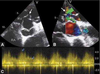
Figs 16.57A to C: Panel A shows aneurysm of right aortic sinus opening into the RVOT. Panel B shows color Doppler interrogation and panel C shows systolic-diastolic continuous flow across the defect.
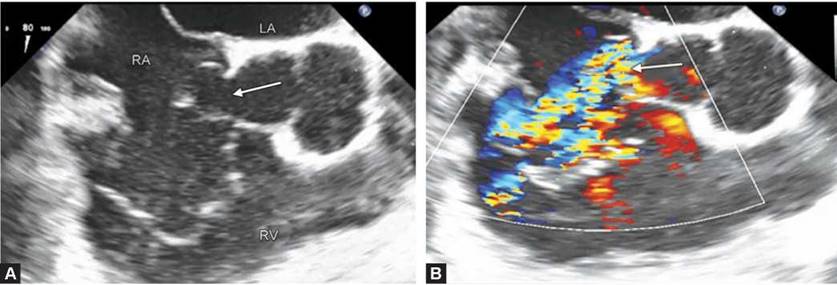
Figs 16.58A and B: Transesophageal echocardiographic view showing noncoronary sinus aneurysm opening into the right atrium (left panel, arrow). The right panel shows color Doppler interrogation of the flow.

Fig. 16.59: Transesophageal echocardiographic long-axis view showing communication of the left aortic sinus with the left atrium (arrows) without any aneurysm formation.

Figs 16.60A and B: Aneurysm of noncoronary aortic sinus communicating with the right atrium. 2D transesophageal echocardiographic color Doppler flow mapping (panel A) compared to 3D transthoracic echocardiographic imaging (arrow).
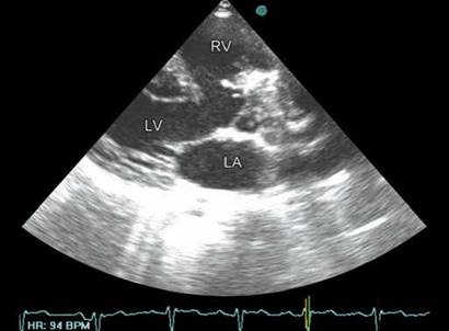
Fig. 16.61: Parasternal long-axis view showing large subaortic ventricular septal defect and override of the aortic root due to anterior deviation of the infundibular septum.

Fig. 16.62: Modified short-axis view showing infundibular obstruction (arrow) and mild hypoplasia of the pulmonary arteries. The peak systolic gradient of 100 mm Hg by continuous wave Doppler is shown in the right panel.
• Aortic insufficiency (20-30%).
• Bicuspid Aortic Valve (10%).
• Coarctation.
Aortic sinus aneurysms are more common among men than women and among Asians than other ethnic groups. Nonruptured aneurysms may be asymptomatic and incidentally discovered, or they may be symptomatic and manifest acutely with mass effect on adjacent cardiac structures. Ruptured Valsalva sinus aneurysms result in an aorto-cardiac shunt and may manifest as acute or insidiously progressive congestive heart failure, severe acute chest pain with dyspnea, or, in extreme cases, cardiac arrest.21
A significant number of patients with TOF survive to adulthood. The proportion of children reaching adulthood is predominantly determined by the magnitude of the hypoxia. TOF, the most common cyanotic malformation in adults, represents a spectrum from mild RVOT obstruction to complete pulmonary atresia.
Tetralogy of Fallot is diagnosed using recognized morphological criteria.24 TOF is a conotruncal defect resulting from anterior malalignment of the infundibular septum. This single morphologic defect gives rise to the four main components of TOF (Figs 16.61 to 16.63).
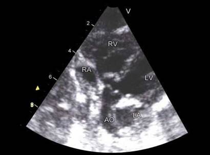
Fig. 16.63: Subcostal four-chamber view showing biventricular origin of the aortic root due to aortic override.

Fig. 16.64: Tetralogy of Fallot with atretic outflow tract.

Fig. 16.65: Complete atrioventricular septal defect with a large right ventricle (RV) in tetralogy of Fallot.

Fig. 16.66: Double-outlet right ventricle (RV) variant of tetralogy of Fallot. Note malalignment ventricular septal defect (arrow) with > 50% aortic override.
1. Anterior deviation of the infundibular septum
2. Ventricular septal defect
3. Obstruction to right ventricular outflow
4. Right ventricular hypertrophy
Malalignment of the infundibular septum to the trabecular septum is present, resulting in a malalignment VSD (Fig. 16.63).
Anterior displacement of the bulbotruncal region has been postulated to cause the infundibular stenosis.
• TOF with pulmonary atresia (Fig. 16.64).
• TOF with atrioventricular septal defects (Fig. 16.65).
• Double-outlet RV variant (Fig. 16.66).
• Absent pulmonary valve syndrome (Fig. 16.67). Double-outlet RV requires at least 50% of each great
vessel to arise from the morphological RV. This broad definition covers lesions from TOF to those with singleventricle physiology. The VSD is typically large and has four potential locations: subaortic, subpulmonic, doubly committed or remote. The insertion and location of the outlet septum define which great vessel is related to the VSD.
Tetralogy of fallot with 'absent pulmonary valve syndrome' is a rare cardiac malformation, usually associated with a number of other defects.25 Congenital absence of the leaflets of the pulmonary valve is

Fig. 16.67: Double-Outlet RV variant of tetralogy of Fallot with absent pulmonary valve. There is narrowing of the pulmonary annulus and pulmonary artery is aneurysmally dilated.

Fig. 16.68: Parasternal long-axis view showing Type 1 truncus arteriosus.

Fig. 16.69: Transthoracic echocardiographic short-axis view showing large ventricular (VSD) and absent right ventricular outflow tract (RVOT) in a case with truncus arteriosus.
less common when the ventricular septum is intact. Characteristic features of the syndrome include dysplasia or absence of the pulmonary valvar leaflets, permitting severe pulmonary regurgitation and aneurysmal dilation of the pulmonary artery (Fig. 16.67).
In TOF adults, evaluation of surgical residua and sequelae includes imaging of:26
• Aortic-to-pulmonary arterial palliative shunts.
• Detection of residual VSD.
• Patch leaks.
• Right ventricular outflow tract obstruction.
• Definition of extracardiac conduit patency.
• Quantitation of ventricular function and valvular regurgitation.
In pulmonary atresia, persistence of descending thoracic branches accounts for the abnormal pulmonary arterial supply in this condition. Major aortopulmonary collateral arteries may anastomose at any site in the pulmonary vascular tree. Most frequently, the right and left pulmonary arteries are patent and maintain free communication with each other; they are termed confluent pulmonary arteries. The pulmonary arteries may also be hypoplastic and nonconfluent. No antegrade blood flow is present from the RV to the pulmonary arteries. The ductus arteriosus is often an important source of blood supply, although it is occasionally absent.
TRUNCUS ARTERIOSUS OR COMMON ARTERIAL TRUNK
Truncus arteriosus or common arterial trunk is a very uncommon lesion in adults, and essentially all adult patients will have had surgical intervention (Figs 16.68 and 16.69).
• It consists of a single arterial trunk giving origin to the pulmonary arteries, coronary arteries and the systemic circulation.27
• It is rare even among congenital heart lesions, occurring in only ~1%.
• The Society of Thoracic Surgeons' classification simply combines Types 1 and 2 (main pulmonary trunk or branches coming off the trunk).
• The common trunk is large, and cystic medial necrosis is usually present, resulting in further dilatation as the patient ages.

Fig. 16.70: Paraternal long-axis view showing subaortic membrane (arrow). The right panel shows peak gradient of 64 mm Hg across the membrane with regurgitation as well.

Fig. 16.71: 3DE en face view of the orifice of the membrane (arrows) in the left ventricular outflow tract (LVOT) (left panel) compared to profile view in 2DE long-axis view (right panel).
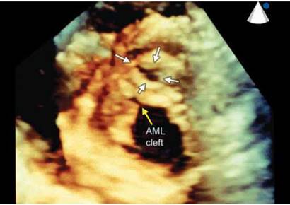
Fig. 16.72: Cleft anterior mitral leaflet (AML) (single yellow arrow) with subaortic membranous orifice (white arrows).
• the common trunk usually overrides the VSD and is committed to both ventricles.
• the VSD is large and nonrestrictive and results from absence of the infundibular septum. It lies between the two limbs of the septomarginal trabeculation.
• the truncal valve may have from 1 cusps to 6 cusps. In 1 study, 61% had a tricuspid truncal valve, 31% a quadricuspid valve and 8% a bicuspid valve.
• Poor valve function (mostly incompetence) usually results in early heart failure and death in infancy.
• the coronary arteries may arise above the truncal leaflets in a variety of positions, although many are normally located.
Subaortic membrane (SAM) is a form of fixed subaortic obstruction in which a fibrous membrane is located below the aortic valve (Fig. 16.70).
Subaortic membrane may involve the ventricular septum, the anterior leaflet of the mitral valve, and the aortic valve itself (Fig. 16.71).28 It may be associated with other structural anomalies of the aortic valve such as:
• bicuspid aortic valve
• abnormalities of the left ventricular outflow tract, such as in atrioventricular canal (Fig. 16.72) or tunnel subaortic stenosis.
Ventricular septal defect can also be associated with SAM (Fig. 16.73).
The subaortic stenosis causes problems in several different ways. It may obstruct left ventricular outflow and cause subaortic stenosis. The resultant effects are essentially the same as valvular aortic stenosis: left ventricular hypertrophy from the pressure overload, myocardial ischemia.
A SAM may alter left ventricular outflow dynamics and harm the aortic valve without any significant obstructive component. the abnormal flows cause aortic insufficiency and permanent structural damage to the aortic valve.
ttere may be other effects from SAM as well, including permanent damage to the mitral valve and direct damage

Fig. 16.73: Subaortic stenosis (white arrow) with subaortic ventricular septal defect (yellow arrow) with color flow map.

Fig. 16.74: Subaortic membrane very close to the aortic valve (arrow) with aortic regurgitation (see inset).
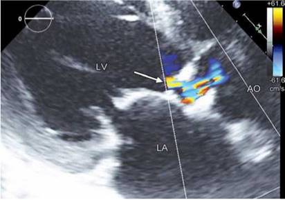
Fig. 16.75: Flow regurgitation from the membrane in diastole.

Fig. 16.76: Real-time 3DE in parasternal long-axis view showing the membrane (arrow) and its relationship to the aortic valve.
to the aortic valve, if the membrane grows up into the aortic valve itself.
The abnormal flow characteristics that initiate the growth of a SAM may allow the regrowth of the membrane, even after complete resection.
Echocardiographic features are:
• Direct visualization of the membrane at varying distance from the aortic valve (Fig. 16.74).
• A high systolic flow velocity in the LVOT.
• Increased turbulence in the LVOT begins proximal to the aortic valve.
• Varying degree of aortic regurgitation (Fig. 16.75). Real-time 3DE has considerably advanced our
understanding and visualization of the subaortic stenosis.29 Note only the rims of the membrane can be seen well
(Fig. 16.76), the membrane can be seen en face as well and the area can be planemetered.
TRANSPOSITION OF GREAT VESSELS
In adults, it is not unusual to see corrected transposition of great vessels and operated complete transpositions.30 This may be asymptomatic or may present with left-sided tricuspid valve regurgitation or heart blocks (Figs 16.77 and 16.78).
In simple transposition, usually such patients are seen with Mustard repair or Senning atrial switch (Figs 16.79 and 16.80).
It is necessary to study atrioventricular and ventriculoarterial relationships to identify the transpositions.
Some patients with single ventricle with malpositions also survive into adulthood (Fig. 16.81).

Fig. 16.77: Atrioventricular discordance.
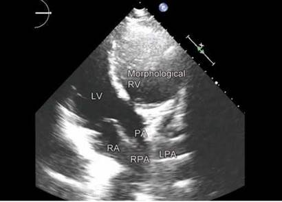
Fig. 16.78: Ventriculoarterial discordance.

Fig. 16.79: Complete transposition of great vessels with Senning operation. Baffle is seen draining pulmonary venous blood into the right-sided circulation.

Fig. 16.80: Direction of pulmonary venous blood flow through the baffle after Senning operation.

Fig. 16.81: Morphologically left ventricle (LV) type of single ventricle.

Fig. 16.82: Same patient as in Figure 16.81. Both great vessels come out off the single ventricle (SV) with infundibular pulmonary stenosis and D-malposition of great vessels.
references
1. Monro J. The changing state of surgery for adult congenital heart disease. Heart. 2005;91(2):139-40.
2. Gatzoulis MA, Webb GD, Daubeney PEF (Eds). Diagnosis and Management of Adult Congenital Heart Disease. Edinburgh, Scotland: Churchill Livingstone; 2003. pp. 299-303.
3. Warnes CA, Liberthson R, Danielson GK, et al. Task force 1: the changing profile of congenital heart disease in adult life. J Am Coll Cardiol. 2001;37(5):1170-5.
4. Burch TM, Mizuguchi KA, DiNardo JA. Echocardiographic assessment of atrial septal defects. Anesth Analg. 2012; 115(4):772-5.
5. Kutty S, Smallhorn JF. Evaluation of atrioventricular septal defects by three-dimensional echocardiography: benefits of navigating the third dimension. J Am Soc Echocardiogr. 2012;25(9):932-44.
6. Marriott K, Manins V Forshaw A, et al. Detection of right- to-left atrial communication using agitated saline contrast imaging: experience with 1162 patients and recommendations for echocardiography. J Am Soc Echocardiogr. 2013;26(1):96-102.
7. Plymale J, Kolinski K, Frommelt P, et al. Inferior sinus venosus defects: anatomic features and echocardiographic correlates. Pediatr Cardiol. 2013;34(2):322-6.
8. Tobis J, Shenoda M. Percutaneous treatment of patent foramen ovale and atrial septal defects. J Am Coll Cardiol. 2012;60(18):1722-32.
9. Kutty S, Sengupta PP, Khandheria BK. Patent foramen ovale: the known and the to be known. J Am Coll Cardiol. 2012;59(19):1665-71.
10. Koppen H, Palm-Meinders IH, Ferrari MD. Right-to-left shunts and micro-embolization in migraine. Curr Opin Neurol. 2012;25(3):263-8.
11. Kaul S. The interventricular septum in health and disease. Am Heart J. 1986;112(3):568-81.
12. Soto B, Becker AE, Moulaert AJ, et al. Classification of ventricular septal defects. Br Heart J. 1980;43(3):332-43.
13. Minethe MS, Sahn DJ. Ventricular septal defects. Circulation. 2006;114(20):2190-7.
14. Badano L, Fox K, Sicari R, et al. Adult congenital heart disease. The EAE Textbook of Echocardiography, 1st edition. Oxford University Press; 2011. pp. 389-92.
15. Tatebe S, Saji Y, Aoki K, et al. Intermediate type atrioventricular septal defect in the elderly. Ann Thorac Surg. 2006;81(5):1888-90.
16. Dearani JA, Danielson GK. Congenital Heart Surgery Nomenclature and Database Project: Ebstein's anomaly
and tricuspid valve disease. Ann Thorac Surg. 2000;69 (4 Suppl):S106-17.
17. Schreiber C, Cook A, Ho SY, et al. Morphologic spectrum of Ebstein's malformation: revisitation relative to surgical repair. J Thorac Cardiovasc Surg. 1999;117(1):148-55.
18. Acar P, Abadir S, Roux D, et al. Ebstein's anomaly assessed by real-time 3-D echocardiography. Ann Thorac Surg. 2006;82(2):731-3.
19. Carpentier A, Chauvaud S, Mace L, et al. A new reconstructive operation for Ebstein's anomaly of the tricuspid valve. J Thorac Cardiovasc Surg. 1988;96(1):92-101.
20. Hubail ZJ, Ramaciotti C. Spatial relationship between the ventricular septal defect and the anomalous muscle bundle in a double-chambered right ventricle. Congenit Heart Dis. 2007;2(6):421-3.
21. Kirklin JW, Barratt-Boyes BG. Congenital aneurysm of the sinus of Valsalva. In: Cardiac surgery. Morphology, diagnostic criteria, natural history, techniques, results, and indications. 2nd Edition. New York: Churchill Livingstone; 1993. p. 826.
22. Zannis K, Tzvetkov B, Deux JF, et al. Unruptured congenital aneurisms of the right and left sinuses of Valsalva. Eur Heart J. 2007;28(13):1565.
23. Moustafa S, Mookadam F, Cooper L, et al. Sinus of Valsalva aneurysms-47 years of a single center experience and systematic overview of published reports. Am J Cardiol. 2007;99(8):1159-64.
24. Anderson RH, Weinberg PM. The clinical anatomy of tetralogy of fallot. Cardiol Young. 2005;15(Suppl 1):38-47.
25. Zucker N, Rozin I, Levitas A, et al. Clinical presentation, natural history, and outcome of patients with the absent pulmonary valve syndrome. Cardiol Young. 2004;14(4): 402-8.
26. Hokanson JS, Moller JH. Adults with tetralogy of Fallot: long-term follow-up. Cardiol Rev. 1999;7(3):149-55.
27. Collett RW, Edwards JE. Persistent truncus arteriosus; a classification according to anatomic types. Surg Clin North Am. 1949;29(4):1245-70.
28. Bharucha T, Ho SY, Vettukattil JJ. Multiplanar review analysis of three-dimensional echocardiographic datasets gives new insights into the morphology of subaortic stenosis. Eur J Echocardiogr. 2008;9(5):614-20.
29. Carr JA, Sugeng L, Weinert L, et al. Images in cardiovascular medicine. Subaortic membrane in the adult. Circulation. 2005;112(23):e347.
30. Warnes CA. Transposition of the great arteries. Circulation. 2006;114(24):2699-709.