
Vincent Di Marino1 and Hubert Lepidi1
(1)
UER Médecine, Aix-Marseille Université, Marseille, France
Abstract
It would be logical to believe that the history of the clitoris is part of the history of the external female genitalia. However, this is not the case. Knowledge of the clitoris was acquired at a later date and remained inaccurate and incomplete for a long time. More surprising still, after having finally been the subject of remarkable anatomical studies, the clitoris was then ignored or unknown during certain periods! It was only in the twentieth century that the significance of this anatomical formation was acknowledged and integrated into an actual apparatus: the bulbo-clitoral apparatus.
1.1 General
It would be logical to believe that the history of the clitoris is part of the history of the external female genitalia. However, this is not the case. Knowledge of the clitoris was acquired at a later date and remained inaccurate and incomplete for a long time. More surprising still, after having finally been the subject of remarkable anatomical studies, the clitoris was then ignored or unknown during certain periods! It was only in the twentieth century that the significance of this anatomical formation was acknowledged and integrated into an actual apparatus: the bulbo-clitoral apparatus.
1.1.1 Foundations of Knowledge
Our prehistorical ancestors could already represent the vulva, such as revealed, on the one hand, by the numerous engraved, drawn or painted pubic triangles, discovered on prehistoric sites of the Palaeolithic era and, on the other hand, by the small statues, “Venus impudicae”, representing female bodies with generous and disproportionate curves, large breasts and exposed vulva. Anatomical details cannot be observed on these various vulvar figurations as the vulvas, represented by these proto-artists, are systematically summarised as triangles whose incomplete bisecting line represents the “large cleft”. Only in rare cases do the edges of the cleft, slightly convex, evoke labia majora.1 However, the anatomical precision does not go further and therefore knowledge of the clitoris and the labia minora cannot be prejudged. Generally, these are not representations (except in cases where the hips are outlined) but figurations, of which many “confine to abstraction”.
Therefore, we may question the meaning of these vulvas, especially when various numbers are represented, at the entrance to a cave or to a room or inside a cave:
· Do they symbolise the reproductive system?
· Do they symbolise the transition from darkness to light?
· Are they an initiation symbol? (Worship glorifying the fertile woman? Worship of the goddess mother?)
As anatomists, we must assist historians in order to provide answers to these questions and must therefore question the general position of vulvar clefts, which have been represented and preferentially placed by the artists of this era, in a pre-pubic ventral position, whereas the men from the Magdalenian era could not be unaware of the fact that the vulvar cleft is almost invisible on an adult woman in a standing position, as it is located in the perineal region.
More close to us, old Egypt remains mysterious as regards knowledge of the labia minora and the clitoris. There is no ostracon specifically representing the external female genitalia. Neither is there any low relief specifically revealing this part of the female anatomy whereas the male apparatus is often represented (representations of ithyphallic god Min,2 in erection, and scenes of male circumcisions on the lower reliefs of tombs or temples, such as those of Karnak). However, a few small statues dating back from the Neolithic era have been found. They represent nude women3 but they are very rare, as opposed to the small phallic statues. Moreover, considering the size of these small statues, no conclusion can be drawn in relation to the anatomical knowledge of their authors, only that they had a more harmonious idea of the female body than their ancestors.
Later, Greek physicians made progress in their knowledge of the female genitalia, even if it is known that this knowledge was not direct. None of these famous physicians performed a vaginal examination or dissection on a woman’s body. It is well established that their knowledge was only based on reports from midwives and women (who became experts in the practice of vaginal self-examination)4 and perhaps also on what they observed during encounters with their own partners.
It is to be recalled that Hippocrates 5 provided a very good description of vaginal transudation, which occurs at the same time as sexual excitation, but was wrongly considered as “female sperm”. He used the term “to aidoïon”, i.e. “the shameful parts”6 to refer to the entire genitalia. He had good knowledge of the matrix and its neck. The vagina does not have a specific name denomination and is considered as part of the “aidoïon”. The vulvar cleft is known as “nature”. According to certain scholars having studied all of his work, he has knowledge of the clitoris but considers it as a simple protrusion, which he refers to as columella or uvula, on the basis of its resemblance with the uvula palatina.
As for Aristotle,7 he denies the existence of female sperm in relation to generation. For him, “when conceiving, the woman provides the matter and this matter consists of the menses”. In chapter XIV of the treatise on the generation of animals,8 he explains that “if some naturalists have believed that women release semen during the act of copulation, it is due to the fact that sometimes women feel as much pleasure as men and that they release a liquid excretion” but “this is an error”. “This liquid is not at all spermatic… It is a special fluid….In women who release this liquid, there is not the same amount of liquid as sperm but sometimes there is much more”. In this same chapter, Aristotle also mentioned clitoral excitation. He wrote “Which also shows that women do not release sperm, only during copulation, they feel pleasure from being touched in the same place as men, but, in their case, there is no liquid emission”.
Even Aristotle has provided an outlined description of what could correspond to the clitoris but with a number of improbabilities. Thus, it is possible to read in the history of animals: “This is how Nature has laid out the pathway followed by sperm in women: They have a canal, such as men have a sex, but it is inside the body. They suction through this canal by means of a small orifice positioned above the location where women urinate. Sperm falls into the uterus from this canal”.9 Overall, Aristotle describes an imaginary canal, but at the same time, he refers to the existence of an anatomical structure, located above the external orifice of the urethra.
As a good observer, Aristotle also describes the modifications of the external female genitalia during sexual excitation. He wrote that “by observing female rats, we can see that the humidified sex of the female swells when she approaches a male”.10
During the three following centuries, progress was made in the acquisition of gynaecological, anatomical and medical knowledge, and we can only marvel when reading the treatise “Diseases in women”, written by Soranos of Ephesus,11 leader of the Methodical School.12 The description of the female genitalia provided by this author in the first pages of his treatise13 is astonishingly precise, even if wild ideas remain, in particular concerning “the seminal tract”, which exits the matrix, crosses each ovary, skirts the sides of this apparatus up to the bladder and then accesses the neck of the latter.
According to him, “the wings (pterygomata) or labia of the vagina, thick and fleshy, are separated from one another by a cleft. Downwards, they end at the 2 thighs. Upwards, they end at what is called the clitoris (kleitoris) or myrton or nymph”. Soranos not only mentions this formation. He also specifies and explains: “The latter, which forms the beginning of the labia, consists of a waffle with a muscular aspect. This small formation is called the nymph because it is hidden underneath the labia such as young brides under their veil”. He even provides the following observations: “Slightly below the nymph, another protuberant wattle is hidden, it is the end of the neck of the bladder; referred to as the urethra”. Therefore, even if Soranos mixes up the internal and external ostia of the urethra, he positions the clitoris or nymph correctly, above the meatus, which is used for the emission of urine.
Rufus of Ephesus,14 a contemporary of Soranos, has good anatomical knowledge based on dissections performed on monkeys. He wrote, only in Greek, a book in which the entire nomenclature of anatomical terms of the time is assembled and specified. Thus, he confirms the term “clitoris”, which he connects to a verb with an erotic meaning: cleitoriaxo. He confirms that the labia majora or myrtle lips (myrtocheïla) must now be referred to as “pterygomata”. As for the term of nymph, it indicates the unit formed by the clitoris and the labia minora. The cleft is called “schisma”. All of the internal and external genitalia form the “shameful parts” or aidoïon.
Neither Soranos nor Rufus will achieve the celebrity of their successor, Galen,15 who is also a Greek physician based in Rome, where he will live until the end of the second century (the date of his death is discussed: 204 AD ?).
Galien recalls the bi-spermatic theory made by Hippocrates by adding the essential role (according to him) of blood “matter”, whose surplus overflows periodically as menstruations.
For Galen, the female genitalia is the reverse copy of the male genitalia. This copy is interiorised whereas man’s genitalia is exteriorised. The male’s penis corresponds, according to him, to the uterus. The scrotum occupies the location of the matrix. The female “testicles” are positioned on either side of the matrix. The prepuce of the penis becomes the vagina in women. The latter represents with the vulva, the female shameful part (aidaïon gunaikeïon). No place is made for the clitoris so that the progress made by Aristotle and the physicians of Ephesus as regards the anatomy of the female sex is truly forgotten.
Despite this fact, Galien became the greatest physician of Western civilisation, even more famous than Hippocrates, such that his findings influenced medical knowledge, especially in relation to human anatomy, until the fifteenth century.
One century and half later, Oribase,16 who was also born, such as Galien, in Pergamon, was put in charge, by the Emperor Julian the Apostate (331–363), of gathering all the knowledge of the eminent Greek physicians, since Hippocrates. Oribase completed this task by compiling a magnificent encyclopaedia (70 books), based on the work of his predecessors.
It refers to the female genitalia as pecten. It calls the vulva, “the big cleft”. The labia majora are “the wings” of this big cleft. The clitoris is obviously well known. He positions it correctly and describes it as “a muscular waffle, located in the centre”. He calls it myrtle or nymph so that the labia minora become “the wings of the myrtle”.
Simultaneously to Greek medicine, foreign medicines (Arab and Persian, in particular) have progressed, while profiting from the knowledge contributed by Greek and Latin work. Two physicians from the tenth century, who distinguished themselves among these foreign physicians, knew about the clitoris: a Persian physician, Avicenna17 (980–1037), which calls the clitoris “el bathr” (penis) and an Arabic physician, Abulcasis18 (936?–1013), which calls it “tentigo” (which is placed under tension) but also “softness of love”, a term which will be used by Colombo.
The main work completed by Avicenna, a book called Canon, includes 5 volumes and the first volume is a panorama of the anatomy and pathology of the various organs.
Abulcasis compiled a medical and surgical encyclopaedia, in 3 parts, and the last part was entirely devoted to surgery with a paragraph concerning clitoridectomy. Abulcasis’ reputation (who practised his art at Cordoue) went beyond the Arab world and the borders of Spain, and his work, a reference for teachers for more than one century, was to be republished several times and successively translated into Latin, English, French, Hebrew and the Provençal language.
Few physicians of the following centuries became known for their work and Galen’s influence lasted until the Middle Ages. Nevertheless, a few anatomical books were published and included superb prints, which were as beautiful as inaccurate. Among these books, the book written by Mondino De Liuzzi 19 entitled Anathomia was a real success. Moreover, many readers, on the basis of the author’s work, in which he mentions that, in 1316, he performed two dissections on female bodies, consider him as the “protodissector”, i.e. the first physician to have restored cadaveric dissection,20 which had disappeared since antiquity. Actually the first cadaveric anatomical dissection was performed in 1313, in Montpellier by Henri de Mondeville21 (who was also Mondino’s professor). Nevertheless, Mondino’s book, based on knowledge from human dissections and not from animal dissections, such as the work completed by Greek authors and Galen’s in particular, was going to initiate the future upheaval of anatomical knowledge of the human body.
Two other books using engraving were to be published a few years later and initiate the expansion of anatomical culture in all Europe of the early Renaissance. This was an indication of the development of this fundamental subject of medicine in the near future:
Firstly, a book written by Jacopo Berengario da Carpi,22 with a promising Latin title Isagogae breves perlucidae ac uberrimae in anatomiam humani corporis a communi medicorum academia usitatam, was published in Bologna in 1527 then a book written by Charles Estienne,23 a contemporary of Vesalius, who published the Dissection des parties du corps humain (Dissection of the parts of the human body), successively in 1545, in Latin, then in 1546, in French. This book, which included even more anatomical charts than Berengario’s book, used such as the latter, the technique of staging human bodies.
Sadly, this staged representation, although pleasant and elegant to look at, outweighed the anatomical reality and made the body structures very difficult to observe as they were too small, inaccurate and generally incorrect. The actual descriptions are inaccurate (e.g. that of woman’s “shameful member”, which seems to more or less refer to a description of the clitoris).
The fact remains that the three above-mentioned authors had established the foundations of modern anatomy, based on the direct observation of the human body and the reproduction of this reality in anatomical charts, which Vésalius rapidly enhanced and developed.
But what happened to the anatomy of the clitoris during the Middle Ages?
In spite of the relative small amount of anatomical discoveries made during this period, the assumed role of the clitoris in fecundation spread in the thatched cottage homes during the Middle Ages. The majority of physicians of the time adopted the theory of the existence of female sperm and considered that this secretion, called cyprine, was produced by the clitoris. The Catholic Church, however, which expressed reservations on the topic of carnal pleasure, had to accept the medical advice favouring fecundation: “before the sexual intercourse, the husband shall delicately rub the button of love with a finger moistened with perfumed oil, in a circular motion”.
1.1.2 Rebirth and “Discovery” of the Clitoris
The Renaissance, a period of renewal and all types of discoveries, was going to the era during which anatomical knowledge, including that of the clitoris, was developed. Little by little, prohibitions concerning cadaveric dissection were going to be finally reduced thus allowing a direct approach of the human body and thus of its knowledge.
It is in this new environment that the Belgian, Andreas Vesalius,24 who was going to become the most famous anatomist, was going to rapidly emerge and define the foundations of modern anatomy, in his masterly exercise at the faculty of Padua:
· By transforming dissections on human bodies from exceptional “public place anatomies”, such as was the case, in Antwerp or Venice25 (Fig. 1.1) into a current practice, carried out in dedicated locations: initially, anatomical theatres, dismountable installations and then rapidly permanent structures26

Fig. 1.1
“Piazza dell’Anatomia” in Venice (a) and his accesses “sottoportego dell’ anatomia” (b): the stairs for going down from the bridge of anatomia (ponte dell’anatomia), and the porch that allows the access to the little river (rio). The first and exceptional public anatomies were carried in that place, hidden from sight! The cadavers and the anatomical remains were transported by gondola across the rio
· By only transmitting to his students’ concepts verified through direct anatomical observations
· By performing the dissection himself during his anatomy lessons, such as shown by the front page of his book
· By being the first to write an exceptional book, De humani corporis fabrica, published in Basle in 1543, which provided a significant anatomical accuracy, enhanced by the presence of an exceptional iconography (charts prepared by students of the school of Titien, which had attended the cadaveric dissections) and, furthermore, which was printed (following the recent discovery of the printing technique by Gutenberg)
Nevertheless, in the De humani corporis fabrica book, libri septem, there is no detailed description of the external female genitalia and even less precisions on the clitoris, which Vesalius seems to ignore (a superb image of the entire genitalia shows a vulva, which is simply represented as 2 greater lips, with a nice representation of combed body hair) and even considers as an anomaly, which is only visible in hermaphrodites.
It is true that Vesalius was only able to study this anatomical region on a small number of specimens, which were only available for a very short time (autopsies).
It was thus necessary to wait for Vesalius’ successors as Professor of Anatomy at the University of Padua, Colombo then Fallopio, who conducted accurate studies on the clitoris, which clarified its function and structure.
Realdo Colombo 27 was successively Vesalius’ assistant (1541), temporary substitute in 1543 and then permanent substitute as from 1544 (when Vesalius left to take up his position at the court of Charles Quint). He became the Professor of Anatomy at the University of Pisa in 1545 and wrote a remarkable book on anatomy De re anatomica, which was only published after his death in 1559.
Sadly, this book included no iconography, except for a superb front page (Fig. 1.2) by Veronese.28 The book (which was extremely documented) was a great success and its fame went beyond the borders of the Republic of Venice, such as represented by a frontispice painting “A Lesson of Anatomy”, commissioned by the famous English anatomist, John Banister, lecturer in anatomy of the Barber Surgeons’ Company (licensed by the king Henry VIII in 1540). On this painting, visible at the Glasgow’s University, Banister is performing an anatomical demonstration in front of his students while reading the book written by Colombo, which is open on his desk. Numerous discoveries are owed to Colombo, an exceptional anatomist, who performed many dissections (apparently, he has personally performed 1,000 dissections). He was also a talented physiologist. One of his most famous discoveries is that of pulmonary circulation29 (which was used as a basis, 30 years later, for the descriptions of the large and small circulation by William Harvey). We also owe him remarkable work on the clitoris, which he claimed to have discovered, whereas, such as we have noted, Greek (but so Arab and Persian) physicians and surgeons already had knowledge thereof, such as proved by their texts.
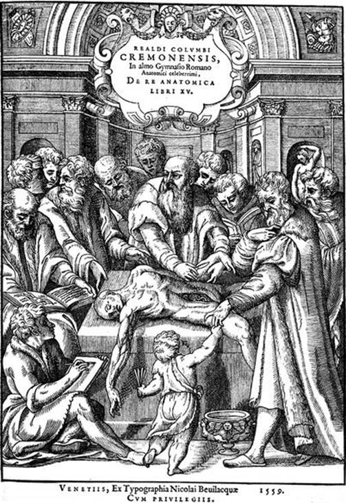
Fig. 1.2
Frontispiece of the book De re anatomica (engraving woodcut of Nicolai Bevilacquae, 1559, probably from a picture of Paolo Veronese). Realdo Colombo is dissecting a human cadaver in front of both his students and the famous physiologist, Andrea Cesalpino
In spite of these obvious facts, Colombo unequivocally stated that he was the first “discoverer” of the clitoris, such was also the case of his homonym and fellow citizen, the famous Italian navigator, Cristoforo Colombo (Christopher Columbus), who discovered America, in 1492! .............
What remains certain is not only that he accurately described the external female genitalia but, in particular, that he highlighted the clitoris in this description and celebrated, for the first time, its physiological properties, thus demonstrating, in terms of quasi-poetic exaltation, the exceptional role played by this formation in the genesis of female pleasure (De Visceribus, Liber XI.243,30 in Fig. 1.3). However, he assigned a (female) sperm secretion function to the clitoris (which he referred to as “Amor Veneris”), which is an idea inherited from former anatomists and which is totally false.

Fig. 1.3
Superior part of the magnificent page of De re anatomica in which Realdo Colombo describes, for the first time, the clitoris as “amor Veneris”, the sublime organ of female pleasure
Gabriele Falloppio,31 also one of Vesalius’ students, rapidly claimed the paternity of the discovery of the clitoris, as he wrote in his book Observationes anatomicae, published in 1561 (but which he is supposed to have written in 1550), “this part is hidden and ignored by anatomists,… it is so hidden that I was the first to discover it and if others speak of it, please know that they learnt about it from me and my students!” In this statement, he is nearly accusing Colombo of plagiarism. Nevertheless, there was an enmity and competition between the 2 students of the Master, which is easily explained by the fact that Fallopio was always the eternal second, as he succeeded Colombo, firstly as Professor at the University of Pisa in 1548 and then as Professor at the University of Padua, in 1551.
During the next century, another anatomist of talent, Thomas Bartholin,32 was going to restore the truth in relation to Vesalius’ 2 successors by writing in chapter XXXIV of book I, “De clitoride”, from his “Anatomia reformata”,33 written in Latin but easily understandable: “Hujus inventionem sibi arrogat Fallopius: Columbus, vero gloriose,… sibi tribuit. cum tamen ejus fecerint mentionem Avicenna, Albucasis, Ruffus, Pollux § alii” thus recalling all of the scientists who had referred to this topic before our dear Venetian anatomists. In addition, in this chapter, a very interesting outline of the description of the clitoris can be found with Schemes.
1.1.3 The Pinnacle of Knowledge
During the seventeenth century, other famous anatomists (among whom we can count Jean Riolan34 from France) have significantly contributed to the study of the anatomy of the clitoris. Undeniably, the biggest contribution was made by Regnier de Graaf,35 who was most successful at compiling all the research work describing this formation, as well as the entire external female genitalia, in his book Histoire anatomique des parties génitales de l’homme et de la femme (Anatomic history of the genitalia of man and woman) published in 1649 in Latin, but which was soon to be translated into French. It is especially the anatomy of the female genitalia which owes a lot to de Graaf, who discovered the spongious bulbs he called the reticular plexus or retiform plexus. He was the first to describe and include these bulbs in a book, which was published in 1672: De Mulierum Organis Generationi Inservientibus Tractatus Novus Cum Figuris, (even if Swammerdam wrongly disputed the priority given to this discovery in his opus, Miraculum Naturae Seu Uteri Muliebris Fabrica, which was published the same year).
Following the example of the anatomists, obstetricians began to describe the female genitalia that the anatomists had self-appropriated. One of these obstetricians, Pierre Dionis,36 a surgeon appointed to the King’s Garden by Louis XIV to teach “anatomy according to the circulatory system and new discoveries”, left a remarkable description of the clitoris in his “Traité Général des Accouchements qui Instruit de Tout ce Qu’il Faut Faire Pour Etre un Habile Accoucheur” (General Treatise on Childbirth for the Training of an Accomplished Obstetrician). For the same period, we can also mention two books including anatomical charts with legends including excellent representations of the external female genitalia, which specify the accurate location of the clitoris: the works of Giulio Casserio and Govard Bidloo.
· A book written by Giulio Casserio37 Tabulae Anatomicae LXXIIX Omnes Novae Nec Ante Hac Visae, published in 1627, 11 years after the author’s death, is a work of great precision. It includes charts which are true paintings and stylised representations of the human body, in a similar manner to Vesalius. The six figures of table XVIII and of book VIII allow the author to represent what can be seen of the clitoris when observing the vulva but also the entire make-up of the clitoris from the two crura (roots) which the author called “nerve bodies”.
· A book written by Govard Bidloo,38 Anatomia de Humani Corporis Demonstrata et Illustrata, was published in 1685 and includes “Centum et quinque tabulis, per artificiossis G. de Lairesse ad vivum delineates”, as well as superb illustrations of dissections sketched in real time. In de Lairesse’s illustrations representing the vulva (Fig. 1.4), the clitoris is perfectly identified and drawn. If one compares, for example, the illustration referred to as “Quinquagesima Primae” from Bidloo’s book to illustration 60 of De Humani Corporis Fabrica of Vesalius, we realise the extent of progress achieved by anatomists in the precise description of the clitoris within less than a century and a half. Other well-known French authors also decided to provide complete descriptions of the clitoris. Similar to de Graaf, the reticular plexus was included in their anatomical descriptions. However, French work had a difficult time crossing the channel and thus we are not to be surprised to read that, during the eighteenth century, John Bell,39 a great English anatomist and author of Anatomy of the Human Body (Volume 3), wrote that the clitoris does not include spongious tissue!

Fig. 1.4
An example of the magnificent drawings of G. de Lairesse (De Lairesse’s drawings illustrated two books of Anatomy, Govard Bidloo’s, in 1685, and William Cowper’s, in 1698) (a) Connections between the urinary apparatus and the female genital apparatus. (b) Magnification of the previous, showing the precision and accuracy of the drawings. Notice the perfect representation of the external genitalia especially the clitoris
The nineteenth century should have been the “clitoris’s century”, thanks to Georg Ludwig Kobelt,40 who in a highly remarkable book, Die männlichen und weiblichen Wollustorgane des Menschen und einiger Säugetiereabout the male and female organs of sexual arousal in man and certain other mammals), superbly illustrated with drawings based on his own dissections, described the complete and precise anatomies of the organs of sexual arousal in both men and women, with a few references to their compared anatomy. If the study of the male genitalia (the first part of the book) does not reveal major medical progress in comparison to previous work, the second part, dedicated to the study of the female genitalia, undoubtedly demonstrates for the first time (with the support of vascular injections) that this region is comprised of two areas, the clitoris and the spongy bulbs, and that the two areas are connected by a vascular network called the “pars intermedia”. After these anatomical chapters, the author, in some remarkable sections that retain their relevance, begins a functional study of the “connection” between the male and female organs of sexual arousal, thus giving an impetus to future studies on the physiology of coitus. Kobelt’s work, some of whose drawings we show (Fig. 1.5), appeared in Freiburg in 1844. It was not an especially successful publication until its French edition in Strasburg in 1851, in a translation by Von H. Kaula.
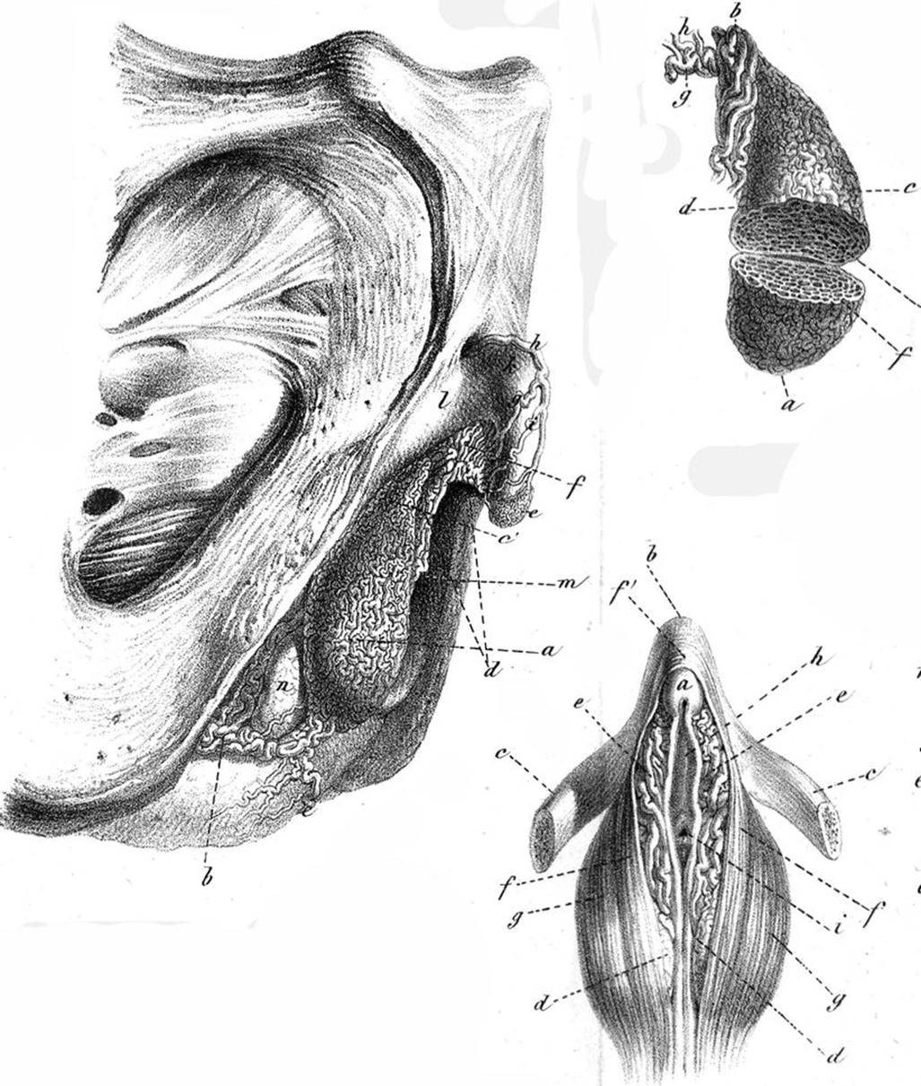
Fig. 1.5
An example of the remarkable illustrations (Notice the accuracy and the precision of the drawings (Taken from the own dissections of the author)) of Georg Ludwig Kobelt’s book: Die männlichen und weiblichen Wollustorgane des Menschen und einiger Säugetiere (part of the third page of illustrations, concerning human female)
From this point on, the anatomy of the clitoris was perfectly known, and one might have expected that this knowledge would spread on a worldwide scale. There were a few physicians who, having read Kobelt’s work, attempted to bear witness to the clitoris’s supremacy. Thus, in France, Dr. Guyot, who, as soon as 1852, had written the surprisingly titled Breviary of Experimental Love, which after “secretly making the rounds” was finally published in 1882 (10 years after the author’s death). In this recently republished work,41 Dr. Guyot insists on the importance of the preludes that must precede all penetration and strongly criticises men who misunderstand the clitoris and its role in women’s sexual excitement: “There exists an immense number of ignorant, egoistic, brutal men who do not bother to study the instrument that God has entrusted to them”. Alas, there were also books, which discredited the clitoris. For instance, as sexologist Sylvie Chaperon reports, Dr. Pierre Garnier, a prolific and successful author, became more and more critical, in each of his books, until in 1891 he wrote: “to claim that this miniscule apparatus, which is most often an insensitive little button so long as it has not been touched or artificially manipulated, is the most active erogenous centre, is to implicitly accuse all girls of once having resorted to masturbation or of one day becoming debauched”.
What’s more, an excessive prudishness took over in scientific work and the clitoris was forgotten in certain anatomical treatises. Kobelt’s discoveries, for their part, were most often considered pointless and occluded.42
The turbulent years afflicting the clitoris did not only affect France!
1.1.4 Decline and Disappearance of Clitoris
It has to be believed that the “magnification” of the clitoris resulting from Kobelt’s work was also ignored or misunderstood by English physicians (including those boasting of being specialists in obstetrics or gynaecology), given that within a few years, the unfounded and monstrous idea that the clitoris was useless spread among them. From uselessness to harmfulness, there was only a small step, and it was soon taken. Henceforth, many English physicians would claim that the clitoris was an absolutely useless apparatus and that in certain cases it should be excised! It was in this context that a renowned obstetrician, Dr. Isaac Baker Brown (see E. Sheehan’s excellent article), founder and director of his own hospital (“The Surgical Home for Women”), elected President of the Medical Society of London in 1865, published, the following year, a book with a strange and frightening title: On the Curability of Certain Forms of Insanity, Epilepsy, Catalepsy, and Hysteria in Females, in which he proposed clitoridectomy as the cure for every one of these afflictions, on the basis that their principal cause was masturbation. He specified that he had been successfully practising such operations in his hospital since 1858. The book became very popular when published and gave rise to few criticisms, such that clitoridectomy spread as a practice across all of Victorian England and also in America. Several months were required for a reaction to set in, for the physicians and surgeons to protest and for female patients to contest the effectiveness of Dr. Brown’s so-called miracle cure (a cure that, what’s more, had to be paid up front with “hard cash”). Finally, a coalition of young gynaecologists denounced Dr. Brown’s practices and obtained that an enquiry of the Obstetrical Society be made into clitoridectomies (December 1866). In February 1867, the Board of the Obstetrical Society, ruling on the notoriety and debates surrounding Brown’s use of clitoridectomy, recommended his expulsion from the society. A strong majority voted in favour of the expulsion in April of the same year, and Brown resigned not only from the Society he had presided but also as director of the hospital he had founded.43 But the harm was done, and clitoridectomy remained a recommended practice in books written only a few years before by renowned gynaecologists, particularly in America, so that it was only abandoned progressively.44 A new blow was struck in relation to the usefulness of the clitoris in 1884, with the discovery of the microscopic process of procreation by the famous Belgian embryologist, Edouard van Beneden.45 His work on fecundation showed that embryos result from the sperm’s penetration of the ovum and that the latter is a living cell that makes its way from the maternal ovary without any help from some substance secreted by the clitoris. It was the end of all the legends claiming that cyprine46 was secreted by the clitoris and played a role in the fecundation process. Henceforth, the clitoris’s “physiological depreciation” was sealed, and even gynaecologists began to think of the clitoris as an apparatus presenting no interest and thus forgot the pertinent pages written by Colombo and later by Kobelt.
Very surprisingly, it was Sigmund Freud 47 who, by seriously depreciating the clitoris, gave the final blow. To serve his own purposes, Freud appropriated the theory proposed by his colleague Dr. Krafft-Ebing, the famous Viennese psychiatrist, that there are two erogenous zones that succeed one another in the construction of the woman individual, the clitoris for virgins and the vagina after being deflowered. And in his “Three Essays on Sexual Theory”, which was published in 1905, Freud integrated into his grand theory of Oedipal evolution the postulate according to which clitoral orgasms represent an infantile stage, while normal adult orgasms are vaginal. This was as much as to say that there were two types of women, the “clitoral”, who had remained sexually immature, and the “vaginal”, with a flourishing adult sexuality. Although contested, Freud’s theory made enough of an impact that the clitoris’s role in female pleasure was forgotten. Despite the Master’s theories, his student, patient and faithful friend, Marie Bonaparte, was going to provide food for thought in relation to the clitoris, for she remained convinced of this organ’s importance, though adding another limiting factor: the distance between the glans clitoridis and the vagina.48 She therefore believed that the frigidity she suffered from was due to a “too great a distance” between these two areas and decided to be operated to reposition her clitoris. Sadly, the operation (though performed by the famous Austrian surgeon, Joseph von Halban) was unsuccessful.
1.1.5 The Return to Favour
Freud’s ideas were also to be discussed, with many authors, who referred back to Kobelt’s work and recalled that the “vaginal canal is not the sensory apparatus”. In 1953, the Freudian theory lost its halo when the Kinsey Report “Human Behaviour in the Human Female”49 stated that “vaginal orgasms do not exist”, but this report was also criticised for several reasons. Finally, the vaginal and clitoral theories concerning female orgasms remain valid to this day, with the clitoris’s predominance being based on the vagina’s poor innervation, compared to the significant innervation of the clitoris. Scientists in favour of the vaginal predominance found new arguments with the discovery of the extremely mediatised G-spot.
The second half of the twentieth century was rich in events that altered people’s sex lives (sexual revolution of the 1960s and 1970s, introduction of oral contraceptives, development of sexology, particularly with the books written by Masters and Johnson,50 feminist movements) and succeeded in putting an end to a certain number of taboos, thus allowing women to live more fulfilling sexual lives.
In this liberal climate, the clitoris’s rehabilitation begun, after being ignored for years, during which it was not considered as an actual part of women’s bodies. It is spoken of more freely. Media (including television) have begun to address this topic. New scientific studies have been devoted thereto. But the road ahead is long, and at the beginning of the twenty-first century, conservatism still predominates, thus preventing the public, and particularly the male public, from truly learning about this “extraordinary jewel” of the female body.
It is for this reason that we have decided to publish this study.
Footnotes
1
This is the case of the most famous of these Venuses, the Venus of Willendorf in Austria (24,000 years B.C.).
2
Min God: one of the oldest divinities in old Egypt (god of fertility and reproduction).
3
The most beautiful of these statuettes was found in a tomb at Badari. It is made out of ivory and dates back to the Predynastic era (4,500 B.C.). Its sexual symbolism is clear (“pubic” vulva, highlighted by a pattern). This statuette is kept at the British Museum.
4
“I only know what women have taught me”, (Des chairs, XIX) in Hippocrates’ complete series of books and written work, Littré, vol.8.
5
Hippocrates (towards 460 c. BC. in the Kos island–towards 370 c. BC at Larissa): Greek physician, “Father of medicine”.
6
(Hippocrates, De naturâ muliebris, Lib II,).
7
Aristotle (384 c. BC in Stagire–322 c. BC in Chalcis): Greek philosopher and scientist, one of Plato’s students. He considerably influenced Hellenistic medicine through his knowledge gained by performing dissections (animal dissections).
8
(Aristotle, Treatise on the generation of animals, first book, chapter XIV, 728 b).
9
(Aristotle, History of animals, Book X, p.637 to 621).
10
(Aristotle History of animals, p 572 a and b).
11
Soranos of Ephesus, a Greek physician, from Ephesus, practised in Rome during the second half of the first century and the beginning of the second century, under the reigns of the emperors Trajan then Adrien.
12
The “methodical” doctrine prohibited, at Soranos, the execution of dissections (whereas dissections will be systematically performed by “dogmatics” such as Galien).
13
(Soranos of Ephesus, Diseases in women 1, tà gynaikeïa, written in Greek and then translated into Latin).
14
Rufus of Ephesus, a Greek physician, also lived during the reign of Trajan and wrote “About the name of the parts of the human body”.
15
Claudius Galenus (Galen) (131 born in Pergamon in Asia Minor, died in 204?, in Rome) was a well-known physician and philosopher. He was the physician of the emperors Marc Aurele and Commode. He was also a great animal anatomist (as the dissection of human cadavers was prohibited in Rome). He is considered as the founding father of pharmacy (oath of Galen….galenics!). His work, which has been translated into Arabic, has largely contributed to the development of Arab medicine and then, in turn, to medieval medicine.
16
Oribase (born around 325 at Pergamon, deceased around 395, in Constantinople) was the last of the famous Greek physicians.
17
Avicenna (980–1037), a Persian physician, scientist and philosopher, wrote a book which has remained well known: Kitab Al Quanûm fi Al-Tibb (Book of the law concerning medicine), known under the abbreviation Quanûm (Canon). This book was written in Persian and was translated, one century and half later, into Latin by Gerard de Crémone, under the title Canon médicinae. It was only printed in Arabic in 1593.
18
Abulcasis (Aboul Kasim Al Zahravi) (936?–1013), a physician and surgeon, rapidly became well known and his reputation reached all of Europe after his death. His major work, Kitab Al-Tasrif (Book of the medical method), is an encyclopaedia: 30 volumes! It was translated into Latin, such as Avicenna’s work, by Gerard de Crémone, during the twelfth century.
19
Mondino de’ Liuzzi, an Italian physician (born in Bologna in 1270 and deceased in the same city in 1326).
20
Until then, cadaveric dissection for gaining anatomical knowledge had been prohibited by the Church. Only a few autopsies were authorised for legal purposes. Thereafter, the progressive transgression of prohibitions was going to finally allow the development of human anatomy.
21
Henri de Mondeville (born in Mondeville in 1260 and deceased in 1320) was a French anatomist, who was the surgeon of kings of France, Philippe le Bel and Louis le Hutin. In 1304, he was named professor in Montpellier (where his students included, among others, the famous Guy de Chauliac). He was also a professor in Bologna.
22
Jacopo Berengario da Carpi, a physician and famous Italian anatomist (born in 1460 in Carpi and deceased in 1530), is one of the first to have included anatomical illustrations with his written work.
23
Charles Estienne, a French physician, anatomist and printer (1504–1564), wrote many books in several fields and [in] especially La Dissection des Parties du corps humain (“the Dissection of the Parts of the human body” (divided into 3 books), published by Simon de Colines (who was his father-in-law), in 1546. This book was the translation of “Dissectione partium corporis humanis”, whose engravings on wood, executed by Estienne de la Rivière, a surgeon and engraver, had been ready since 1539 but whose publication had been interrupted due to a lawsuit for plagiarism. Charles Estienne was a great anatomist who especially discovered the nutrient foramina of the bones and their role.
24
Andreas Vesalius, a physician, humanist and especially a famous anatomist (born in 1514 in Brussels and deceased in 1564 on the Island of Zakynthos, in Greece). He was a Professor of Anatomy and Surgery at Padua and also taught at Pisa and Bologna. It is in 1543 that he published his major work De humani corporis fabrica, in which he dared expose nearly 200 errors made by Galien, which generated considerable enmities. One year later, he left his position at Padua to become the surgeon of the emperor Charles Quint then of Philippe II of Spain. He died on the Island of Zakynthos from typhus when returning from a pilgrimage to the Holy Land.
25
In Venice, a free city where nobody feared the anger of the Inquisition, we can still see the “Place de l’anatomie” (“Anatomy square”), where “public anatomies” took place (Fig. 1.1). This square is adjacent to a small canal were a gondola would come at night time to recover the leftovers from the dissection and evacuate them further at sea.
26
The first “permanent anatomical amphitheatres” with a central dissection table, concentric observation circles for the students and a pulpit from which the Master anatomist gave his lesson, were only built in 1550 (Salamanca). The very beautiful and very famous amphitheatre of Padua, was only built in 1584, on the initiative of the anatomist Girolamo Fabrizi d’Acquapendente.
27
Matteo Realdo Colombo (1515/1516–1559) was a student of Vesalius, who he replaced to teach anatomy at Padua. He then rowed with Vesalius, who learned that in his absence, his pupil had criticised one of his publications! Colombo then became a permanent Professor of Anatomy at the University of Pisa (in the service of Cosimo de Medici) then a Professor of Anatomy in Rome, at the papal University of Sapienza (in the service of the Pope Jules III). His discoveries (clitoris, stapes bone, pulmonary circulation, position of the kidneys) were all disputed by critics, on the basis that they had already been made by other anatomists. He also completed important research work on the pleura and the peritoneum. He recorded all of his “discoveries” in a remarkable book entitled De re anatomica, which sadly, was only published in 1559, after his death, and which did not contain any diagrams (which were to be completed by his friend, Michelangelo, with whom he shared a passion for cadaveric dissection in Rome). In 1550, he was appointed surgeon of the pope Jules III and remained in his service until his death. Among other things, he performed a dissection on a saint (such as proclaimed by the Church): Ignace de Loyola, deceased in Rome in 1556.
28
This front page represents Colombo performing a dissection in the presence of the famous naturalist-physiologist, Andrea Cesalpino.
29
Similarly to Colombo, Michael Servetus (1511–1553), a Spanish physician and theologist, also described pulmonary circulation, before being burnt to death for being a heretic, in Geneva, in 1553. However, it seems that the original discovery of this small circulation took place beforehand, such as mentioned in the work completed by Ibn al-Nafis (1210–1288), a Syrian physician and anatomist, which had been translated by a friend of Colombo’s.
30
This caused two serious problems for him. He had to appear before the court of the Inquisition and only owed his salvation to his vehement defence, which he ensured himself, supported by his anatomical discoveries. At the end of his inquisitorial lawsuit, he was declared innocent but the publication of his book De re anatomica was prohibited (it was only to be published after his death, thanks to the tenacity of his 2 sons).
31
Gabrielle Falloppio (Fallopius), known in France as Gabriel Fallope (1523–1562), was not only a great anatomist (professor at both Pisa and Padua) but was also a talented botanist. We owe him numerous discoveries, some of which refer to his name: the facial canal (aquaductus Fallopi), the uterine tubes (fallopian tubes) and also the “vagina” (a designation he introduced). He also made several important discoveries in the internal ear region.
32
Thomas Bartholin (1619–1680), a Danish doctor and anatomist, was one of a long line of scientists in his family and is best known for his work on the lymphatic system. It was his son Caspar (the Younger), who first described the greater vestibular gland, which is now referred to as: “Bartholin’s gland”.
33
Anatomia Reformata, which was first published in 1655, was in fact a revised and annotated edition of the work of his father, Caspar (the Elder), Anatomiae Istitutiones Corporis Humani, published in 1611.
34
Jean Riolan the Younger (1577–1657) was doctor to Marie de Medicis and Louis XIII. In 1618, as a professor at the Faculty of Medicine in Paris, he published Anthropographia and Osteologia. The book, which was written in Latin, covered the anatomical description of man. A second complete edition was published in 1626. The second edition was translated into French in 1629 by G. Constant, who published it under the title Les œuvres anatomiques de Monsieur Jean Riolan (The Anatomical Works of Mr Jean Riolan).
35
Regnier de Graaf, a Dutch doctor and anatomist (1641–1673) was taught by Sylvius in Paris and then worked at Delft. The year of his death, he and his colleague, Stenon, discovered the ovarian follicle, which adopted his name, “De Graaf follicle”. He also wrote the Histoire anatomique des parties génitales de l’homme et de la femme qui servent à la génération (Anatomical History of the Male and Female Genitalia employed for Generation) published in 1649, at the same time as a suc-pancreatic treatise.
36
Pierre Dionis (1643–1718), obstetrician, was named by Louis XIV as surgeon responsible for teaching anatomy at the King’s Garden in 1672. In 1680 he became surgeon in ordinary to Queen Marie Thérèse, before being named first surgeon to the children of France in 1712. In 1713, he became the Duchess of Berry’s obstetrician. In 1715, he was called upon to assist Louis XIV in his last moments.
37
Casserio Giulio (Julius Casserius Placentinus), whose date of birth is unknown (1552 ?) and who died in 1616, was a remarkable anatomist. He was a Professor of Anatomy, Physics and Surgery at the University of Padua, and like his great predecessors, he left numerous works on anatomy enriched with superb illustrations. His works on the sensory organs and the larynx are still relevant today.
38
Govard Bidloo (1649–1713), Dutch doctor and anatomist, was a professor at Leiden and published an anatomical treatise, “Ontleding des menschelyken lichaams” in 1685, which contained 105 plates of anatomical drawings engraved from G. de Lairesse’s remarkable original drawings, which had themselves been published by their author by 1680 (Fig. 1.4). These drawings were reused by English anatomist William Cowper in his Anatomy of the human bodies of 1698, which earned this author an accusation of plagiarism and a violent quarrel with his colleague Bidloo.
39
John Bell (1763–1820) was not only an excellent anatomist and a great vascular surgeon (he contributed to this specialisation’s beginnings) but also a talented artist. He was responsible for many of the illustrations to his own works. He wrote “Anatomy of the human body” in collaboration with his younger brother, Charles Bell, who became a celebrated neuroanatomist.
40
Georg Ludwig Kobelt (1804–1857), a German anatomist, only joined the Faculty of Medicine after abandoning his legal studies that held no interest for him. He became a Professor of Anatomy at the University of Freiburg and also taught at Heidelberg. A keen dissector, he left some remarkable anatomical specimens, preserved in the anatomical collections of the two cities. He died of tuberculosis.
41
The Breviary of Experimental Love (Payot) was written “on the occasion of the marriage of Prince Napoleon and Princess Clothilde of Savoy. It was discretely read by numerous physicians and a privileged few (Prince Napoleon, Claude Bernard, the actress Virginie Dejazet, Georges Masson, Georges Sand, Sainte-Beuve and the archbishop of Rheims.)
42
Thus, today we are puzzled when consulting Bourgery and Jacob’s magnificent book from 1866 to 1867, in which an isolated clitoris is described, without the spongy bulbs (which are described later in the book, under their ancient name of “retiform plexus”).
43
Brown died miserably, paralysed after several heart attacks and had to be financially assisted by a “Baker Brown Charitable Fund” contributed to by physicians and rather non-vindictive former patients, which had been operated.
44
Some cases of “therapeutic” clitoridectomies, thankfully exceptional, were published until 1940.
45
Edouard Joseph Louis Marie van Beneden (1846–1910), a renowned embryologist, was a Professor of Zoology, Anatomy and Comparative Physiology and Embryology at the University of Liège. He provided evidence not only for the fecundation process but also for the meiosis and mitosis processes.
46
It is recalled that previous physicians referred to the liquid, which moistens the vagina during sexual excitation, “cyprine”. They believed it to be secreted by the clitoris and that this secretion played a role during fecundation.
47
Sigismund Schlomo Freud (1856–1939) simplified his name in 1877 and was thereafter called Sigmund Freud. As neurologist and physician, and student of Meynert and Charcot, he became Professor and Director of Neurology at the “Clinique des Enfants malades” (Sick Children’s Clinic). He then settled in Vienna, which he only left in 1938 for London, after the Nazi invasion of Austria. He was the founder and uncontested Master of psychoanalysis. He was first diagnosed with oral cancer in 1923 and eventually died despite going through at least thirty different operations.
48
Relationships of the bulbo-clitoral organ, connections with the Urethra, Chap. 13, p. 114–120.
49
Dr. Alfred Charles Kinsey, Professor of Entomology and Zoology, published two books on human sexual behaviour with an enormous impact: Sexual Behaviour in the Human Male in 1948 and Sexual Behaviour in the Human Female in 1953. The latter was translated into French in 1954 as Comportement sexuel de la femme.
50
William Howell Masters, a gynaecologist, and Virginia Eshelman Johnson, a psychologist, are known for their remarkable work on human sexuality in 1957–1965. Most notably, they described the various phases of sexual intercourse (see Chap. 15) and studied treatments for sexual disorders and dysfunctions.