
Vincent Di Marino1 and Hubert Lepidi1
(1)
UER Médecine, Aix-Marseille Université, Marseille, France
Abstract
This innervation is extremely important, not only due to its density but also due to the major role it plays in sexual life. It consists of a double innervation, which is, on the one hand, cerebrospinal and, on the other hand, vegetative. The pudendal nerve (shameful nerve), cerebrospinal contingent, is the key component and has therefore been referred to as “king of the perineum” (R. Robert et al.). However, it is not the only nerve structure to be involved. The other key nerve contingent is the vegetative contingent, originating from the inferior hypogastric plexus: the cavernous nerve of the clitoris. Such as will be noted in the following sections, the two components converge and meet to ensure the extraordinary innervation of the bulbo-clitoral organ.
8.1 General
This innervation is extremely important, not only due to its density but also due to the major role it plays in sexual life. It consists of a double innervation, which is, on the one hand, cerebrospinal and, on the other hand, vegetative.1 The pudendal nerve (shameful nerve), cerebrospinal contingent, is the key component and has therefore been referred to as “king of the perineum” (R. Robert et al.). However, it is not the only nerve structure to be involved. The other key nerve contingent is the vegetative contingent, originating from the inferior hypogastric plexus: the cavernous nerve of the clitoris. Such as will be noted in the following sections, the two components converge and meet to ensure the extraordinary innervation of the bulbo-clitoral organ.
8.1.1 Pudendal Nerve
The anatomy of the pudendal nerve (shameful nerve according to former authors) is better known since a number of specialists, and a team from Nantes2 (already mentioned above) in particular, have focused their attention on the investigation of the causes of positional perineal pains and the discovery of neuralgia by “pudendal nerve entrapment syndrome”.
The pudendal nerve is the terminal branch of the pudendal nerve plexus dedicated to external genitals and the perineum. The pudendal plexus is most generally plexiform and formed by the fusion of the fibres from the ventral branches of spinal nerves S2, S3 and S4. This plexus originates from the true pelvis and ends as a single trunk: the pudendal nerve.
As soon as it has formed, the nerve leaves the true pelvis, crosses the greater ischiatic incisure (greater sciatic foramen) in the infrapiriformis canal (sub-pyramidal canal) and reaches the gluteal region (buttocks region). It rapidly leaves the latter and passes through the true pelvis again by circumventing the ischial spine (sciatic spine) and by crossing the lesser ischiatic incisure (lesser sciatic foramen). It passes through the true pelvis, with the artery and the pudendal veins (shameful internal vessels), against the internal surface of the ischial tuberosity, and follows the lateral wall of the ischio-rectal fossa by engaging through a fibrous tunnel, Alcock’s pudendal canal (shameful canal). This canal consists of the fascia of the obturator internus muscle and arched fibres stretched between this fascia and the falciform process of the sacrotuberous ligament (greater sacro-sciatic ligament). It is in the first centimetres of this fibre canal that the pudendal nerve forms an important collateral branch: the inferior rectal nerve. It then divides into its 2 terminal branches: the perineal nerve and the dorsal nerve of the clitoris.3
The perineal nerve will, in turn, divide at the posterior edge of the anterior perineum (urogenital perineum), into a superficial perineal nerve and a deep perineal nerve:
· The superficial perineal nerve is a sensory nerve. It is dedicated to the teguments of the perineum (posterior labial nerves dedicated to the posterior part of the labia majora) and to the external orifice of the urethra.
· The deep perineal nerve, once it has circumvented the posterior edge of the deep transverse muscle, is successively routed in the deep space and then in the superficial space of the anterior perineum. Its motor contingent supplies the ischiocavernosus and bulbospongiosus muscles, muscles of the erectile bodies of the bulbo-clitoral organ. Its sensory fibres are dedicated to the major vestibular gland, the spongy bulb and partly to the vestibule.
The dorsal nerve of the clitoris extends in the direction of the pudendal nerve and remains in the pudendal canal, along the ischio-pubic branch, which it will follow to join the clitoris.
Such as we have just observed, all is clear for the anatomy of the pudendal nerve. So much cannot be said for the anatomy of the dorsal nerve of the clitoris. Not many studies have been conducted on this nerve until recently.
Considering the importance of this nerve and the observations that we have been able to make during our dissections, its specific study proves to be essential and will be conducted later.
8.1.2 Cavernous Nerve of the Clitoris
It is this nerve which leads the contingent of fibres of the autonomic nervous system (vegetative nerve fibres) to the erectile bodies. Its fibres originate from the uretro-vaginal part of the inferior hypogastric plexus (Fig. 8.7), plexus pair located in the sub-peritoneal cellular tissue, which coats the lateral surfaces of the female pelvis.
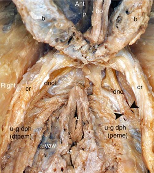
Fig. 8.1
The dorsal nerves of the clitoris in their ischio-pubic initial segment (dissection of the female anterior perineum, gynaecological position). The bulbs and the clitoris were folded against the pubis. The crura were released from their fibrosis attachments and pushed slightly outside. b bulb, bo clitoral body (descending part), cr crus of clitoris, dncl dorsal nerve of clitoris, p pubic symphysis, u external urethral orifice, u-g dph uro-genital diaphragm, dtpem deep transverse perineal muscle (visible on the right, after resection of the perineal membrane), u-g dph (peme) perineal membrane, va vaginal orifice, vaw vaginal wall, black arrowheads they line the banks of the incision of the perineal membrane making the nerve, which still remains in the deep perineal space, visible. Note the oblique course of the dorsal nerve of the clitoris, going forwards and within
Each plexus is formed by the plexiform convergence of ortho- and parasympathetic fibres.4 The following fibres are included in its composition:
· Orthosympathetic nerve fibres: They are the sacral splanchnic nerves—they come from the pelvic sympathetic ganglia (sacral sympathetic ganglia).
· Parasympathetic nerve fibres, an important contingent from the pelvic splanchnic nerves (Eckard’s erectile nerves), collateral of the pudendal plexus, originating from the ventral roots of sacral nerves S2, S3 and S4.
· Mixed nerve rami from the superior hypogastric plexus (inter-iliac plexus or presacral nerve of Latarjet), which conveys both the orthosympathetic and parasympathetic fibres.
The efferent branches of the inferior hypogastric plexus are dedicated to the rectum (medium rectal nerves), the uterus, the vagina, the bladder, the clitoris and the bulbs.
Thus, the vegetative nerve fibres dedicated to the clitoris are routed thereto via the 2 cavernous nerves, which originate from this inferior hypogastric plexus.
8.1.3 Descriptive and Topographic Anatomy of the Clitoral Innervation
8.1.3.1 Dorsal Nerve of the Clitoris
The dorsal nerve of the clitoris has, up until recent times, rarely been studied and therefore is badly known, especially since few authors have had the curiosity to reread the remarkable work conducted by Kobelt and, even more exceptionally, the will to study the said nerve by dissection. However, respecting the integrity of this nerve guarantees that the functionality of the clitoris will not be compromised and, therefore, that an essential and key part of female sexuality will not be affected.
This nerve is at the centre of many surgical acts: treatment of fractures of the pelvis, reduction of symphysis disjunctions, surgery of the urethra, repair surgery of certain lower urogenital congenital anomalies (Baskin L.S.), clitoridoplasties, surgery of tumours of the pubis, installation of trans-obturator tape for treating urinary incontinence (Achtari C. et al.), “transgender” plastic surgery, etc. Major iatrogenic consequences, which are generally final, can only be avoided through perfect knowledge of the anatomy. It is necessary to identify, for each of the 2 dorsal nerves of the clitoris, several segments, which each have a specific significance: the initial ischio-pubic segment, the ascending clitoral segment, the pre-symphysis (prepubic) segment, the corporeal clitoral dorsal segment and the termination at the glans. These various segments are separated by direction changes and actual kinking, which also deserve to be analysed in a specific study, especially the first during which we will observe how the cavernous nerve meets the dorsal nerve of the clitoris.
· The initial or ischio-pubic segment of the dorsal nerve of the clitoris is the superior division branche of the pudendal nerve of which it extends the direction, thus remaining in the fibrous pudendal canal with the artery and pudendal veins. It should be noted that this dorsal nerve of the clitoris is difficult to release in this initial segment, where it is routed against the ischio-pubic branch, below the fascia of the obturator internus, above the insertions of the deep fascia (superior fascia) of the urogenital diaphragm. It provides, at this level, the nerve of the external sphincter of the urethra, which will cross the above-mentioned fascia to join the sphincter fibres in the deep space of the perineum. Beyond the pudendal canal, the dorsal nerve of the clitoris will cross the pudendal artery, which will be in the more medial position. It surfaces by crossing through the superior fascia of the urogenital diaphragm and penetrates the deep space of the perineum, where it becomes accessible for perineal dissection. It may then, depending on the situation, remain above the inferior fascia of the urogenital diaphragm or, on the other hand, resurface by crossing through this fascia (Fig. 8.1). This penetration has been the subject of recent studies (A. Vaze et al.): according to these authors, it is located, on average, at 2 cm, 7 from the external orifice of the urethra. In both cases, the nerve will continue its forward and inward diagonal route towards the inferior branch of the pubis, which shelters and protects it and to which it is closely connected. At the inferior edge of this branch, a more or less deep groove can be observed. This groove has been described by Sedy J et al. as follows: the groove of the dorsal nerve of the clitoris (nearly 70 % of cases) is more or less marked, 15.8 mm long on average and even extends, according to these authors, at the inferior part of the ventral surface of the pubis. According to Sedy, this protective groove, which contains the nerve (which is often covered with a fibrous tissue), is as important as the pudendal canal because, in some cases, such as the latter, it can be too narrow and become a site of nerve compression (for cyclists in particular!), the consequence being a “syndrome of the dorsal nerve” (slightly different from the “pudendal nerve entrapment syndrome” and involving evocative symptoms such as an erectile malfunction). In order to meet the clitoris, the nerve finally reaches the inferior edge of the body of the pubis and its very dense prepubic fibrous coating. More precisely, it appears in the lateral part of the infra-pubic region, in front of the transverse perineal ligament (Henlé’s or Krause’s ligament), below the caudal symphysis edge (inferior edge), coated with the significant fibrous layers of the arcuate pubic ligament (Lauth’s ligament). Therefore, the nerve is in a very dense fibrous atmosphere, on which no author has insisted (except for Hruby S. et al., who described it in men) and which, however, plays a major protection role. Each of the 2 nerves will be wrapped at this level, in an actual protective sheath, a fibrous sleeve (with smooth internal wall), formed by the numerous fibrous components which surround the infra-pubic region. In some cases, certain nerves are wrapped in a fibrous canal formed by the duplication of the arcuate pubic ligament or even in a tunnel duplicated from the pre-symphysis tissue. The incision of the most superficial lamina of this envelope releases the nerve, which then seems to sprout out from an actual fibrous cone (Fig. 8.2). It should be noted that the homologous pudendal artery is also well protected, inside an underlying fibrous sleeve. The above details can be perfectly well observed by slightly tilting the clitoris, released from its pubic attachments, ventrally and backwards (Fig. 8.2). The thin subpubic urethral plate, which spreads out laterally up to the 2 nerves (which can be observed by transparency) must also be resected (Fig. 13.4).
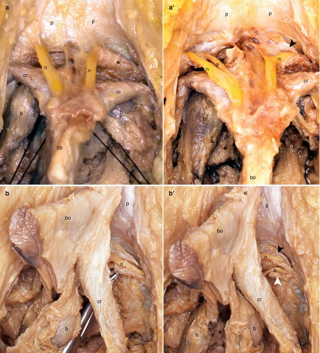
Fig. 8.2
The infra-pubic protective fibrous sheaths of the dorsal nerve and dorsal artery of clitoris. (a, a′) Views of the prepubic area (clitoral body retracted backwards and downwards). (b, b′) Side views of the bulbo-clitoral organ (detached from the pubic symphysis). (a) Note the left fibrous cone-shaped sheath , protecting the left dorsal nerve of clitoris. (a′) Note the opened fibrous sheaths. The opening of the left fibrous sheath (black arrowhead) shows the dorsal nerve of clitoris before it receives the cavernous nerve. (b) Note the opened fibrous sheath for the left dorsal nerve of clitoris (tracted by a forceps). (b′) Note the left opened fibrous sheaths of the nerve (black arrowhead) and of the artery (white arrowhead). a dorsal artery of clitoris, b bulb, bo body of clitoris, cr crus clitoridis, n dorsal nerve of clitoris, p pubis, sl suspensory ligament
· The first elbow or pubic elbow (Fig. 8.3): Each nerve will then modify its trajectory to rise up in front of the pubic symphysis, against the dorsal surface of the clitoris. However, beforehand, it will be considerably densified by the arrival of the cavernous nerve, which converges towards it while supplying the vegetative contingent. Due to this contribution, the dorsal nerve of the clitoris (beyond this anastomosis) will become a particularly exceptional nerve, a true “mixed nerve”, belonging both to the cerebrospinal nervous system and to the vegetative nervous system, as a motor component for the deep transverse muscles and external sphincter of the urethra, a sensory component for the corpora cavernosa, the glans and prepuce, and a vegetative component for the cavernous and spongious bodies. It is thus our aim to evoke, with the description of this first elbow of the dorsal nerve of the clitoris, the descriptive anatomy of the cavernous nerve.
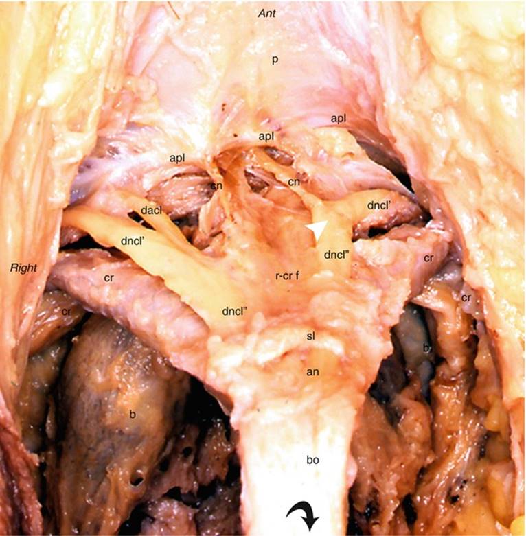
Fig. 8.3
Dissection of the prepubic area. It shows the constitution of the dorsal nerve of clitoris just before entering clitoris (the clitoral body has been retracted back and down: black curved arrow). anangle of the clitoral body, apl arcuate pubic ligament, b bulb, bo clitoral body (descending part), cn cavernous nerve, cr crus clitoridis, dacl dorsal artery of clitoris, dncl′ dorsal nerve of clitoris (before receiving cavernous nerve), dncl″ dorsal nerve of clitoris (after incorporation of the cavernous nerve), p pubic symphysis, r-cr f retro-crural fascia (partially resected), sl suspensory ligament, white arrowhead it shows the change of direction of the dorsal nerve of clitoris, successively oblique forward and inward, then vertical and ascending, after receiving the cavernous nerve
· The ascending clitoral segment (Fig. 8.2): After having received and incorporated the autonomous fibres of the cavernous nerve, the dorsal nerve of the clitoris only remains visible for a short moment. It is routed medially and then becomes vertical as soon as it meets the inflection of the terminal portion of the crura, i.e. at the level where the crura will be supported. It then penetrates below the retro-crural fascia (Fig. 8.4) (fibrous structure that we have studied previously), which will, once again, provide an efficient protection.5 The nerve is then in the retro-crural space (Fig. 5.9c) . It is routed with the dorsal artery of the clitoris, whose pathway is more medial. The veins essentially emerge from the centre line. It should be recalled that the retro-crural fascia is placed against the fibrous coating, which covers the pubic symphysis. It can be detached therefrom without much difficulty. The vertical pathway will make it possible for the nerve to pass behind the ascending portion of the clitoral body and rise up to the angular top of the body to penetrate the base of the suspensory ligament of the clitoris.
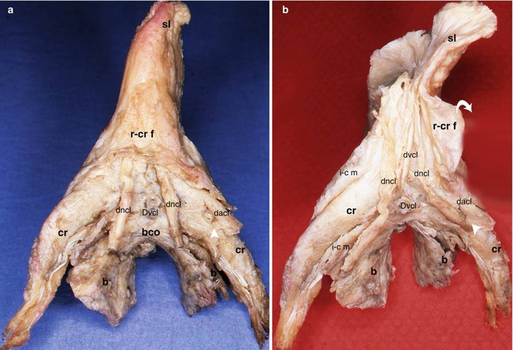
Fig. 8.4
Dorsal aspects of the bulbo-clitoral organ showing the second part (ascending clitoral segment) of the dorsal nerves of clitoris. (a) Both dorsal nerves of clitoris go into the retro-crural space (dorsally limited by the retro-crural fascia). (b) The retro-crural fascia has been cut medially and retracted outside (white curved arrow), showing the ascending course of the nerves. b bulb, bcobulbar commissure, cr crus clitoridis, dacl dorsal artery of clitoris, dncl dorsal nerve of clitoris, dvcl dorsal vein of clitoris, Dvcl deep vein of clitoris, i-c m ischio-cavernosus muscle, r-cr f retro-crural fascia, sl suspensory ligament, white arrowhead deep artery of clitoris
· The second elbow or clitoral elbow: The elbow of the nerve will follow the change of direction of the clitoral body by adopting an angulation identical to that made by the clitoral body to pass from its ascending portion to its descending portion. In order to adapt to the elbow of the clitoris, each dorsal nerve passes in the basal part of the suspensory ligament, which, in turn, plays its role of protective component. The nerve is routed very deeply, against the external wall of the albuginea, at a lower level than that of the large dorsal vein of the clitoris (which is on the centre line), on either side of the small dorsal veins (which are almost median and also against the albuginea). The 2 nerves are located at 11 a.m. and 1 p.m., respectively. The microscopic examination perfectly shows the pluri-fascicular structure of each nerve and the relations of these bundles with the numerous components, which pass via the base of the suspensory ligament of the clitoris (Fig. 12.3).
· The clitoral descending segment (Figs. 4.5 and 8.6): It is corporeal. It successively concerns the descending segment of the body, then the glans (to which it is distributed). While progressing along the descending segment and emerging beyond the suspensory ligament, the nerve slips between the 2 superficial and deep lamina of the clitoridis fascia, which are still outside the thick corporeal albuginea. At this level, the nerve is no longer really dorsal. It becomes lateral and tends to go deeper towards the end of the body. The protection of the nerve persists in the corporeal trajectory: It is no longer fibrous and now consists of a thick preputial coat, such as demonstrated by dissections (Fig. 8.5). Along this corporeal trajectory, the nerve provides very thin branches, perpendicular to its longitudinal axis (including several branches dedicated to the corpora cavernosa and perforating the albuginea). We can only be surprised by the considerable diameter of the nerve respect to the diameter of the clitoral body. In order to have an idea of the size ratio, the comparison with the diameter of the collateral digital nerves and that of a woman’s little finger can be referred to! This underlines, in both cases, the significance of innervated components! The numerous undulations of the nerve trunk should also be noted in this corporeal trajectory (Fig. 8.5) as they correspond to as many extension reserves for an erection. When the nerve arrives near the glans, it develops an important collateral branch (Fig. 8.6) dedicated to the latter and then goes deeper. It initially passes under the superficial lamina of the clitoral fascia and then crosses the deep lamina to penetrate the glans.
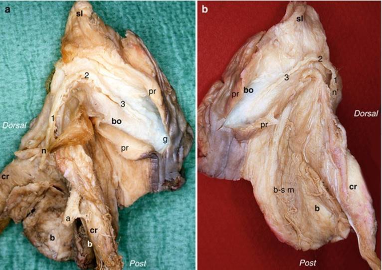
Fig. 8.5
Courses of the two dorsal nerves of clitoris. (a) Right nerve; (b) left nerve. a right dorsal artery of the clitoris, b bulb, b-s m bulbo-spongiosus muscle, bo clitoral body (descending part), cr crus clitoridis, g glans clitoridis, n right dorsal nerve of clitoris, n′ left dorsal nerve of clitoris, (1) prepubic part, (2) angle, (3) descending part of the nerve, pr prepuce (opened on midline and pulled outside), sl suspensory ligament. Note that the nerve starts superficial, then goes deeper when it reaches the glans
· The nerve ending: The final development of the nerve is not immediate. Large subdivisions of the nerve are observed. They are routed relatively superficially in the direction of the apex of the glans. It is even possible to observe terminal branches of remarkable dimensions arriving in contact with the external layers of the glans, in the area of the corpuscles (Fig. 8.6). It is only with specific colouring, which shows innumerable “sensory terminations” and a great amount of corpuscular receptors and demonstrate the significant innervation of the glans, that it is possible to understand why the terminal branches of the nerve have such considerable and almost unusual dimensions (Fig. 8.6), resolving in innumerable microscopic rami only in the immediate vicinity of the Krause-Finger corpuscles. It should also be noted that the glans is not the only “nerve terminal”. The microscopic observation of cross sections within the axis of the clitoral body shows that branches of the dorsal nerve of the clitoris are directed laterally to supply the clitoral hood (Fig. 9.10b) and contribute to the very dense innervation of the labia minora (Fig. 9.8). In addition, the observation of frontal sections of the bulbo-clitoral organ, on which the nerves have been identified by the S-100 protein, has shown branches of the dorsal nerve of the clitoris, which descend against the lateral walls of the cavernous albuginea to laterally reach the spongy structures and contribute, from the centre line, to the remarkable innervation of each lateral wall of the vestibule.
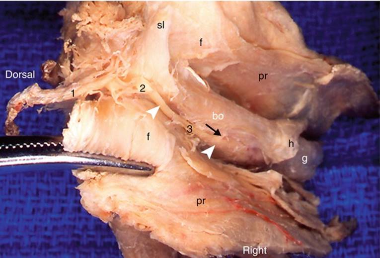
Fig. 8.6
Bird’s-eye view of a dissection of the right dorsal nerve of the clitoris. The right half of the preputial sheath (sectioned medium) is retracted outside by a pulling clamp. bo body of clitoris, fclitoral fascia, g glans clitoridis, pr prepuce (or foreskin), sl suspensory ligament, black numbers distal parts of the dorsal nerve of clitoris, 1 pre-pubic part, 2 angle, 3descending part, black arrow it indicates a nerve branch to the corpus cavernosum, white arrowheads they show the path of the nerve. Note that the nerve becomes less superficial when approaching the glans in which it will give its terminal branches. Note also that the path of the nerve is more lateral than dorsal
8.1.3.2 Cavernous Nerve
The cavernous nerve that, as we have observed, joins the 1st elbow of the dorsal nerve of the clitoris, deserves to be studied due to its considerable significance. This nerve, such as we have recalled previously, therefore exits the inferior hypogastric plexus (Fig. 8.7) by passing under the middle part of the pubic symphysis, or more laterally, under the para-symphysis part of the body of the homolateral pubis. The nerve can be free but, in most cases, it penetrates the fibres of the arcuate pubic ligament. It is relatively short and does not exceed 2 cm as it is also small (approximately 1/6 of the diameter of the pudendal nerve). It is shortly routed under the thin subpubic urethral plate, on either side of the urethra and clitoral veins (which will join the Santorini’s venous plexus), and then becomes straighter to slide in front of the inferior edge of the pubis and meet the dorsal nerve of the clitoris. Our dissections have perfectly demonstrated this convergence (Fig. 8.3). It is interesting to note that certain authors have reported similar conclusions on foetuses of 14–34 weeks old, by using layered and serial sections, histochemical staining and 3D reconstructions (S. Yucel et al.). The layered sections of the dorsal nerve of the clitoris are nNOS negative throughout the entire ischio-pubic trajectory. However, they become the site of intense nNOS activity from the pubic arch, which is a sign confirming that a positive nNOS contingent (in fact that of the cavernous nerve) has joined the dorsal nerve of the clitoris. On the other hand, other authors seem to ignore the existence of the cavernous contingent and believe that the function of the dorsal nerve of the clitoris is not clear!
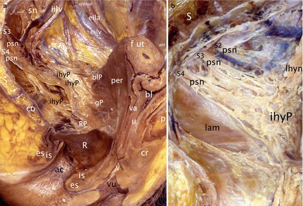
Fig. 8.7
The inferior hypogastric plexus (from which the autonomic nerves of the bulbo-clitoral organ originate) viewed on two dissections of the lateral wall of female left hemi-pelvis (after resection of the upper 2/3 of the rectum). (a) Overview; (b) details after dissection of the interlacing nerves. ac anal canal, bl bladder, blP bladder plexus, co coccyx, cr crus clitoridis, eila external iliac artery, eilv external iliac vein, es external sphincter (sphincter ani externus), f ut fundus of uterus, gP genital plexus (utero-vaginal plexus), ihyP inferior hypogastric plexus, iilv internal iliac vein, is internal sphincter, lamlevator ani muscle, lhyn left hypogastric nerve (from superior hypogastric plexus), per peritoneum (anterior peritoneum of the utero-rectal recess), psn pelvic splanchnic nerve (nervi erigentes); they originate in the ventral rami of the pudendal plexus (S2, S3, S4), R rectum (remaining part after resection of the upper 2/3), RP rectal plexus, S sacrum, sn one sacral nerve, S2, S3, S4 ventral rami of nerves, u urethra, V vestibule, va vagina, vu vulva
8.1.4 Descriptive and Topographic Anatomy of the Innervation of the Spongy Part
We want to devote a paragraph to the corpus spongiosum due to its specificity and to the fact that it is generally unknown. Such as for the “clitoral” parts, this innervation is a dual-type innervation as it is both somatic and autonomic. It has a few alternatives according to the parts being considered:
8.1.4.1 Innervation of the Spongious Bulbs
· The sensory innervation passes via the bulbar nerve, collateral of the deep perineal branch of the perineal nerve, which itself originates from the pudendal nerve (Fig. 13.2). This bulbar nerve approaches the bulb at the level of the lateral edge of its posterior half, providing a thin collateral for the greater vestibular gland (Bartholin’s gland). The bulbar nerve also contributes to the sensory innervation of the vestibule. As for its motor branches, they mainly ensure the motor function of the homolateral ischiocavernosus and bulbospongiosus muscles.
· Over the last years, vegetative innervation has been the subject of many work projects employing 3D reconstruction methods with serial sections of coloured embryos, specifically according to the method of Yucel, taken up by the French authors G. Benoit, F. Peschaud and D. Moszkowicz. This is therefore how it was possible to describe a group of nerve fibres, which are mainly parasympathetic and NOS positive. They originate from the posterior part of the vaginal plexus (itself efferent from the inferior hypogastric plexus) and reach the internal surface of each bulb, “improperly”6 called bulbar nerves. These nerves directly reach the spongy bulb such that it is possible to say that, at the level of the bulb, somatic and vegetative terminal innervations are separate. There probably exist distal anastomoses between the 2 innervations. The bulbar branches of the deep perineal nerve undoubtedly receive, which is impossible for us to affirm, vegetative nerves from the bulbar autonomic contingent (similar to what has been observed for the dorsal nerve of the clitoris)! In addition, our own research, using standard dissection techniques combined with the histological sections stained with the S100 anti-protein, showed us that the cords of the dorsal nerve of the clitoris also contribute to the bulbar innervation: These rami are routed, such as we have observed previously, against the lateral surfaces of the corpora cavernosa of the clitoris and reach, along the “pars intermedia”, the tapered anterior ends of the bulbs, thus contributing to their innervation. It is highly probable that a communicating network develops between the anterior and posterior vegetative fibres, i.e. between the vegetative fibres provided by the cavernous nerve and the “bulbar vegetative fibres”.
8.1.4.2 Innervation of the Infra-corporeal Residual Spongy Part
The innervation of the infra-corporeal residual spongy part is ensured by the lateral collaterals of the dorsal nerve of the clitoris, in its descending segment.
8.1.4.3 Innervation of the Glans
The innervation of the glans is ensured, such as has been shown during the study of the dorsal nerve of the clitoris, by the terminal branches of this nerve, which cross through the clitoral fascia and penetrate the conjunctivo-vascular parenchyma, where they ramify and develop to ensure the transmission of information received by the multiple highly specialised receptors of the glans and its cutaneous environment. The nerves of the glans essentially include sensory somatic cords. They also carry vegetative nerve rami, dedicated to the smooth muscle fibres (of the vessels in particular) of this formation. This innervation is extremely dense, such as that of the preputial hood and labia minora.
8.1.5 Functional Anatomy of Female Sexual Arousal
It is better known since the current practice of MRI. The stimuli are multiple, whether they are tactile stimuli (especially in erogenous zones), or stimuli involving other sensory apparatus (especially visual apparatus but also hearing, olfactory and gustatory apparatus), even direct conceptual stimuli (erotic reading) or indirect conceptual stimuli belonging to erotic imagination (dreams, souvenirs, fantasies or even erotic conversations). The various types of above-mentioned stimuli can also cumulate, interfere and be reinforced. Tactile stimuli will be conveyed via the pudendal nerve and pass via the spinal relay before reaching the specific sensory brain areas, via the spino-thalamic and thalamo-cortical pathways. Stimuli directly acting on specific cortical areas and the brain in general (amygdaloid body, nucleus accumbens, hypothalamus, limbic system, etc.) will, in turn, act on the spinal centres to trigger somatic and vegetative reactions representing sexual arousal in women. It should be noted that, at any moment, the brain (frontal areas in particular) can exert its control on reactions induced in this manner by playing a reinforcing or inhibiting role. It should however be recalled that, in women, as well in men, the spinal effector centres are significantly independent as, during periods of cortical rest (during sleep periods), sexual arousal with erection is possible!
It is therefore at the level of the spinal cord that the vegetative centres are located. The protoneurons (preganglionic neurons) of the ortho- or parasympathetic vegetative pathways are developed from these vegetative centres. The parasympathetic centres are located at the level of the sacral cord (from S2 to S4), while the sympathetic centres are at the level of the thoraco-lumbar cord (from T11 to L2).7
The parasympathetic pathways form the splanchnic pelvic nerves (Eckard’s erector nerves), which detach from the spinal ventral roots from S2 to S4. These nerves reach the inferior hypogastric plexus. They contain the axons of the parasympathetic protoneurons, which cross through this plexus without acting as a relay. They then pass via the cavernous nerves or bulbar vegetative nerves to reach, such as we have observed previously, the erectile bodies, where the synapse with the effector deutoneuron (postganglionic neuron) occurs.
The orthosympathetic pathways develop from the intermedio-lateral zone of the spinal cord and from its lateral horn, from T11 to L2. The axons of these protoneurons depart from the ventral ramus of the corresponding spinal nerves, pass via the latero-vertebral sympathetic trunk and are then routed in the sacral splanchnic nerves to reach the inferior hypogastric plexus, where they act as a relay. The effector deutoneuron will then pass via the cavernous nerves and the bulbar vegetative nerves to reach the erectile bodies.
The physiology of sexual arousal can be thus represented. The various stimuli will involve local responses (responses at the level of the bulbo-clitoral organ):
1.
2.
Other phenomena will occur at the same time as sexual arousal and may precede an orgasm: genital secretions (glandular in particular), cutaneous vasomotor phenomena, various mammary reactions (nipple erection, areolar turgescence) and contractions of the levator ani muscles. We mention them in this section despite the fact that they do not directly involve the subject of our study, the bulbo-clitoral organ. When the sexual excitation ceases, the local and general phenomena will be reversed: the action of the parasympathetic nerve will be inhibited, the tonicity of the sympathetic nerve will be resumed and clitoral flaccidity will be restored.
Footnotes
1
The central nerve system (whose centres are cortical) is dependent on the conscience and will. The vegetative or autonomous system, which is independent of the conscience and will, ensures the operation of the organs.
2
The potential compression areas are located in the space between the sacrotuberous and sacro-spinous ligaments, at the level of the falciform process and, lastly, in the pudendal canal. (Alcock’s canal). See R. Robert et al, Neurosurgery, 55, 453–469.
3
The termination of the pudendal nerve can also be located before the penetration in the pudendal canal, above the ischial tuberosity.
4
The ortho- and parasympathetic fibres (whose actions are antagonistic) especially control the motricity of the vessels, the smooth muscles, the viscera and the glands.
5
When the bulbo-clitoral organ is unstuck from the pubis, the 2 dorsal nerves of the clitoris remain covered by their “fibrous cape” and there is no risk of lesion. On the other hand, the cavernous nerves, which benefit from a reduced protection at this level, are likely to be injured due to stretching if surgical operations are not carried out with precaution and care.
6
The “bulbar nerve”, term used by many authors, is an inadequate term because it leads to confusion: It is normally used to refer to the somatic branches of the deep perineal nerve, dedicated to the spongious bulb. Moreover, the term of nerve evokes a well-structured nerve structure, such as is the case for the cavernous nerve, whereas it actually concerns, for these bulbar vegetative fibres, superimposed thin rami.
7
The pathways of the vegetative nervous system, whether they belong to the ortho- or parasympathetic contingent, are all bi-neuronal, with a first neuron, the protoneuron, which departs from the spinal centre and is articulated with a second neuron, the deutoneuron, which ends at the effector organ. The synapse between the 2 neurons occurs at a different site for each of the 2 contingents.