
Vincent Di Marino1 and Hubert Lepidi1
(1)
UER Médecine, Aix-Marseille Université, Marseille, France
Abstract
The bulbo-clitoral organ is, such as we have seen previously, provided with a significant innervation. The sensory somatic fibres form a very important contingent in this innervation. They also make it possible to permanently inform the overlying nerve centres of any stimulus occurring at the level of this organ. Very specialised and specific receptors (“corpuscles”), positioned at the ends of some of these sensory fibres, receive the stimuli corresponding to “sensuous sensations” and transmit them to the nerve fibres, which convey them to the sensitive and specialised cerebral areas. Nerve endings are extremely numerous at the level of the bulbo-clitoral organ, especially the glans, which explains the extreme sensitivity of the latter. It should be noted from the start that although the clitoral glans has the same number of sensory “terminations” and genital corpuscles as the penile glans, assessed at around 8,000, the density of these receptors, with respect to the size of each of these organs, is 50 times higher for the female glans. This means that the sensitivity of the clitoral glans is extreme compared to that of the male glans, which is already very high!
9.1 General
The bulbo-clitoral organ is, such as we have seen previously, provided with a significant innervation. The sensory somatic fibres form a very important contingent in this innervation. They also make it possible to permanently inform the overlying nerve centres of any stimulus occurring at the level of this organ. Very specialised and specific receptors (“corpuscles”), positioned at the ends of some of these sensory fibres, receive the stimuli corresponding to “sensuous sensations” and transmit them to the nerve fibres, which convey them to the sensitive and specialised cerebral areas. Nerve endings are extremely numerous at the level of the bulbo-clitoral organ, especially the glans, which explains the extreme sensitivity of the latter. It should be noted from the start that although the clitoral glans has the same number of sensory “terminations” and genital corpuscles as the penile glans, assessed at around 8,000, the density of these receptors, with respect to the size of each of these organs, is 50 times higher for the female glans. This means that the sensitivity of the clitoral glans is extreme1 compared to that of the male glans, which is already very high!
These terminations and corpuscles make the clitoris, and especially the glans, an extraordinary organ, which is very specialised, and exclusively dedicated to female pleasure, whereas the penis and the penile glans are “multifunctional” as they are also used for urinating purposes, penetration and spermatic emission during the coitus act.
It should be noted that clitoral sensory corpuscles have been observed on animals (studies conducted by J.F. Tello on female mice, rats and ewes and by D. Ohmori on female rabbits, bitches and cats). However, these authors acknowledge that the genital corpuscles of the females under study are infinitely simpler than those of the human clitoris!
Several types of nerve endings (sensory nerve “terminations”) can be identified in the human clitoris:
· Some are not specific to the clitoris and are similar to tactile terminations, the same nerve endings as those identified over all of our skin and more particularly in areas dedicated exteroception: palms of the hands, soles of the feet and labia majora.
Among these terminations, some do not have any corpuscles: These are free terminations, “free nerve endings” and there exist others, which end at a receptor corpuscle or a tactile corpuscle; these terminations are referred to as encapsulated terminations, “corpuscular nerve endings”. In this case, it is the unit consisting of the nerve fibre termination and the corpuscle, in which this termination has developed, which forms the receptor. Such as over the entire skin surface, the corpuscles observed at the clitoris are standard tactile corpuscles: Meissner’s corpuscles, Ruffini’s corpuscles and Pacini’s corpuscles.
· Other hyper-specialised corpuscles, specific to the clitoris, Krause-Finger’s corpuscles or pleasure corpuscles.
All these nerve endings have a certain number of similar characteristics:
· They are part of the general context referred to by J.F. Tello as “sensory terminations of external genitals”, which are the “mucocutaneous specialised sensory end organs” of the English authors (R.K. Winkelmann).
· They only appear at late stage and develop at puberty (Winkelmann), which suggests a hormonal influence. The free nerve endings and Pacini’s corpuscles are an exception to this rule as they can be observed and are already perfectly formed (J.F. Tello) in newborn children (R.K. Winkelmann) and even in foetuses (K.E. Krantz).
· Their degree of organisation and their complexity tend to increase with age (R.K. Winkelmann) and the following issue can arise (such as mentioned by H. Jaeger) “certain repercussions of the intensity of sexual life on the development of these formations”.
· They are more or less numerous according to the part of the external genitalia being considered, the clitoris being incontestably the organ which has the greatest number.
· Their number and their distribution vary according to individuals.
We will successively study non-specific nerve endings and specific nerve endings.
9.1.1 Non-specific Nerve Endings
Among the non-specific nerve endings, terminations in direct connection with the skin’s epidermis and terminations located in the dermis can be mentioned.
· Nerve endings connected to the epidermis: These include, on the one hand, free nerve endings and, on the other hand, terminations with Merkel discs.
The free nerve endings correspond to axons, which exit their myelin sheath (Schwann’s cell envelope), ramify and end in the dermis, in contact with the epidermis (Fig. 9.1), or in the epidermis itself, if they have crossed the lamina basalis, which remains exceptional (H. Jaeger). Small terminal swelling or bulbs may be present at the end of the ramifications (K.E. Krantz). They can anastomose to form, at the most superficial part of the dermis, a subepidermic nerve reticulum (H. Jaeger). They are distributed as thermoreceptors (sensitive to thermal variations, especially if they are sudden) and nociceptors (sensitive to pain). The extreme density of the distal axons and their arborisation and ramifications, which can be observed on histological preparations, account for the abundance of these free nerve endings, not only at the level of the clitoris but also of the labia minora.
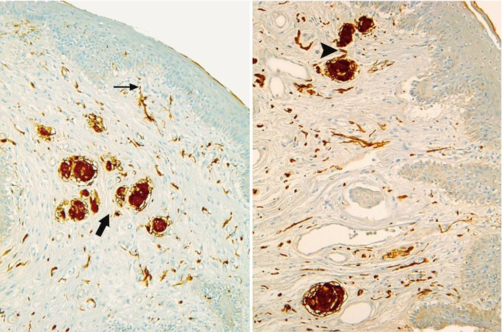
Fig. 9.1
Free nerve endings and bulbous corpuscles (PS100 staining). Note: A free nerve ending at the top of a dermal papilla (thin black arrow). A group of corpuscles (thick black arrow). The axon penetrating into the corpuscle (black arrowhead)
The meniscoid corpuscles (discs of Merkel or “tactile menisci”) are very superficial, in contact with the basal epithelium of the glabrous skin’s epidermic ridges. They consist of axon ramifications, whose dilated ends come in contact with a particular meniscoid cell: the Merkel cell. C.J. Cold and J.R. Taylor observed them on the glabrous part of the external epithelium of the prepuce, i.e. at the level of the clitoral hood. Some are also present at the level of the labia minora. These discs detect localised light pressures and remain sensitive throughout the duration of this pressure. They therefore are rapidly adapting mechanoreceptors with an extended action.
· Dermic nerve endings: They are Meissner’s corpuscles and Ruffini’s corpuscles.
The encapsulated tactile corpuscles (Meissner’s corpuscles) are also rapidly adapting mechanoreceptors. They have an alveolar aspect and are located in the top part of the dermis of the glabrous skin, in the middle of the dermis papillae. As they are sensitive to the slightest touch (they are capable of appreciating the speed at which a pressure is applied!) and therefore to brushing, they are also capable of detecting the slightest inequalities. They consist of spiral nerve ramifications, within a stack of lamellar cells, the lemnocytes, which are Schwann’s cells and which are flattened and superimposed like piles of plates. They have a fibrous capsule (collagen fibres). They are especially observed on the medial surface of the labia minora and, in a smaller number, at the level of the clitoral hood.
Ruffini’s corpuscles are located at a deeper level, in the dermis of the prepuce and of the labia minora. They are at both mechanoreceptors and proprioceptors. They are ovoid formations consisting of collagen fibres, between which axon ramifications having penetrated the corpuscle have developed. A nerve fibre exiting the corpuscle to reach the epithelium, as a free termination, or to end in a nearby Meissner’s corpuscle (K.E. Krantz) is sometimes observed at their level. They are positioned parallel to the skin. They are slow adapting corpuscles, sensitive to cutaneous stretching (e.g. during shaving) or to an extended pressure (as they appreciate the related intensity and duration).
· Dermo-hypodermic nerve endings (present in the deep dermis and the hypodermis): These are the corpuscles discovered by Vater and then studied by Pacini,2 a few years later (Vater-Pacini’s corpuscle), which are now referred to as lamellar corpuscles. Their presence in significant numbers at the level of the clitoris deserves an in-depth description. These corpuscles are often large (transverse diameter of 5–6.5 μm) and are both mechanoreceptors and proprioceptors. They have an ellipsoidal shape, and their section is generally oval or circular even if we have observed irregular aspects (Fig. 9.2), polygonal on certain sections. They can be isolated and positioned at a certain distance from each other. They are often routed in groups of two, in similar sizes, or on the contrary, in very different sizes. They can also be grouped in clusters, in a half circle or even in a single line (Fig. 9.3). On a structural level (Fig. 9.4) the corpuscle consists of several cellular rings “the lamellae”, concentric as an onion bulb and surrounding a central axon, which has lost its myelin sheath by penetrating into the axial space. The peripheral conjunctive envelope, as well as the external lamellae (capsule), is within the extension of the perineurium of the axon. The internal lamellae (teloglial lamellae), forming the intermediate growth area, consist of flattened cells, resembling fibroblasts but originating from Schwann’s cells, which are more closely applied than the external laminae. Even more internal and more closely arranged lamellae form the “central club” of the corpuscle (“core” according to the Anglo-Saxon authors), in which is located the receptor nerve fibre, ending with a bulge. In electronic microscopy, these internal lamellae are only half-lamellae facing each other and separated by a radial slit. The total number of lamellae in a corpuscle is variable (20–30 on average in an adult). However, around 30 or more is frequent (Figs. 9.2 and 9.4). There are 5–10 lamellae in a foetus; this number increases with time (growth in the area of the teloglial lamellae) and, in particular, during puberty. As of birth, the lamellar corpuscles are already formed (J.F. Tello): It is already possible to identify the external lamella/strips separated by wide spaces and the internal lamellae, which are very close to each other, especially when approaching the centre. For this author, at puberty, the corpuscles will increase in volume, the number of lamellae will increase, the lamellae will tend to become closer to each other and the central area will generally tend “to be reduced to its simplest expression”. However, corpuscles with a larger core can be observed in adults. It should be recalled that the inter-lamellar spaces of a mature corpuscle are not empty but filled with a viscous liquid film, in which collagen fibrils can be observed by electronic microscopy (C. Cavallotti et al.). The central axon, which has penetrated the corpuscle, is routed up to the core end and often ends in by one or more bulges. Along its route in this core, irregularities, varicosities and thickness inequalities are also observed on this axon (H. Jaeger). For J.F. Tello, the presence of corpuscles as of birth and the fact that they are formed in foetuses as of the 6th month, in contact with the nerve components, should be explained by their role in the early acquisition of protopathic sensitivity.

Fig. 9.2
Pacinian corpuscles in the hypodermis of the female prepuce (HPS staining). Observe: The nerve trunk (N) adjoining one of the two lamellated corpuscles (left picture). The numerous concentric in shape successive lamellae of each corpuscle. The central naked axon in each core. The adipose tissue in which you can find the lamellated corpuscles
In the clitoris, lamellar corpuscles can be found in various locations: Firstly, in the vicinity of nerve cords (nerve ramus developed from the branches of the pudendal nerve), which they follow more or less closely, sometimes even inside the epineurium of the nerve (Fig. 9.5), especially to transmit the slightest vibratory excitation. They may even be positioned alongside the nerve endings to form a sort of sensitive pack. It is in the retro-crural small space (in the cell tissue limited by the retro-crural fascia) and in the latero-cavernous areas of the descending part of the body (Fig. 9.3) that their presence is the most obvious. They are also found in the dermis (especially the deep dermis) but the favoured sites are the subcutaneous cell tissue and the hypodermis, in contact with fatty tissue (especially at the level of the prepuce), and the suspensory ligament of the clitoris (see Chap. 12). A small number also exists in the albuginea of the cavernous bodies and more exceptionally in the septula of the cavernous bodies (K. Yamada). Such as we have observed, it is primarily the clitoral part of the bulbo-clitoral organ, which is equipped with lamellar corpuscles. However, it is not the only part to be provided as such. Lamellar corpuscles are identified at the end of the residual spongy part and glans, which form the distal part thereof (E. Lastly); the presence of lamellar corpuscles in the epithelium of the prepuce and at the labia minora (K.E. Krantz) is also to be recalled. It should be noted that all the above-mentioned formations have a greater amount of lamellar corpuscles than the penile glans or the male prepuce (C.J. Cold and J.R. Taylor).
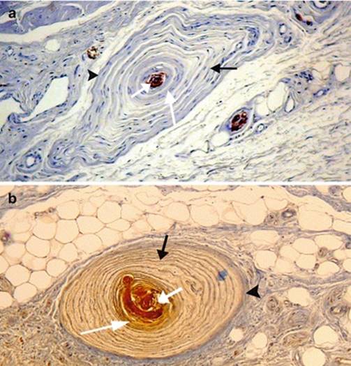
Fig. 9.3
Usual places of the clitoral lamellated corpuscles (The blue arrows show the corpuscles). (a) A corpuscle inside the epineurium (connective sheath of the nerve). The corpuscle occupies the place of a bundle of axons and is surrounded, just like it, by a sort of perineurium confounded with its peripheral connective layer. (b) Three lamellated corpuscles inside the cellular tissue around the tunica albuginea (Alb) of the corpora cavernosa (CC). (c) Lamellated corpuscles at the level of the junction of hypodermis with dermis. (d) A lamellated corpuscle inside the adipose tissue of the preputial hypodermis. (e) Several corpuscles in single file, inside the basis of the glans clitoridis. Note the different shapes of the corpuscles: Classical, oval-shaped (a), rounded (b, e), triangular (c), bludgeon-shaped or “mussel-shaped” (d), (evocative forms of seafood are frequent)

Fig. 9.4
Structure of the lamellated corpuscles of clitoris. (a, b Two examples of corpuscles located behind the angle (elbow) of the clitoral body) PS100 Immuno-staining for (b). black arrowexternal lamellae (capsule), long white arrow internal lamellae (intermediate growth zone), short white arrow core of the corpuscle (granular substance + naked axon, wrapped by joined lamellae), black arrowhead layer of connective tissue wrapping the corpuscle
It is well known that lamellar corpuscles are rapidly adapting mechanoreceptors and that they are not only very sensitive to pressures (they perceive deformations of a few microns!) but also particularly sensitive to vibrations!3They also detect, with the free nerve endings, the sensory information related to tickling.
Their activity is biphasic: production of an impulse when the compression occurs and production of a new impulse when the compression stops (however, they are indifferent to a constant pressure). Another particularly important concept: Certain lamellar corpuscles could, according to K.E. Krantz, be integrated into the group of the corpuscles involving sexual activity; they are the corpuscles, which are positioned at the site of a nerve fascicle inside the epineurium (Figs. 9.3 and 9.5). According to this author, when sexual excitation occurs in the genital sphere, the blood supply to the vasa nervorum (arteries of the nerves) increases, which involves a pressure increase inside the entire surface circumscribed by the epineurium. “This pressure increase acts on the nerve endings of the lamellar corpuscles, which immediately lower the threshold necessary for a nerve discharge”.

Fig. 9.5
Relationships between the lamellated corpuscles and the nervous ramifications inside the clitoris. 1 Large intradermal nervous ramifications (N) accompagnied by 2 lamellated corpuscles (white arrows). One of the corpuscles is stuck on a nervous branch. 2 A lamellated corpuscle (white arrow) located in the immediate neighbourhood of several nervous ramifications. 3 Assembling of nervous ramifications with 2 lamellated corpuscles in the hypodermis. 4 Three lamellated corpuscles (white arrows) whose 2 in single file, parallel to close nervous ramifications. Note on the photomicrographs 3and 4, the presence of adipose tissue close to the lamellated corpuscles
9.1.2 Specific Nerve Endings
They are all specialised nerve endings, characteristic of areas dedicated to generating sexual excitation in women. Many are observed in the clitoral part of the bulbo-clitoral organ. They are nerve endings provided with particular corpuscles. It is Krause and then Dogiel, who described them first as “Genitalkörperchen” or “endkolben”. Other authors have also studied these formations, which are described as Golgi-Mazzoni corpuscles. Finger specified, by means of a graphic representation (in terms of pear, pea, heart shapes, etc.), the morphology of certain Krause corpuscles, which seem to be more specifically dedicated to the excitation of specific sexual erogenous areas,4 which explains their name, the Krause-Finger corpuscles.
These “wollustkörperchen”, due to their key function in women, deserve this terminology, which is translated into French by the term “corpuscules de la volupté” (“corpuscles of pleasure”). The term of genital corpuscles, by which they are often referred to, indicates that they are also observed in extreme genitalia (male as well as female). Their name is related to their globular shape: bulbous corpuscles. Such as we have observed during our study, the distribution of these corpuscles is not identical in all women but their numbers and density seem to be extremely high in all specimens, which have been examined. Nevertheless, for K.E. Krantz, there are significant variations in terms of the quantity, quality and localisation of these structures that could explain the differences in sensitivity and excitability of the clitoris. In addition, certain authors have mentioned the possibility of an involution of these formations after the menopause (J.F. Tello). For Tello, signs of involution should be visible, at the level of the free terminations: broken or segmented fibres, terminal bulges with highly increased volumes, as well as at the level of the corpuscles—lower content in fibres and more stroma. In our research, no histological evidence supported the idea of a possible involution and we found similar corpuscles in young and old women. As opposed to lamellar corpuscles, bulboid corpuscles are not present in newborn babies or foetuses.
These corpuscles have especially been studied by many authors over the last years. They all agree to describe a general common structure (Fig. 9.6):
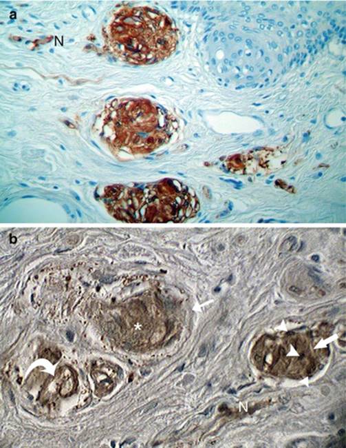
Fig. 9.6
Structure of the genital corpuscles (“corpuscles of pleasure”) of the glans clitoridis (Immuno-staining by PS100). (a) Three large bulbous corpuscles superimposed, under epithelium. (b) Three corpuscules examined under high-magnification factor. N afferent nerve fibre, asterisk granular contents, white thin arrow capsule, white large arrow corpuscular syncitium, white curved arrow it shows a large and coiled intra-corpuscular nerve fibre, white arrowheads syncitial nuclei
· Spherical or ovoid aspect.
· Large size (which can reach 150 μm in length on sections, in our studies).
· Lamellar conjunctive peripheral capsule (frequent but “not mandatory”).
· Important central cavity filled with a fine granular substance, in which bathe the cores forming a syncytium, without cellular limits. This cavity is occupied by a nerve fibre (or by several nerve fibres), which has lost its myelin sheath and which ramifies or is wound up and whose end includes club-type bulges.
· Dermic location (surface or deep).
This explains the excellent definition suggested by F. Sfameni for this type of corpuscle: “nerve system organ, covered or deprived of a connective ‘involucrum’ and consisting of one or more nerve fibres, which, once they have been stripped of their myelin sheath, develop in the centre region and around a granular and nucleated substance”.
· Primary location at the glans clitoridis.
· Multiplicity (great number of corpuscles in the same area).
Particularities are to be noted concerning this general diagram:
(a)
(b)
(c)
(d)
With K. Yamada, who used the previous research work carried out by Ikui, 3 types of genital corpuscles can be identified5:
1.
2.
3.
According to K. Yamada: If type-1 corpuscles are primarily located in the dermis of the glans clitoridis, they can also be found in the external and internal layers of the prepuce and even in the cavernous bodies. Type-2 corpuscles are located, in order of frequency, especially in the dermis of the glans, in the internal layer of the prepuce, in the cavernous body of the glans and its surrounding area, in the external layer of the prepuce, at the periphery of the cavernous bodies and in the tunica albuginea itself. Still according to K. Yamada, type-3 corpuscles prevail in the thick conjunctive tissue and therefore in the tunica albuginea, in the cavernous septula and under the glans.
For our part, we have essentially highlighted type-1 and type-2 corpuscles, not only in the clitoris, in the prepuce and its hood but also in the dermis of the vestibule (Fig. 9.7) and in that of the labia minora (Fig. 9.8). However, in these two last locations, these formations are scattered and never have the density, which can be observed in the clitoris. As for type-3 corpuscles, we have observed them not only in the clitoral conjunctive tissue but also in the dermal papillae of the external epithelium of the labia minora (Fig. 9.8).
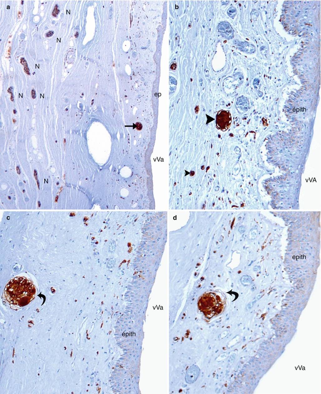
Fig. 9.7
Microscopic aspects of the vestibular corpuscles (PS100 immuno staining). (a) Right wall of the vestibule (low magnification); (b–d) morphology of the corpuscles (high magnification). Epith (ep) epithelium of the vestibule, vVa vestibule of the vagina, N nerve of the right wall of the vestibule, black right arrow (a) it shows a bulbous corpuscle (isolated, on the observed area), black arrowheads (b) they show two bulbous corpuscles, curved black arrow (c, d) they show the capsules of two large bulbous corpuscles. Note: scarcity of the vestibular bulbous corpuscles and their typical morphology. Note also the nerve wealth of the vestibular walls with many nerve endings under the epithelium, in the dermal papillae (visible under high magnification)

Fig. 9.8
Microscopic aspects of the genital corpuscles of the labia minora (PS100 immuno staining). (a) Transversal section of a labium minus (low magnification); (b) observation of the area in which several large corpuscles are visible; (c, d) high magnification of the genital corpuscles. ep l epithelium of the lateral wall of the labium minus, ep m epithelium of the medial wall, hb hair bulb, t2 large rounded corpuscles, of type 2, t3 elongated corpuscule, of type 3, red arrowheads they show the genital tubercles, little red arrow it identifies the draft capsule around the corpuscle, visible in (d). Note: Most corpuscles under the epithelium of the labium minus’ lateral wall. The intradermal position of the corpuscles, housed at the bottom of the dermal papillae. On (d), the nerve afferent fibre of the corpuscle is clearly visible
In the dermis of the glans clitoridis, the location of the corpuscles is as variable as their frequency (Figs. 9.9 and 9.10). Similarly to H. Jaeger, the glans clitoridis can be divided into 5 areas: a central median area, the deepest, 2 medium intermediate areas and 2 more superficial lateral areas, which are subdermic (the point is part of the superficial areas but has a particular specificity). In most cases, the corpuscles are observed in the medium and lateral areas. A central location is relatively exceptional. As for the point, it may be provided with corpuscles or not. When it contains some corpuscles, the site of these corpuscles can obviously favour sexual excitation in a woman (Fig. 9.11). Otherwise, the frequency of these tubercles, along and at the rear of the preputial plate (epithelial preputial lamina), on both sides of the hidden part of glans should also be underlined (Fig. 9.9). It is interesting also to observe the corpuscles in the end of the residual spongy part, at the lower half of the glans (Fig. 9.12).
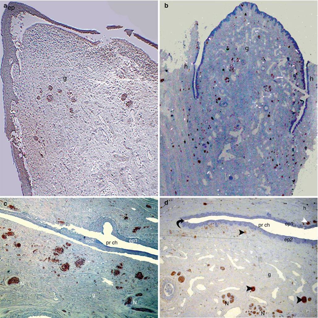
Fig. 9.9
Morphological aspects of the genital corpuscles (corpuscules of pleasure) of the glans clitoridis (PS100 immuno staining). (a) Corpuscles (dark purple) grouped at the end of the glans clitoridis. (b) Scattered corpuscules (brown-black) not only occupying the whole glans but also the distal part of the clitoral body; (c) many corpuscles of wide diameter, arranged under the epithelium of the glans, and along the preputial chamber too; (d) some big deep bulbous corpuscles and a multitude of punctated superficial corpuscules (light brown under the epithelium of the glans). ep epithelium, ep1 epithelium of the medial surface of the hood, ep2 epithelium of the glans, g glans, h hood, N nerve rami, black curved arrow neck of the glans, pr ch preputial chamber, Arrowheads (black for the glans and white for the hood) show some corpuscles
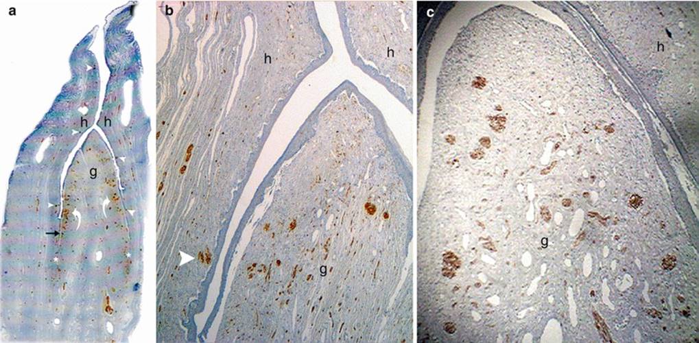
Fig. 9.10
Examples of distribution of the genital tubercles (corpuscles of pleasure) in the glans clitoridis. (a, b) Predominance of lateral distribution (curved arrows on a); (c) harmonious distribution without predominance. Three transverse sections of the glans clitoridis (PS100 immuno staining). The corpuscles of pleasure are stained dark brown. (a) Low-power view (note the presence of corpuscles* below the neck of the glans; actually, on both sides of the hidden part of glans). (b, c) High-power views. g glans clitoridis, h hood of the glans, black arrow epithelial preputial plate. Note on Sections (a) and (b), some corpuscles located on the hood (white arrowheads)

Fig. 9.11
Location of genital corpuscles (corpuscles of pleasure) in the glans clitoridis. Longitudinal section of the glans; PS100 immuno staining. Arrowheads distal superficial corpuscles (“corpuscles of the extremity”), thin right arrows lateral superficial corpuscles, short large arrows corpuscles of the middle part, curved arrow deep corpuscles. Note: The lack of tubercle on the middle part of the glans. The presence of corpuscles at the neck of the glans
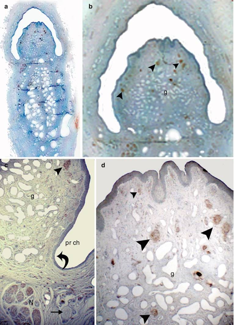
Fig. 9.12
The corpuscles of pleasure in the residual spongy part (lower half of the glans). (a–d) Transverse sections through the RSP, showing the corpuscles of pleasure in the glans (PS100 immuno staining). (a) photomicrograph (low power view), (b) photomicrograph (intermediate power view), (c, d) microscopic aspects (high power view). g glans, N terminal branch of the dorsal nerve of clitoris, pr chpreputial chamber, curved black arrow neck of the glans, right black arrow it shows a corpuscule of Pacini, black arrowheads corpuscules of pleasure (note on d, the different sizes of these corpuscles)
It should also be noted that some authors have classified bulboid tubercles according to their dimensions as very large, large and small tubercles. According to our observations, it appears that the very large tubercles, including some which can reach significant dimensions (Fig. 9.10), are more readily located in the medium intermediate part. The large tubercles occupy medium areas as well as surface areas. Lastly, the small tubercles are generally located in the surface area and many of them are in a subepidermic position (this could mean that it is not necessary for a corpuscle to have a large diameter to be effective, if it is located near to the surface of the receptor area!). The median central area, on the other hand, either has no bulbous corpuscles or a very low content thereof.
At the end of this study, it will be necessary for us to analyse the functional role of these bulbous corpuscles. To do so, we must gather the major observations which we could make:
1.
2.
3.
4.
5.
6.
7.
Overall, our entire study demonstrates, once again, the extraordinary complexity and abundance of sensory receptors in the clitoris, the exceptional richness of this organ in nerve terminations and the incredible density of genital corpuscles over a very small surface.
The clitoris is the most elaborate sensory organ of the female body. It is provided with “specific tactile sensitivity receptors”, which are highly specialised, in connection with the sexual function. With its invaluable sensors, the clitoris has only one function: the reception of pleasure stimuli for a single purpose: to generate sexual excitation in women in all types of contexts (masturbation, foreplay, sexual intercourse).
Footnotes
1
This hypersensitivity is an element, which must be taken into account by a male partner during the prelude to a sexual act, as certain women support direct clitoral stimulation very badly as it generates a truly painful sensation when it is intended to provide pleasure!
2
It is Filippo Pacini (1812–1883), an Italian anatomist, who first described the lamellar corpuscles. However, they had already been observed by Abraham Vater (1684–1751) (a German anatomist)!
3
The lamellar corpuscles have an extreme sensitivity: they are capable of perceiving slight skin deformations (of around a few microns). They are also very sensitive to high-frequency vibrations (around 300 Hz) but have a “vibratory range” from 30 to 1,500 Hz, which is well known by “sex-toys” designers!
4
Two types of erogenous areas are observed in women (R.K. Winkelmann):
· The specific erogenous areas, which are areas whose stimulation produces the strongest sensations and which are necessary to generate an orgasmic reflex: prepuce, clitoris, vulva, perianal skin, labia, oral cavity and breasts (areolas and nipples, which also contain lamellar corpuscles and bulboid corpuscles).
· The non-specific erogenous areas include cutaneous areas with many non-specific receptors and whose simple stimulation can generate sexual excitation with intumescence but which is not sufficient to generate an orgasmic reflex: sides of the neck and nape of the neck, armpits, internal surface of the arms, lateral walls of the thorax, internal surface of the thighs, infra-umbilical area, inguinal region, perinea, fingers, toes, scalp, etc.
5
A slightly different classification was established per H. Jaëger: This author describes 5 types but, on the one hand, his type 1 concerns corpuscles organised in a bundle, comparable to Meissner’s corpuscles, and on the other hand, his types 2 and 3 are comparable to Yamada’s type-2 corpuscles. Finally, his type 4 is identical to Yamada’s type 3 (corpuscle placed in the middle of conjunctive fibres without their own capsule). Lastly, Jaeger isolates a type 5, which he calls the “interposed corpuscles” or intermediate corpuscles, thus named due to the fact that the nerve fibre does not end there and penetrates them to become the terminal fibre of another corpuscle.
6
This multi-innervation concept has been discussed by certain authors, including J.F. Tello “due to the difficulty of distinguishing if they are separate fibres or branches from the same fibre”.
7
In addition to this theory of nerve substance concentration, there is another particularly interesting theory (made by F. Sfameni), whereby the function of each bulbous corpuscle would be to modify the stimulus received by the peripheral free nerve endings, to which it is connected, and transform it into a sensation of pleasure. The granular substance of the corpuscles would contain, for this purpose, an endocrine secretion, produced by the nuclear syncytium, and would therefore play a major part in this modulation of the sensory message.
8
According to D. Perhaps Ohmori, “corpuscles organised in packs may be responsible for the sensations produced by friction or sliding, circumstances which occur with the greatest intensity in the external genitals”. For our part, we believe that the possibility of spontaneous orgasms occurring during sport exercises, such as pedalling on a bicycle or climbing a rope, is due to the extreme abundance of genital corpuscles at the level of the clitoris associated with the corpuscles (scattered but present) of the external genitalia. An interesting study concerning “sexual pleasure induced during exercise (PSIE) and possible orgasms induced during exercise (OIE), in a group of women of all ages”, was published recently and concluded that these situations were very frequent. The authors (D. Herbenick) have called this type of orgasm, induced by exercise, “coregasm”.