
Robert S. Robinson1 and Kathryn J. Woad1
(1)
School of Veterinary Medicine and Science, University of Nottingham, Sutton Bonington Campus, Loughborough, Leicestershire, LE12 5RD, UK
Robert S. Robinson (Corresponding author)
Email: bob.robinson@nottingham.ac.uk
Kathryn J. Woad
Email: katie.woad@nottingham.ac.uk
Abstract
The structure and function of the corpus luteum (CL) is dependent on the development of an intricate vasculature via the process of angiogenesis. The establishment of the luteal vascular network begins in the preovulatory follicle and is ultimately stimulated by the luteinizing hormone (LH) surge. Following ovulation, the corpus luteum undergoes extremely rapid growth and intense angiogenesis that is tightly regulated by a balance achieved between pro-angiogenic and anti-angiogenic factors. This review summarizes what is known about the critical control of luteal angiogenesis and the complex interplay between numerous factors, the functions of which are only just beginning to be elucidated.
Keywords
Corpus luteumLutealAngiogenesisVasculatureFGF2VEGFAEndothelialPericyte
1.1 Critical Importance of Luteal Angiogenesis
The principal function of the corpus luteum (CL) is to produce vast quantities of progesterone , which is absolutely essential for the establishment and maintenance of mammalian pregnancy. Groundbreaking work by the research labs of Hamish Fraser and Dick Stouffer in the early 2000s elegantly demonstrated the wholly crucial role of angiogenesis and the extensive vascularization required for the development and function of the CL. Namely, the inhibition of vascular endothelial growth factor-A (VEGFA, pro-angiogenic factor) with targeted antibody or soluble VEGF receptor isoforms completely prevented luteal vascularization and progesterone production [1–3] in primates. Similarly, in cows, the local neutralization of either VEGFA or fibroblast growth factor-2 (FGF2) also suppressed luteal development and progesterone production, albeit to a lesser extent [4]. Additional evidence of the crucial role of luteal angiogenesis for CL functions has been demonstrated using transgenic, knockout or knock-in mice models in which angiogenesis has been directly or indirectly targeted. Table 1.1summarizes such studies where ovulation appeared normal but CL development and function was disrupted. Overall, compromised plasma progesterone levels were associated with disrupted luteal vascularization and reduced fertility, providing further evidence for the close relationship between luteal angiogenesis and function.
Table 1.1
Summary of knockout mouse studies in which ovulation occurred but corpus luteum (CL) function was disrupted
|
Gene |
Plasma P4 levels |
Steroidogenic enzyme and/or StAR expression |
Luteal vascularization |
Fertility |
Reference |
|
Brain and muscle ARNT-like protein 1 (Bmal1) |
↓ |
↓ ↑ |
↑ VEGFA |
Infertile |
[115] |
|
Cyclin-dependent kinase 4 (cdk4) |
↓ |
Unaffected |
n.d. |
Infertile |
[116] |
|
Endothelial nitric oxide synthase (eNOS) |
↑ |
n.d. |
n.d. |
n.d. |
[117] |
|
Fibroblast growth factor-2 (FGF2) |
n.d. |
n.d. |
n.d. |
Normal |
[117] |
|
Frizzled4 (Fzd4) |
↓ |
↓ |
Fragmented and punctate vascular network ↓ VEGFA |
Infertile |
[118] |
|
Neat1 |
↓ |
↓ |
↓ VEGFA |
Subfertile |
[119] |
|
Nr5a2 (Lrh1) |
↓ |
↓ |
↓ VEGFA |
Infertile |
[120, 121] |
|
Plasminogen |
↓ |
Unaffected |
Unaffected |
n.d. |
[122] |
|
Prolactin |
↓ |
↓ VE-cadherin |
Infertile |
[123, 124] |
|
|
Scavenger receptor-B1 (SCARB1) |
↓ |
n.d. |
No effect |
n.d. |
[125] |
|
Superoxide dismutase (SOD1) |
↓ |
↓ |
↓ Vasculature |
Infertile |
[126] |
|
Tissue inhibitor of metalloproteinase 1 (TIMP1) |
↓ |
n.d. |
n.d. |
n.d. |
[127] |
|
Transforming growth factor-B1 (TGFB1) |
↓ |
↓ |
n.d. |
Infertile |
[128] |
n.d. not determined
The formation of the CL is a truly remarkable biological process with numerous and integrated events occurring in a relatively short timeframe, including (1) the luteinization/differentiation of granulosa and theca cells into luteal cells; (2) a steroidogenesis shift from estradiol to progesterone production that is accompanied with a massive upregulation in steroidogenic output (up to 1000 fold in the cow); (3) extensive remodeling, intermingling of cells (particularly in ruminants), and proliferation of the follicular tissue into a fully developed CL [5]. The bovine CL undergoes rapid growth (Fig. 1.1a), developing from less than 5 mm in size at ovulation to more than 20 mm within 10 days; this equates to a 60- to 100-fold increase in cellular volume, and the luteal growth rate is only matched by the fastest growing tumors [6]. Intriguingly, in the cow the postovulatory rise in progesterone lags behind the growth of the CL by 2–3 days (Fig. 1.1a). It has been speculated that the relatively slower rise in progesterone in ruminants compared to primates might be related to differences in luteal tissue remodeling and vascularization processes [7]. All these processes are totally dependent on intense angiogenesis, or formation of new blood vessels. This intensity is exemplified by the 15-fold increase in the total luteal volume of endothelial cells (EC) during formation of a mature bovine CL (Fig. 1.1b). Not surprisingly, the majority of the proliferating cells in the early CL are of vascular origin, with proliferation indices greater than 25 % [8, 9].

Fig. 1.1
Relationship between corpus luteum (CL) growth, plasma progesterone concentrations , and vasculature in the developing bovine corpus luteum. (A) Rapid increase in CL size after ovulation as determined by transrectal ultrasonography (n = 15). There is a similar increase in plasma progesterone concentrations, but this is initiated 2–3 days later compared to CL growth. (B) Tenfold increase in total luteal vasculature (EC; von Willebrand factor immunohistochemistry) during the formation of the CL (P < 0.01). There is a similar pattern of increase in the total area covered by pericytes (PC: smooth muscle actin immunohistochemistry). Data adapted from references [8, 114]
1.2 Follicular Programming of Luteal Angiogenesis
1.2.1 Preovulatory Follicular Vascular “Seed”
The structural and functional framework for CL development is provided by the preovulatory follicle, which leads to the concept of follicular programming of luteal function [5, 10, 11]. Although less is known about the role of follicular vascularization in this programming, it is entirely feasible that the degree of vascularization within the theca layer of the follicle could determine the rate of subsequent luteal vascularization. It has been proposed that during the follicle–luteal transition endothelial cells form ‘vascular initiation points .’ These points then create a scaffold from which endothelial cells can migrate and proliferate during the reconstruction of the vascular bed [7, 12]. As technologies for measuring microvascular blood flow develop, it will be important to determine the association between follicular vascularization and subsequent luteal function.
The second likely role of the preovulatory follicle in luteal angiogenesis is the active accumulation during folliculogenesis of various pro-angiogenic growth factors [e.g., fibroblast growth factor-2 (FGF2) and vascular endothelial growth factor-A (VEGFA)]. Typically, concentrations of these factors in follicular fluid reach 1 ng/ml for FGF2 [13] and 2–5 ng/ml for VEGFA [14, 15]. It is further likely that these factors are sequestered (e.g., by perlecan) within the follicular basement membrane through their heparin-binding properties [16] and are released following the LH surge-induced activation of proteases during ovulation [17]. Indeed, addition of human follicular fluid to human umbilical vein endothelial cells (HUVECs) dose-dependently increased EC proliferation [18]. Intriguingly, however, heat inactivation or immuno-neutralization of FGF2/VEGFA failed to block this stimulation. Further experiments revealed that the mediator of follicular fluid-induced angiogenesis was the lipid molecule sphingosine-1-phosphate [18]. An alternative candidate is the biologically active phospholipid, lysophosphatidic acid (LPA) , which is produced by granulosa cells and is present in greater concentration in follicular fluid (up to 25 μM) compared to serum (~0.6 μM) [19]. Moreover, 10 μM LPA-treated granulosa-lutein-conditioned media stimulated migration, permeability, and proliferation of HUVECs, and this was mediated through increased interleukin (IL)-6 and -8 production [20]. LPA has also been shown to increase bovine aortic endothelial cell proliferation as well as progesterone production by bovine luteal cells [21].
1.2.2 Initiation by the LH Surge
The LH surge, as well as stimulating the ovulatory process, is also crucial in the upregulation of a plethora of genes, many of which are involved in regulating angiogenesis. Indeed, the size of the endogenous LH surge was positively associated with the degree of luteal vascularization in the developing CL [5]. Hence, the LH surge is often considered to initiate luteal angiogenesis [5]. For example, follicular FGF2 mRNA and protein concentrations are dramatically increased shortly after the LH surge in cows [13, 22, 23]. However, VEGFA expression appears to be less affected by the LH surge [13, 24]. It is still unclear as to the exact effect of the LH surge on VEGFA expression in luteinizing granulosa cells of other species, with some reports of significant stimulation [25, 26], whereas others reported decreased expression [27, 28].
The development of next-generation sequencing has enabled detailed profiling of the effect of the LH surge on the follicular transcriptome. Ingenuity pathway analysis has revealed that the expression of a number of angiogenesis-related genes are regulated by the LH surge: a summary of these genes is shown in Table 1.2. This approach has confirmed several factors already known to stimulate the angiogenic process and/or endothelial cell function (e.g., FGF2, fibronectin, and ephrin B2). It has also revealed several genes associated with tissue remodeling that also affect angiogenesis (e.g., secreted acidic cysteine-rich glycoprotein and transforming growth factor-β1), discussed later. The final and surprising observation was the identity of several upregulated factors classically considered as anti-angiogenic factors (e.g., pentraxin 3, semaphorin 3A/C, thrombospondin 1). It is feasible that this reflects a suppression of angiogenesis in the immediate post-LH surge period that is important until after ovulation has occurred.
Table 1.2
Identification of key angiogenesis-related genes by transcriptomic analyses that are affected by luteinizing hormone (LH) surge/gonadotropin stimulation
|
Gene |
Gene name |
Fold change |
Function |
Reference |
|
ADAM10 |
ADAM metallopeptidase domain 10 |
↑ 2.5-fold (b; GC/TC) |
Cleaves VE-cadherin, enabling EC migration and increasing vasculature permeability |
[129] |
|
ADAMTS1 |
ADAM metallopeptidase with thrombospondin type 1 motif; 1 |
↑ (m) ↑ 9-fold (b) |
Cleaves versican and aggrecan, enabling EC invasion |
[52, 78] |
|
ANGPT1 |
Angiopoietin 1 |
↑ 3-fold (h) |
Promotes vascular stabilization |
[28] |
|
CD24 |
CD24 molecule |
↑ 80-fold (h) |
Pro-angiogenic, and promotes cell invasion |
[28] |
|
CD36 |
CD36 molecule |
↑ 3-fold |
Thrombospondin receptor |
[129] |
|
EFNB2 |
Ephrin B2 |
↑ 16-fold (h) |
Regulates cell adhesion and migration during angiogenesis |
[28] |
|
FGF2 |
Fibroblast growth factor 2 |
↑ 22-fold (b) |
Promotes EC proliferation, migration, and sprouting |
[22] |
|
FN1 |
Fibronectin 1 |
↑ 5-fold (m) ↑ 3-fold (b) ↑ 5-fold (h) |
Component of ECM that promotes EC migration and proliferation |
[28, 52, 78] |
|
ITGA5 |
Integrin, alpha 5 |
↑ 10-fold (h) |
Adhesion molecule; acts as the fibronectin receptor |
[28] |
|
IGFB1 |
Integrin, beta 1 |
↑ 6-fold (m) |
Adhesion molecule; acts as fibronectin receptor |
[52] |
|
PTX3 |
Pentraxin 3 |
↑ 700-fold (b) |
Inhibitor of angiogenesis |
[78] |
|
PDGFBB |
Platelet-derived growth factor BB |
Upstream regulator |
Expressed in EC tip cells and stimulates pericyte recruitment |
[130] |
|
SEMA3A/C |
Semaphorin 3A and 3C |
↑ 16-fold (h) |
Anti-angiogenic and induces EC apoptosis |
[28] |
|
SPARC |
Secreted acidic cysteine-rich glycoprotein |
↑ 7-fold (m) |
Pro- and anti-angiogenic properties |
[52] |
|
THBS2 |
Thrombospondin 2 |
↑ 10-fold (b, GC) ↑ 5-fold (b, TC) |
Potent inhibitor of angiogenesis/VEGFA; inhibits EC proliferation and migration; Induces EC apoptosis |
[78] |
|
TGFB1 |
Transforming growth factor-B1 |
↑ 6-fold (h) Upstream regulator |
Pro- and anti-angiogenic properties |
[28] |
|
VEGFA |
Vascular endothelial growth factor-A |
↓ 2-fold (h) |
Pro-angiogenic |
[28] |
It should be noted that the different studies have collected tissue at different times relative to the LH surge, ranging from 1 to 36 h depending on the study design. Species: b bovine, h human, m mouse; cell type: GC granulosa cell, TC theca cell, EC endothelial cell
Ovulation requires the production of prostaglandins (PG), and treatment with cyclooxygenase-2 (COX-2) inhibitors impairs ovulation [29]. The synthesis of prostaglandins from the granulosa cells is upregulated by the LH surge, reaching peak concentrations just before ovulation [30, 31], and PGE2 has been identified as the key ovulatory prostaglandin, at least in primates [32]. There is increasing evidence that PGE2 is not only crucial for ovulation but that it is also an important stimulator of luteal angiogenesis. Sakurai et al. [33] showed that treatment of rats with the COX-2 inhibitor NS-398 suppressed the formation of luteal vasculature in the newly formed CL as well as decreasing progesterone production. Furthermore, the coadministration of PGE2 reversed this effect. It is likely that PGE2 is acting through the PGE type 2 receptor (PTGER2) , because the PTGER2 antagonist AH6809 suppressed luteal EC tube formation in rats [34]. The PTGER1-4 co-localized with EC within the luteinising granulosa layer 36 h after hCG treatment in primates [32]. Furthermore, intrafollicular injection of PTGER1 and PTGER2 agonists promoted vascularization of the luteinizing granulosa layer, via stimulating branching angiogenesis [32]. An additional complexity is the potential interplay between PGE2 and the key pro-angiogenic growth factors FGF2 [35] and VEGFA [34]; this concept warrants further investigation.
1.3 Control of Luteal Angiogenesis
1.3.1 Local Regulation of Luteal Angiogenesis
1.3.1.1 Fibroblast Growth Factor-2 and Vascular Endothelial Growth Factor-A
Luteal angiogenesis requires the highly coordinated and orchestrated interplay between endothelial and steroidogenic cells as well as fibroblasts and pericytes to create an extensive and complex vascular network that is absolutely essential for luteal function. The best characterized pro-angiogenic regulators of luteal angiogenesis are FGF2 and VEGFA, which are both potent mitogens of vascular endothelial cells (EC) as well as stimulating EC migration and survival. FGF2 is a more potent stimulator of luteal EC proliferation than VEGFA [36], whereas VEGFA also induces vascular permeability [37, 38]. The critical importance of VEGFA for luteal angiogenesis is emphasized in studies where inhibition of VEGFA action massively reduced luteal vascularization and progesterone production in primates [2].
In the developing CL, VEGFA protein is localized predominantly to steroidogenic cells in cows [39] and humans [40], and this is thought to direct angiogenesis toward the hormone-producing cell [41]. Intriguingly, in the very early CL, there are transiently very high levels of FGF2, which return within a few days to basal levels [13, 36]. At the same time, FGF2 protein localization shifts from EC to steroidogenic cells and back again [23], which led us to speculate that FGF2 has a dynamic role in the initiation of luteal angiogenesis in the cow [5].
We have further dissected the function of FGF2 and VEGFA in regulating bovine luteal angiogenesis using a physiologically relevant in vitro system in which multiple luteal cell types (steroidogenic cells, ECs, fibroblasts, pericytes) isolated from a recently ovulated CL are co-cultured in a specialized EC media [42]. Importantly, in this system EC form an intricate network, and the degree of network formation is highly responsive to angiogenic stimuli [42, 43]. Image analysis of these EC networks revealed that there are multiple branch points and interconnections that develop with time in culture [44, 45]. Simultaneously, progesterone production increases over time, and this is responsive to an LH challenge.
The formation of EC networks was suppressed with the addition of specific VEGF receptor (VEGFR) and FGF receptor (FGFR) inhibitors, with EC appearing most sensitive to FGFR inhibition [43]. Using a different approach, the treatment of bovine luteal cells with either FGF2 or VEGFA antibody more or less completely inhibited the formation of EC networks, even when the other angiogenic factor was present (Fig. 1.2). Similarly, FGF2-induced EC proliferation and migration were inhibited by treatment of bovine luteal cells with small molecule 27 (a fragment of thrombospondin that sequesters FGF2) [36]. Further investigation revealed that EC were most sensitive to FGFR inhibition during the time in which islands of EC are starting to sprout/branch [45]. More importantly, FGF2 promoted the precocious transition of undeveloped EC islands into organized EC networks, which was associated with an increased number of EC branch points [44]. The crucial process for EC sprouting is the formation of a specialized EC tip cell within the established vasculature that is capable of migrating toward the angiogenic stimulus. Thus, it is likely that FGFR signaling is crucial for endothelial tip cell formation and vascular sprouting [46]. However, knowledge is still limited about the way FGF2 induces EC tip cells. The effects of intraluteal infusion of FGF2 post ovulation on luteal vasculature and function in vivo certainly warrant investigation.
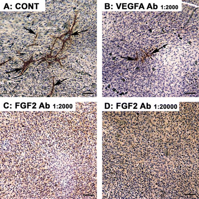
Fig. 1.2
Effect of immuno-neutralization of FGF2 and VEGFA on luteal endothelial cell (EC) networks in a physiologically relevant culture system that mimics luteal bovine angiogenesis and function. Dispersed bovine luteal cells were cultured on fibronectin-coated wells in specialized EC media for 9 days. Immunohistochemistry for von Willebrand factor was performed to identify the EC. (A) Bovine luteal cells treated with control showed extensive EC networks/islands (brown staining, arrows) that resembled a capillary bed. (B) Bovine luteal cells treated with VEGFA antibody at (Ab) 1:2000 dilution. Some reduced EC networks are present. Bovine luteal cells treated with FGF2 antibody at 1:2000 (C) and 1:20,000 (D) had no EC networks. However, there was extensive proliferation of other cell types. Bars 100 μm
The regulation of vascular permeability is a key function of the endothelial cell and is important in the supply of nutrients/hormones to the luteal tissue. As mentioned earlier, VEGFA is a potent stimulator of EC permeability, which is controlled by adherens (e.g., VE-cadherin) and tight junctions (e.g., claudins). Herr et al. [47] showed that hCG induced a VEGFA-dependent downregulation of VE-cadherin and claudin 5 expression, which was associated with increased endothelial cell permeability in a human granulosa-endothelial co-culture system. Additionally, treatment of marmoset monkeys with VEGFA Trap during the mid-luteal phase increased the degree of claudin 5 staining in the CL [48].
1.3.1.2 Interleukins
Interleukin 8 (IL-8) is a neutrophil-specific chemoattractant and pro-angiogenic cytokine that is highly expressed in the early CL, coincident with the abundance of polymorphonuclear neutrophils (PMNs). Moreover, conditioned media from culture of early luteal cells, but not those from a mid-luteal phase CL, stimulated PMN migration in vitro that was blocked with an IL-8 antibody. Importantly, recombinant bovine IL-8 as well as PMN supernatant stimulated luteal EC proliferation and tube formation in vitro [49]. However, Talbott et al. [50] found that IL-8 had no effect on purified luteal endothelial cells derived from early pregnant CL. It is feasible that there is interplay with lysophosphatidic acid (LPA) and IL-8 as LPA induced IL-8 expression in granulosa-lutein cells [20].
1.3.1.3 Secreted Protein Acidic Rich in Cysteine (SPARC)
SPARC is a matrix-associated glycoprotein that regulates cell differentiation, migration, and cell–cell communication. Importantly, the function of SPARC is changed by targeted proteolytic degradation, with the mature SPARC protein being anti-angiogenic and its proteolytic fragments generally being pro-angiogenic [51]. Transcriptomic analysis of hCG-regulated genes in murine granulosa cells revealed that SPARC mRNA was upregulated sevenfold by hCG [52]. The precocious expression of SPARC was dose-dependently induced by transforming growth factor-beta (TGFβ) and fibronectin in bovine luteinizing granulosa cells [53]. Furthermore, SPARC protein expression was abundantly present in luteal and endothelial cells of the developing bovine CL [13, 54]. Functionally, a plasmin proteolytic fragment of SPARC (KGHK) increased EC network formation in vitro and also stimulated progesterone production (to a greater extent than LH) in bovine luteal cells [53]. Thus, SPARC or KGHK-containing peptides could be novel targets for the treatment of luteal inadequacy.
1.3.1.4 Hypoxia
A key driver of angiogenesis during tumor development is the hypoxia-induced upregulation of VEGFA. Cells respond to hypoxia through the activation of hypoxia-inducible factor 1 (HIF1), which is a transcription factor that binds to hypoxia response elements (HRE) in the promoter region of hypoxia-regulated genes. HIF1 is a heterodimer consisting of the oxygen-regulated HIF1A and the constitutively expressed HIF1B [55]. Oxygen concentrations in follicular fluid decrease in the latter stages of antral follicle development [56]. The remodeling of vasculature at ovulation and during early luteal development lowers tissue oxygen concentrations and therefore higher HIF1A would be expected. Indeed, HIF1A is upregulated around the ovulatory period in pigs [57], humans [58], cows [59], and mice [60]. Moreover, echinomycin, a small molecular inhibitor of HIF1 binding to HRE, blocked ovulation in mice [26]. Culturing luteal cells in hypoxic conditions (2–3 % O2) or chemically induced hypoxia increases HIF1A expression and VEGFA [59, 60]. There is also some evidence that hCG/LH directly upregulates HIF1A in granulosa-lutein cells [25, 60]. The relationship between hypoxia and FGF2 expression remains largely unknown. In HUVECs, hypoxic conditions increased FGF2-induced proliferation and tube formation in comparison to normoxia [55]. The exact function of hypoxia in regulating luteal angiogenesis and EC network formation remains to be elucidated.
1.3.1.5 Notch System
The Notch signaling pathway plays an integral role in EC tip formation with the membrane-bound delta-like ligand 4 (DLL4) being expressed in tip cells [61]. When DLL4 binds to its receptor, Notch, on adjoining EC, it converts them into stalk cells [62]. DLL4 and Notch1–4 have been detected in endothelial and steroidogenic cells of developing CL in mice [63, 64]. In mice, treatment with a γ-secretase inhibitor (to block downstream Notch signaling) impaired preovulatory follicle development: this was associated with the theca layer having a disorganized EC framework and an increased vascular smooth muscle cell density. Furthermore, plasma estradiol concentrations were nearly threefold lower [65]. In the same model, treatment with a DLL4-blocking antibody had minimal effects on follicular appearance, although in marmosets, inhibition of DLL4 during the periovulatory period caused the CL to be hypervascularized and decreased progesterone production [66]. This EC patterning has several similarities to that induced by high concentrations of FGF2 in bovine luteal cells [44]. Recent observations showed that the treatment of luteal cells with a γ-secretase inhibitor reduced progesterone concentrations in rats [67]. The potential interplay between FGF2, VEGFA, and the Notch system warrants investigation.
1.3.2 Role of Anti-Angiogenic Factors
1.3.2.1 Thrombospondin (THBS)
Thrombospondin-1 and -2 are large glycoproteins secreted by several cell types that bind to the extracellular matrix (ECM). THBS, acting through its receptor CD36, regulates several processes in EC including migration, adhesion, and apoptosis. Contrary to expectation, the expression of THBS1, THBS2, and CD36 was greatest during the early luteal phase in rats [68, 69]. FGF2 and THBS1 had opposing actions on bovine luteal EC in vitro, with THBS1 reducing cell numbers by inducing EC apoptosis [70]. Furthermore, THBS1 inhibited FGF2-induced luteal EC migration and proliferation [36] and THBS/FGF2 downregulate the opposing gene [70]. These in vitro observations are in agreement with the effect of the thrombospondin-mimetic peptide, ABT898 , on follicular angiogenesis in marmoset monkeys [71]. However, in the same study ABT898 had no effect on ovulation or plasma progesterone concentrations [71].
1.3.2.2 Vascular Endothelial Growth Factor A : ‘b’ Isoform
The alternative splicing of exon 8 in the VEGFA gene yields two different families of VEGFA isoforms. The first family is the classic pro-angiogenic isoform with exon 8a, and the other is the anti-angiogenic isoform with exon 8b present. This isoform is termed VEGFAxxxb [72]. Relatively little is known about this additional complexity, and most VEGFA antibodies do not distinguish between these two different isoform families. Recently, it was reported that the VEGFA120b isoform was expressed in parallel to VEGFA120a, with expression increasing with CL age, whereas VEGFA164b was not detected in the ovine CL [73]. Importantly, mice overexpressing VEGFA164b had reduced fertility, CL number, and degree of vascularization within the CL [74], further emphasizing the importance of the balance between pro- and anti-angiogenic growth factors during luteal development.
1.3.2.3 Vasohibin 1
Vasohibin 1 (VASH1) and IGF-binding protein 7 (IGFBP7) are another two recently identified negative feedback regulators of vascularization. VASH1 is constantly expressed throughout bovine luteal development in luteal endothelial cells, and its expression is upregulated by VEGFA [75]. In the cow, VASH1 suppressed VEGFA-induced luteal EC tube formation [75], suggesting that it could prevent overstimulation of angiogenesis. The high-affinity insulin, low-affinity IGF1-binding protein, IGFBP7, regulates cell proliferation, adhesion, and angiogenesis. It has been detected in the follicular fluid and corpora lutea of rats [76], but to date has not been reported in other species. Recently, IGFBP7 (at 160 ng/ml) was shown to reduce VEGFA- and LH-stimulated luteal EC tube formation, but it had no effect under basal conditions [77]. Lower concentrations of IGFBP7 had no effect [77], and the physiological role of IGFBP7 in regulating luteal angiogenesis remains to be determined.
1.3.2.4 Pentraxin 3
Pentraxin 3 (PTX3) is a 45-kDa glycosylated protein that is produced by endothelial cells and activated phagocytes. It is known to bind FGF2 with a high affinity and thus prevents FGF2 from binding to FGFR, blocking its angiogenic actions [37]. The massive upregulation of PTX3 in bovine granulosa cells following the LH surge [78] shortly before a period of intense angiogenesis seems counterintuitive, especially because PTX3 inhibited FGF2 action on bovine luteal ECs [36]. Interestingly, PTX3 expression is greatly increased in the mature CL following PGF2α administration , suggesting that PTX3 has an anti-angiogenic function in luteolysis [36].
1.4 Establishment of Luteal Vasculature
1.4.1 VE-Cadherin
VE-cadherin is an endothelial-specific molecule that forms adherens junctions between adjacent endothelial cells. VE-cadherin not only maintains vascular integrity but also regulates cellular processes such as EC proliferation, apoptosis, and VEGFR function [79]. It was recently discovered that Notch/VEGFR signaling alters the dynamics of VE-cadherin junctions that drive endothelial rearrangements during sprouting [80]. The immuno-neutralization of VE-cadherin with E4G10 antibody in mice reduced CL development, degree of vascularization, and plasma progesterone concentrations [81], highlighting the critical importance of VE-cadherin for luteal angiogenesis. Treatment of bovine CLENDO cells with TGFB1 caused the loss of VE-cadherin from cell junctions, reduced cell–cell contacts, and increased EC permeability [82]. Conversely, FGF2 signaling promotes VE-cadherin expression and maintains vascular integrity [83].
1.4.2 Pericytes
Pericytes (mural cells) form intimate contacts with endothelial cells and are an integral component of the microvasculature. Classically, pericytes are considered to be involved in the latter stages of angiogenesis by stabilizing newly formed EC tubes. It is increasingly evident that pericytes are also active in the early stages of angiogenesis [84]. The induction of platelet-derived growth factor receptor-B (PDGFRB) expression activates pericytes and stimulates their recruitment to endothelial cells. Indeed, intraovarian PDGFR blockade reduced the number of CL and progesterone production [85, 86] as well reducing the microvessel EC density by nearly 50 %, as well as pericyte coverage of those vessels [86]. It has been observed that pericytes appear to migrate into the luteinizing-granulosa layer “ahead” of EC in the developing sheep [87] and cattle CL [7, 88]. Furthermore, in the developing CL, pericytes are in abundance (Fig. 1.1b) and form a large proportion of proliferating cells [87]. Pericytes could have several functions to support the intense luteal angiogenesis, including (1) laying down fibronectin strands along which EC can migrate [7]; (2) increased migratory phenotype through the suppression of contractile vascular smooth muscle cell [89]; and (3) making multiple contacts between EC that assist with their stabilization and thus co-ordinating the luteal vascularization. This idea is supported by the observation that smooth muscle actin (SMA) -positive mural cells are often localized in close proximity to EC islands and are often an integral component of these islands [5]. Furthermore, one particular mural phenotype has several finger-like projections that connect to EC and other mural cells [5]. Importantly, Woad et al. [43] demonstrated that in vitro PDGFRB blockade with a receptor tyrosine kinase inhibitor greatly attenuated the ability of EC to develop into networks with the earliest stages most sensitive to PDGFRB inhibition.
1.4.3 Insulin-Like Growth Factor (IGF) System
Insulin-like growth factor-1 (IGF1) is a well-established endocrine and paracrine growth factor that is known to be important in regulating follicular and luteal function. IGF1 is locally expressed in the CL [90] and has a stimulatory effect on progesterone secretion [91]. It is also known to have pro-angiogenic properties [92, 93], particularly in respect to developmental angiogenesis and neovascularization [94]. Recent evidence has shown that IGF1 promotes angiogenesis by stabilizing endothelial cell tubes and nascent blood vessels in the retina in response to VEGFA [95]. The latter might be particularly important for luteal angiogenesis because the neovascularization of the CL initially involves the destabilization of the vasculature within the theca layer. Knowledge is limited about the role of IGF1 in regulating ovarian angiogenesis; however, IGF1 decreased thrombospondin 1 expression in porcine granulosa cells [96].
1.5 Clinical Opportunities
Appropriate vascularization is critical to normal ovarian function. Dysregulated vascular growth has been implicated in the origin or development of several ovarian pathologies and is therefore a promising target for the treatment of disease.
1.5.1 Ovarian Cancer
Ovarian cancer , primarily epithelial in type, is a leading cause of female cancer death and the most aggressive of the gynecological cancers. More than 200,000 women develop epithelial ovarian cancer worldwide each year, with most cases diagnosed in women over 55 years of age. It is characterized by a high death rate, largely attributed to the late presentation of many cases and hence associated metastatic disease. Most cases of advanced disease also recur, with disease-free intervals becoming progressively shorter [97]. Standard clinical management is by surgery and platinum-based cytotoxic chemotherapy; however, new therapeutic strategies are emerging, including anti-angiogenic treatment [98].
Tumor blood vessels exhibit multiple abnormalities of structure and function and are especially dynamic [99]. Tumor growth and eventual metastasis require active angiogenesis, resulting from both the upregulation of pro-angiogenic factors and downregulation of endogenous inhibitors, thereby presenting multiple opportunities for therapeutic intervention.
Advanced epithelial ovarian carcinoma is associated with raised serum VEGFA levels [100], and VEGFA has been the primary therapeutic target to date: approaches include using antibodies against VEGFA or VEGFR, soluble decoy receptors (VEGF Trap), or VEGFR tyrosine kinase inhibitors. Phase 3 trials have demonstrated increased progression-free survival in women with ovarian cancer treated with VEGF inhibition (bevacizumab) in addition to standard chemotherapy [101, 102]. The benefit of anti-VEGFA therapy was evident as first-line treatment, maintenance therapy, or at recurrence, although the impact on overall survival is less clear. Further advances are expected in the use of multi-angiokinase inhibitors that target tumor angiogenesis at multiple levels, for example, via combined VEGF, FGF, and PDGF signaling inhibition [103, 104].
Another promising target is the angiopoietin/Tie2 pathway [105, 106]. Treatment with Trebananib (AMG 386), which binds Angpt1 and -2, thereby preventing Tie2 activation, prolonged progression-free survival in women with ovarian cancer [107]. Opportunities to exploit the endogenous inhibition of angiogenesis are also being explored. For example, thrombospondin-mediated inhibition of angiogenesis enhanced the clinical effectiveness of chemotherapy in a mouse model of ovarian cancer by altering vascular morphology, facilitating drug uptake, and increasing apoptotic cell death [108].
1.5.2 Ovarian Hyperstimulation Syndrome
Ovarian hyperstimulation syndrome (OHSS) is a rare but potentially life-threatening complication of ovarian stimulation for fertility treatment. It occurs during the luteal phase or early pregnancy [109] and is characterized by a systemic increase in vascular permeability, thought to result from the ovarian secretion of vasoactive peptides such as VEGFA. VEGFA is implicated because of an association of OHSS with raised luteal and follicular fluid VEGFA expression and bioavailability [110] and the stimulation of vascular permeability in response to VEGFA [111]. Despite the potential importance of VEGFA in mediating OHSS, targeting VEGFA directly led to undesirable side effects in animal models, and hence most preventative treatments have targeted the stimulation protocol itself, such as reducing the duration of the LH surge. An attractive alternative clinical strategy to reduce the incidence and severity of OHSS is the use of dopamine agonists acting as VEGF inhibitors [112]. Treatment of high-risk women with dopamine agonists lowered the incidence of OHSS without detrimental effects on implantation or pregnancy outcome following assisted reproduction [113], despite the clear need for ovarian and endometrial angiogenesis.
1.6 Conclusion
Angiogenesis is critical to support the dramatic growth and development of the CL. Although VEGFA and FGF2 may be seen as the primary regulators of luteal angiogenesis, it is increasingly evident that numerous other factors are expressed by several luteal cell types and that these also have important modulatory functions. Transcriptomic studies are shedding new light on the complexities of luteal angiogenesis at the molecular level, and a better understanding of the control of luteal angiogenesis will also highlight potential new therapeutic opportunities to tackle angiogenesis-dependant ovarian disease.
Acknowledgments
This work has been funded by BBSRC, Pfizer and University of Nottingham. We greatly appreciate the technical assistance of staff at the University of Nottingham without which this work would not have been possible.
References
1.
Fraser HM, Dickson SE, Lunn SF, Wulff C, Morris KD, Carroll VA, et al. Suppression of luteal angiogenesis in the primate after neutralization of vascular endothelial growth factor. Endocrinology. 2000;141:995–1000.PubMed
2.
Wulff C, Wiegand SJ, Saunders PTK, Scobie GA, Fraser HM. Angiogenesis during follicular development in the primate and its inhibition by treatment with truncated Flt-1-Fc (vascular endothelial growth factor Trap(A40)). Endocrinology. 2001;142:3244–54.PubMed
3.
Hazzard TM, Xu FH, Stouffer RL. Injection of soluble vascular endothelial growth factor receptor 1 into the preovulatory follicle disrupts ovulation and subsequent luteal function in rhesus monkeys. Biol Reprod. 2002;67:1305–12.PubMed
4.
Yamashita H, Kamada D, Shirasuna K, Matsui M, Shimizu T, Kida K, et al. Effect of local neutralization of basic fibroblast growth factor or vascular endothelial growth factor by a specific antibody on the development of the corpus luteum in the cow. Mol Reprod Dev. 2008;75:1449–56.PubMed
5.
Robinson RS, Woad KJ, Hunter MG, Sinclair KD, Laird M, Joseph C, et al. Corpus luteum development and angiogenesis. In: Juengel JL, Miyamoto A, Price C, Reynolds LP, Smith MF, Webb R, editors. Reproduction in domestic ruminants VIII. Ashby-de-la-Zouch: Context Products; 2014. p. 327–86.
6.
Reynolds L, Redmer D. Growth and development of the corpus luteum. J Reprod Fertil Suppl. 1999;54:181–91.PubMed
7.
Robinson RS, Woad KJ, Hammond AJ, Laird M, Hunter MG, Mann GE. Angiogenesis and vascular function in the ovary. Reproduction. 2009;138:869–81.PubMed
8.
Robinson RS, Hammond AJ, Nicklin LT, Schams D, Mann GE, Hunter MG. Endocrine and cellular characteristics of corpora lutea from cows with a delayed post-ovulatory progesterone rise. Domest Anim Endocrinol. 2006;31:154–72.PubMed
9.
Hojo T, Al-Zi'Abi MO, Skarzynski DJ, Acosta TJ, Okuda K. Changes in the vasculature of bovine corpus luteum during the estrous cycle and prostaglandin F2 alpha-induced luteolysis. J Reprod Dev. 2009;55:512–7.PubMed
10.
Inskeep EK. Preovulatory, postovulatory, and postmaternal recognition effects of concentrations of progesterone on embryonic survival in the cow. J Anim Sci. 2004;82(E-Suppl):E24–E39.
11.
Lonergan P. Influence of progesterone on oocyte quality and embryo development in cows. Theriogenology. 2011;76:1594–601.PubMed
12.
Kaessmeyer S, Plendl J. Angiogenesis and vasculogenesis in the corpus luteum in vitro. Clin Hemorheol Microcirc. 2009;41:83–101.PubMed
13.
Robinson RS, Nicklin LT, Hammond AJ, Schams D, Hunter MG, Mann GE. Fibroblast growth factor 2 is more dynamic than vascular endothelial growth factor A during the follicle-luteal transition in the cow. Biol Reprod. 2007;77:28–36.PubMed
14.
Berisha B, Schams D, Kosmann M, Amselgruber W, Einspanier R. Expression and localisation of vascular endothelial growth factor and basic fibroblast growth factor during the final growth of bovine ovarian follicles. J Endocrinol. 2000;167:371–82.PubMed
15.
Kaya A, Atabekoglu CS, Kahraman K, Taskin S, Ozmen B, Berker B, et al. Follicular fluid concentrations of IGF-I, IGF-II, IGFBP-3, VEGF, AMH, and inhibin-B in women undergoing controlled ovarian hyperstimulation using GnRH agonist or GnRH antagonist. Eur J Obstet Gynecol Reprod Biol. 2012;164:167–71.PubMed
16.
Rodgers RJ, Irving-Rodgers HF, Russell DL. Extracellular matrix of the developing ovarian follicle. Reproduction. 2003;126:415–24.PubMed
17.
Curry TE, Smith MF. Impact of extracellular matrix remodeling on ovulation and the folliculo-luteal transition. Semin Reprod Med. 2006;24:228–41.PubMed
18.
von Otte S, Paletta JRJ, Becker S, König S, Fobker M, Greb RR, et al. Follicular fluid high density lipoprotein-associated sphingosine 1-phosphate Is a novel mediator of ovarian angiogenesis. J Biol Chem. 2006;281:5398–405.
19.
Tokumura A, Miyake M, Nishioka Y, Yamano S, Aono T, Fukuzawa K. Production of lysophosphatidic acids by lysophospholipase D in human follicular fluids of in vitro fertilization patients. Biol Reprod. 1999;61:195–9.PubMed
20.
Chen SU, Chou CH, Lee HY, Ho CH, Lin CW, Yang YS. Lysophosphatidic acid up-regulates expression of interleukin-8 and-6 in granulosa-lutein cells through its receptors and nuclear factor-kappa B dependent pathways: implications for angiogenesis of corpus luteum and ovarian hyperstimulation syndrome. J Clin Endocrinol Metab. 2008;93:935–43.PubMed
21.
Skarzynski DJ, Piotrowska-Tomala KK, Lukasik K, Galvao A, Farberov S, Zalman Y, et al. Growth and regression in bovine corpora lutea: regulation by local survival and death pathways. Reprod Domest Anim. 2013;48:25–37.PubMed
22.
Gilbert I, Robert C, Dieleman S, Blondin P, Sirard MA. Transcriptional effect of the LH surge in bovine granulosa cells during the peri-ovulation period. Reproduction. 2011;141:193–205.PubMed
23.
Berisha B, Steffl M, Amselgruber W, Schams D. Changes in fibroblast growth factor 2 and its receptors in bovine follicles before and after GnRH application and after ovulation. Reproduction. 2006;131:319–29.PubMed
24.
Berisha B, Steffl M, Welter H, Kliem H, Meyer HHD, Schams D, et al. Effect of the luteinising hormone surge on regulation of vascular endothelial growth factor and extracellular matrix-degrading proteinases and their inhibitors in bovine follicles. Reprod Fertil Dev. 2008;20:258–68.PubMed
25.
van den Driesche S, Myers M, Gay E, Thong KJ, Duncan WC. HCG up-regulates hypoxia inducible factor-1 alpha in luteinized granulosa cells: implications for the hormonal regulation of vascular endothelial growth factor A in the human corpus luteum. Mol Hum Reprod. 2008;14:455–64.PubMed
26.
Kim J, Bagchi IC, Bagchi MK. Signaling by hypoxia-inducible factors is critical for ovulation in mice. Endocrinology. 2009;150:3392–400.PubMedPubMedCentral
27.
Xu F, Stouffer RL, Müller J, Hennebold JD, Wright JW, Bahar A, et al. Dynamics of the transcriptome in the primate ovulatory follicle. Mol Hum Reprod. 2011;17:152–65.PubMed
28.
Wissing ML, Kristensen SG, Andersen CY, Mikkelsen AL, Høst T, Borup R, et al. Identification of new ovulation-related genes in humans by comparing the transcriptome of granulosa cells before and after ovulation triggering in the same controlled ovarian stimulation cycle. Hum Reprod. 2014;29:997–1010.PubMed
29.
Duffy DM (2015) Novel contraceptive targets to inhibit ovulation: the prostaglandin E2 pathway. Hum Reprod Update 21(5):652–670. doi:10.1093/humupd/dmv026.
30.
Fortune JE, Willis EL, Bridges PJ, Yang CS. The periovulatory period in cattle: progesterone, prostaglandins, oxytocin and ADAMTS proteases. Anim Reprod Sci. 2009;6:60–71.
31.
Duffy DM, Stouffer RL. The ovulatory gonadotrophin surge stimulates cyclooxygenase expression and prostaglandin production by the monkey follicle. Mol Hum Reprod. 2001;7:731–9.PubMed
32.
Trau HA, Davis JS, Duffy DM. Angiogenesis in the primate ovulatory follicle is stimulated by luteinizing hormone via prostaglandin E2. Biol Reprod. 2015;92(15):1–2.
33.
Sakurai T, Tamura K, Kogo H. Stimulatory effects of eicosanolds on ovarian angiogenesis in early luteal phase in cyclooxygenase-2 inhibitor-treated rats. Eur J Pharmacol. 2005;516:158–64.PubMed
34.
Sakurai T, Suzuki K, Yoshie M, Hashimoto K, Tachikawa E, Tamura K. Stimulation of tube formation mediated through the prostaglandin EP2 receptor in rat luteal endothelial cells. J Endocrinol. 2011;209:33–43.PubMed
35.
Finetti F, Donnini S, Giachetti A, Morbidelli L, Ziche M. Prostaglandin E(2) primes the angiogenic switch via a synergic interaction with the fibroblast growth factor-2 pathway. Circ Res. 2009;105:657–66.PubMed
36.
Zalman Y, Klipper E, Farberov S, Mondal M, Wee G, Folger JK, et al. Regulation of angiogenesis-related prostaglandin F2-alpha-induced genes in the bovine corpus luteum. Biol Reprod. 2012;86:92.PubMed
37.
Presta M, Dell’Era P, Mitola S, Moroni E, Ronca R, Rusnati M. Fibroblast growth factor/fibroblast growth factor receptor system in angiogenesis. Cytokine Growth Factor Rev. 2005;16:159–78.PubMed
38.
Ferrara N, Chen H, Davis-Smyth T, Gerber HP, Nguyen TN, Peers D, et al. Vascular endothelial growth factor is essential for corpus luteum angiogenesis. Nat Med. 1998;4:336–40.PubMed
39.
Berisha B, Schams D, Kosmann M, Amselgruber W, Einspanier R. Expression and tissue concentration of vascular endothelial growth factor, its receptors, and localization in the bovine corpus luteum during estrous cycle and pregnancy. Biol Reprod. 2000;63:1106–14.PubMed
40.
Sugino N, Kashida S, Takiguchi S, Karube A, Kato H. Expression of vascular endothelial growth factor and its receptors in the human corpus luteum during the menstrual cycle and in early pregnancy. J Clin Endocrinol Metab. 2000;85:3919–24.PubMed
41.
Fraser HM, Lunn SF. Regulation and manipulation of angiogenesis in the primate corpus luteum. Reproduction. 2001;121:355–62.PubMed
42.
Robinson RS, Hammond AJ, Mann GE, Hunter MG. A novel physiological culture system that mimics luteal angiogenesis. Reproduction. 2008;135:405–13.PubMed
43.
Woad KJ, Hammond AJ, Hunter M, Mann GE, Hunter MG, Robinson RS. FGF2 is crucial for the development of bovine luteal endothelial networks in vitro. Reproduction. 2009;138:581–8.PubMed
44.
Laird M, Woad KJ, Hunter MG, Mann GE, Robinson RS. Fibroblast growth factor 2 induces the precocious development of endothelial cell networks in bovine luteinising follicular cells. Reprod Fertil Dev. 2013;25:372–86.PubMed
45.
Woad KJ, Hunter MG, Mann GE, Laird M, Hammond AJ, Robinson RS. Fibroblast growth factor 2 is a key determinant of vascular sprouting during bovine luteal angiogenesis. Reproduction. 2012;143:35–43.PubMed
46.
De Smet F, Segura I, De Bock K, Hohensinner PJ, Carmeliet P. Mechanisms of vessel branching filopodia on endothelial tip cells lead the way. Arterioscler Thromb Vasc Biol. 2009;29:639–49.PubMed
47.
Herr D, Fraser HM, Konrad R, Holzheu I, Kreienberg R, Wulff C. Human chorionic gonadotropin controls luteal vascular permeability via vascular endothelial growth factor by down-regulation of a cascade of adhesion proteins. Fertil Steril 2013;99:1749–1758
48.
Rodewald M, Herr D, Fraser HM, Hack G, Kreienberg R, Wulff C. Regulation of tight junction proteins occludin and claudin 5 in the primate ovary during the ovulatory cycle and after inhibition of vascular endothelial growth factor. Mol Hum Reprod. 2007;13:781–9.PubMed
49.
Jiemtaweeboon S, Shirasuna K, Nitta A, Kobayashi A, Schuberth HJ, Shimizu T, et al. Evidence that polymorphonuclear neutrophils infiltrate into the developing corpus luteum and promote angiogenesis with interleukin-8 in the cow. Reprod Biol Endocrinol. 2011;9:79.PubMedPubMedCentral
50.
Talbott H, Delaney A, Zhang P, Yu Y, Cushman R, Cupp AS, et al. Effects of IL8 and immune cells on the regulation of luteal progesterone secretion. Reprod 2014; 148:21–31.
51.
Yan Q, Sage EH. SPARC, a matricellular glycoprotein with important biological functions. J Histochem Cytochem. 1999;47:1495–506.PubMed
52.
McRae RS, Johnston HM, Mihm M, O’Shaughnessy PJ. Changes in mouse granulosa cell gene expression during early luteinization. Endocrinology. 2005;146:309–17.PubMed
53.
Joseph C, Hunter MG, Sinclair KD, Robinson RS. The expression, regulation and function of secreted protein, acidic, cysteine-rich in the follicle-luteal transition. Reproduction. 2012;144:361–72.PubMed
54.
Wiltbank MC, Salih SM, Atli MO, Luo W, Bormann CL, Ottobre JS, et al. Comparison of endocrine and cellular mechanisms regulating the corpus luteum of primates and ruminants. Anim Reprod. 2012;9:242–59.PubMedPubMedCentral
55.
Meidan R, Klipper E, Zalman Y, Yalu R. The role of hypoxia-induced genes in ovarian angiogenesis. Reprod Fertil Dev. 2013;25:343–50.PubMed
56.
Nishimura R, Okuda K. Multiple roles of hypoxia in ovarian function: roles of hypoxia-inducible factor-related and -unrelated signals during the luteal phase. Reprod Fertil Dev. 2015. doi:10.1071/RD15010.
57.
Boonyaprakob U, Gadsby JE, Hedgpeth V, Routh PA, Almond GW. Expression and localization of hypoxia inducible factor-1 alpha mRNA in the porcine ovary. Can J Vet Res. 2005;69:215–22.PubMedPubMedCentral
58.
Duncan WC, van den Driesche S, Fraser HM. Inhibition of vascular endothelial growth factor in the primate ovary up-regulates hypoxia-inducible factor-1 alpha in the follicle and corpus luteum. Endocrinology. 2008;149:3313–20.PubMed
59.
Nishimura R, Okuda K. Hypoxia is important for establishing vascularization during corpus luteum formation in cattle. J Reprod Dev. 2010;56:110–6.PubMed
60.
Tam KK, Russell DL, Peet DJ, Bracken CP, Rodgers RJ, Thompson JG, et al. Hormonally regulated follicle differentiation and luteinization in the mouse is associated with hypoxia inducible factor activity. Mol Cell Endocrinol. 2010;327:47–55.PubMed
61.
Gridley T. Notch signaling in vascular development and physiology. Development (Camb). 2007;134:2709–18.
62.
Eilken HM, Adams RH. Dynamics of endothelial cell behavior in sprouting angiogenesis. Curr Opin Cell Biol. 2010;22:617–25.PubMed
63.
Murta D, Batista M, Silva E, Trindade A, Mateus L, Duarte A, et al. Differential expression of Notch component and effector genes during ovarian follicle and corpus luteum development during the oestrous cycle. Reprod Fertil Dev. 2015;27(7):1038–1048. doi:10.1071/RD13399.
64.
Vorontchikhina MA, Zimmermann RC, Shawber CJ, Tang HY, Kitajewski J. Unique patterns of Notch1, Notch4 and Jagged1 expression in ovarian vessels during folliculogenesis and corpus luteum formation. Gene Expr Patterns. 2005;5:701–9.PubMed
65.
Jovanovic VP, Sauer CM, Shawber CJ, Gomez R, Wang X, Sauer MV, et al. Intraovarian regulation of gonadotropin-dependent folliculogenesis depends on notch receptor signaling pathways not involving Delta-like ligand 4 (Dll4). Reprod Biol Endocrinol. 2013;11:43.PubMedPubMedCentral
66.
Fraser HM, Hastings JM, Allan D, Morris KD, Rudge JS, Wiegand SJ. Inhibition of delta-like ligand 4 induces luteal hypervascularization followed by functional and structural luteolysis in the primate ovary. Endocrinology. 2012;153:1972–83.PubMedPubMedCentral
67.
Accialini P, Hernandez SF, Bas D, Pazos MC, Irusta G, Abramovich D, et al. A link between Notch and progesterone maintains the functionality of the rat corpus luteum. Reproduction. 2015;149:1–10.PubMed
68.
Petrik JJ, Gentry PA, Feige JJ, LaMarre J. Expression and localization of thrombospondin-1 and-2 and their cell-surface receptor, CD36, during rat follicular development and formation of the corpus luteum. Biol Reprod. 2002;67:1522–31.PubMed
69.
Bagavandoss P, Sage EH, Vernon RB. Secreted protein, acidic and rich in cysteine (SPARC) and thrombospondin in the developing follicle and corpus luteum of the rat. J Histochem Cytochem. 1998;46:1043–9.PubMed
70.
Farberov S, Meidan R. Functions and transcriptional regulation of thrombospondins and their interrelationship with fibroblast growth factor-2 in bovine luteal cells. Biol Reprod. 2014;91:58.PubMed
71.
Garside SA, Henkin J, Morris KD, Norvell SM, Thomas FH, Fraser HM. A thrombospondin-mimetic peptide, ABT-898, suppresses angiogenesis and promotes follicular atresia in pre- and early-antral follicles in vivo. Endocrinology. 2010;151:5905–15.PubMed
72.
Bates DO, Harper SJ. Anti-angiogenic isoforms of VEGF-A: key to anti-angiogenic therapy. Anticancer Res. 2008;28:3207–8.
73.
Guzman A, Macias-Valencia R, Fierro-Fierro F, Gutierrez CG, Rosales-Torres AM. The corpora lutea proangiogenic state of VEGF system components is turned to antiangiogenic at the later phase of the oestrous cycle in cows. Animal. 2015;9:301–7.PubMed
74.
Qiu Y, Seager M, Osman A, Castle-Miller J, Bevan H, Tortonese DJ, et al. Ovarian VEGF(165)B expression regulates follicular development, corpus luteum function and fertility. Reproduction. 2012;143:501–11.PubMedPubMedCentral
75.
Shirasuna K, Kobayashi A, Nitta A, Nibuno S, Sasahara K, Shimizu T, et al. Possible action of vasohibin-1 as an inhibitor in the regulation of vascularization of the bovine corpus luteum. Reproduction. 2012;143:491–500.PubMed
76.
Tamura K, Matsushita M, Endo A, Kutsukake M, Kogo H. Effect of insulin-like growth factor-binding protein 7 on steroidogenesis in granulosa cells derived from equine chorionic gonadotropin-primed immature rat ovaries. Biol Reprod. 2007;77:485–91.PubMed
77.
Tamura K, Yoshie M, Hashimoto K, Tachikawa E. Inhibitory effect of insulin-like growth factor-binding protein-7 (IGFBP7) on in vitro angiogenesis of vascular endothelial cells in the rat corpus luteum. J Reprod Dev. 2014;60:447–53.PubMedPubMedCentral
78.
Christenson LK, Gunewardena S, Hong X, Spitschak M, Baufeld A, Vanselow J. Research resource: preovulatory LH surge effects on follicular theca and granulosa transcriptomes. Mol Endocrinol. 2013;27:1153–71.PubMedPubMedCentral
79.
Vestweber D. VE-cadherin: the major endothelial adhesion molecule controlling cellular junctions and blood vessel formation. Arterioscler Thromb Vasc Biol. 2008;28:223–32.PubMed
80.
Bentley K, Franco CA, Philippides A, Blanco R, Dierkes M, Gebala V, et al. The role of differential VE-cadherin dynamics in cell rearrangement during angiogenesis. Nat Cell Biol. 2014;16:309–21.PubMed
81.
Nakhuda GS, Zimmermann RC, Bohlen P, Liao F, Sauer MV, Kitajewski J. Inhibition of the vascular endothelial cell (VE)-specific adhesion molecule VE-cadherin blocks gonadotropin-dependent folliculogenesis and corpus luteum formation and angiogenesis. Endocrinology. 2005;146:1053–9.PubMed
82.
Maroni D, Davis JS. TGFB1 disrupts the angiogenic potential of microvascular endothelial cells of the corpus luteum. J Cell Sci. 2011;124:2501–10.PubMed
83.
Murakami M, Nguyen LT, Zhuang ZW, Moodie KL, Carmeliet P, Stan RV, et al. The FGF system has a key role in regulating vascular integrity. J Clin Invest. 2008;118:3355–66.PubMedPubMedCentral
84.
Ozerdem U, Stallcup WB. Early contribution of pericytes to angiogenic sprouting and tube formation. Angiogenesis. 2003;6:241–9.PubMedPubMedCentral
85.
Sleer LS, Taylor CC. Platelet-derived growth factors and receptors in the rat corpus luteum: localization and identification of an effect on luteogenesis. Biol Reprod. 2007;76:391–400.PubMed
86.
Kuhnert F, Tam BYY, Sennino B, Gray JT, Yuan J, Jocson A, et al. Soluble receptor-mediated selective inhibition of VEGFR and PDGFR beta signaling during physiologic and tumor angiogenesis. Proc Natl Acad Sci USA. 2008;105:10185–90.PubMedPubMedCentral
87.
Redmer DA, Doraiswamy V, Bortnem BJ, Fisher K, Jablonka-Shariff A, Grazul-Bilska AT, et al. Evidence for a role of capillary pericytes in vascular growth of the developing ovine corpus luteum. Biol Reprod. 2001;65:879–89.PubMed
88.
Amselgruber WM, Schafer M, Sinowatz F. Angiogenesis in the bovine corpus luteum: an immunocytochemical and ultrastructural study. Anat Histol Embryol. 1999;28:157–66.PubMed
89.
Papetti M, Shujath J, Riley KN, Herman IM. FGF-2 antagonizes the TGF-beta1-mediated induction of pericyte alpha-smooth muscle actin expression: a role for myf-5 and Smad-mediated signaling pathways. Invest Ophthalmol Vis Sci. 2003;44:4994–5005.PubMed
90.
Woad KJ, Baxter G, Hogg CO, Bramley TA, Webb R, Armstrong DG. Expression of mRNA encoding insulin-like growth factors I and II and the type 1 IGF receptor in the bovine corpus luteum at defined stages of the oestrous cycle. J Reprod Fertil. 2000;120:293–302.PubMed
91.
Schams D, Kosmann M, Berisha B, Amselgruber WM, Miyamoto A. Stimulatory and synergistic effects of luteinising hormone and insulin like growth factor 1 on the secretion of vascular endothelial growth factor and progesterone of cultured bovine granulosa cells. Exp Clin Endocrinol Diabetes. 2001;109:155–62.PubMed
92.
Plendl J. Angiogenesis and vascular regression in the ovary. Anat Histol Embryol. 2000;29:257–66.PubMed
93.
Bach LA. Endothelial cells and the IGF system. J Mol Endocrinol. 2015;54:R1–13.PubMed
94.
Hellstrom A, Carlsson B, Niklasson A, Segnestam K, Boguszewski M, De Lacerda L, et al. IGF-1 is critical for normal vascularization of the human retina. J Clin Endocrinol Metab. 2002;87:3413–6.PubMed
95.
Jacobo SMP, Kazlauskas A. Insulin-like growth factor 1 (IGF-1) stabilizes nascent blood vessels. J Biol Chem. 2015;290:6349–60.PubMedPubMedCentral
96.
Grado-Ahuir JA, Aad PY, Ranzenigo G, Caloni F, Cremonesi F, Spicer LJ. Microarray analysis of insulin-like growth factor-I-induced changes in messenger ribonucleic acid expression in cultured porcine granulosa cells: Possible role of insulin-like growth factor-I in angiogenesis. J Anim Sci. 2009;87:1921–33.PubMed
97.
Jayson GC, Kohn EC, Kitchener HC, Ledermann JA. Ovarian cancer. Lancet. 2014;384:1376–88.PubMed
98.
Ma J, Waxman DJ. Combination of antiangiogenesis with chemotherapy for more effective cancer treatment. Mol Cancer Ther. 2008;7:3670–84.PubMedPubMedCentral
99.
Baluk P, Hashizume H, McDonald DM. Cellular abnormalities of blood vessels as targets in cancer. Curr Opin Genet Dev. 2005;15:102–11.PubMed
100.
Kraft A, Weindel K, Ochs A, Marth C, Zmija J, Schumacher P, et al. Vascular endothelial growth factor in the sera and effusions of patients with malignant and nonmalignant disease. Cancer (Phila). 1999;85:178–87.
101.
Perren TJ, Swart AM, Pfisterer J, Ledermann JA, Pujade-Lauraine E, Kristensen G, et al. A phase 3 trial of Bevacizumab in ovarian cancer. N Engl J Med. 2011;365:2484–96.PubMed
102.
Burger RA, Brady MF, Bookman MA, Fleming GF, Monk BJ, Huang H, et al. Incorporation of Bevacizumab in the primary treatment of ovarian cancer. N Engl J Med. 2011;365:2473–83.PubMed
103.
Ledermann JA, Hackshaw A, Kaye S, Jayson G, Gabra H, McNeish I, et al. Randomized phase II placebo-controlled trial of maintenance therapy using the oral triple angiokinase inhibitor BIBF 1120 after chemotherapy for relapsed ovarian cancer. J Clin Oncol. 2011;29:3798–804.PubMed
104.
du Bois A, Floquet A, Kim JW, Rau J, del Campo JM, Friedlander M, et al. Incorporation of pazopanib in maintenance therapy of ovarian cancer. J Clin Oncol. 2014;32:3374–82.PubMed
105.
Karlan BY, Oza AM, Richardson GE, Provencher DM, Hansen VL, Buck M, et al. Randomized, double-blind, placebo-controlled phase II study of AMG 386 combined with weekly paclitaxel in patients with recurrent ovarian cancer. J Clin Oncol. 2012;30:362–71.PubMed
106.
Marchetti C, Gasparri ML, Ruscito I, Palaia I, Perniola G, Carrone A, et al. Advances in anti-angiogenic agents for ovarian cancer treatment: the role of trebananib (AMG 386). Crit Rev Oncol Hematol. 2015;94:302–10.PubMed
107.
Monk BJ, Poveda A, Vergote I, Raspagliesi F, Fujiwara K, Bae D-S, et al. Anti-angiopoietin therapy with trebananib for recurrent ovarian cancer (TRINOVA-1): a randomised, multicentre, double-blind, placebo-controlled phase 3 trial. Lancet Oncol. 2014;15:799–808.PubMed
108.
Campbell NE, Greenaway J, Henkin J, Moorehead RA, Petrik J. The thrombospondin-1 mimetic ABT-510 increases the uptake and effectiveness of cisplatin and paclitaxel in a mouse model of epithelial ovarian cancer. Neoplasia. 2010;12:275–83.PubMedPubMedCentral
109.
Delvigne A, Rozenberg S (2002) Epidemiology and prevention of ovarian hyperstimulation syndrome (OHSS): a review. Hum Reprod Update 8:559–577.
110.
Pietrowski D, Szabo L, Sator M, Just A, Egarter C. Ovarian hyperstimulation syndrome is correlated with a reduction of soluble VEGF receptor protein level and a higher amount of VEGF-A. Hum Reprod. 2012;27:196–9.PubMed
111.
Bates DO, Harper SJ. Regulation of vascular permeability by vascular endothelial growth factors. Vascul Pharmacol. 2002;39:225–37.PubMed
112.
Naredi N, Talwar P, Sandeep K. VEGF antagonist for the prevention of ovarian hyperstimulation syndrome: current status. Med J Armed Forces India. 2014;70:58–63.PubMed
113.
Youssef MA, van Wely M, Hassan MA, Al-Inany HG, Mochtar M, Khattab S, et al. Can dopamine agonists reduce the incidence and severity of OHSS in IVF/ICSI treatment cycles? A systematic review and meta-analysis. Hum Reprod Update. 2010;16:459–66.PubMed
114.
Robinson RS, Hammond AJ, Hunter MG, Mann GE. The induction of a delayed post-ovulatory progesterone rise in dairy cows: a novel model. Domest Anim Endocrinol. 2005;28:285–95.PubMed
115.
Boden MJ, Varcoe TJ, Voultsios A, Kennaway DJ. Reproductive biology of female Bmal1 null mice. Reproduction. 2010;139:1077–90.PubMed
116.
Moons DS, Jirawatnotai S, Tsutsui T, Franks R, Parlow AF, Hales DB, et al. Intact follicular maturation and defective luteal function in mice deficient ford cyclin-dependent kinase-4. Endocrinology. 2002;143:647–54.PubMed
117.
Ortega S, Ittmann M, Tsang SH, Ehrlich M, Basilico C. Neuronal defects and delayed wound healing in mice lacking fibroblast growth factor 2. Proc Natl Acad Sci USA. 1998;95:5672–7.PubMedPubMedCentral
118.
Hsieh M, Boerboom D, Shimada M, Lo Y, Parlow AF, Luhmann UFO, et al. Mice null for Frizzled4 (Fzd4(−/−)) are infertile and exhibit impaired corpora lutea formation and function. Biol Reprod. 2005;73:1135–46.PubMed
119.
Nakagawa S, Shimada M, Yanaka K, Mito M, Arai T, Takahashi E, et al. The lncRNA Neat1 is required for corpus luteum formation and the establishment of pregnancy in a subpopulation of mice. Development (Camb). 2014;141:4618–27.
120.
Bertolin K, Gossen J, Schoonjans K, Murphy BD. The orphan nuclear receptor nr5a2 is essential for luteinization in the female mouse ovary. Endocrinology. 2014;155:1931–43.PubMed
121.
Labelle-Dumais C, Pare JF, Belanger L, Farookhi R, Dufort D. Impaired progesterone production in Nr5a2(+/−) mice leads to a reduction in female reproductive function. Biol Reprod. 2007;77:217–25.PubMed
122.
Wahlberg P, Boden I, Paulsson J, Lund LR, Liu K, Ny T. Functional corpora lutea are formed in matrix metalloproteinase inhibitor-treated plasminogen-deficient mice. Endocrinology. 2007;148:1226–34.PubMed
123.
Bachelot A, Beaufaron J, Servel N, Kedzia C, Monget P, Kelly PA, et al. Prolactin independent rescue of mouse corpus luteum life span: identification of prolactin and luteinizing hormone target genes. Am J Physiol Endocrinol Metabol. 2009;297:E676–84.
124.
Binart N, Helloco C, Ormandy CJ, Barra J, Clement-Lacroix P, Baran N, et al. Rescue of preimplantatory egg development and embryo implantation in prolactin receptor-deficient mice after progesterone administration. Endocrinology. 2000;141:2691–7.PubMed
125.
Jimenez LM, Binelli M, Bertolin K, Pelletier RM, Murphy BD. Scavenger receptor-B1 and luteal function in mice. J Lipid Res. 2010;51:2362–71.PubMedPubMedCentral
126.
Noda Y, Ota K, Shirasawa T, Shimizu T. Copper/zinc superoxide dismutase insufficiency impairs progesterone secretion and fertility in female mice. Biol Reprod. 2012;86(16):1–8.PubMed
127.
Nothnick WB. Tissue inhibitor of metalloproteinase-1 (TIMP-1) deficient mice display reduced serum progesterone levels during corpus luteum development. Endocrinology. 2003;144:5–8.PubMed
128.
Ingman WV, Robker RL, Woittiez K, Robertson SA. Null mutation in transforming growth factor beta 1 disrupts ovarian function and causes oocyte incompetence and early embryo arrest. Endocrinology. 2006;147:835–45.PubMed
129.
Li QL, Jimenez-Krassel F, Ireland JJ, Smith GW. Gene expression profiling of bovine preovulatory follicles: gonadotropin surge and prostanoid-dependent up-regulation of genes potentially linked to the ovulatory process. Reproduction. 2009;137:297–307.PubMed
130.
Sirard M-A. Toward building the cow folliculome. Anim Reprod Sci. 2014;149:90–7.PubMed