
Kiyoshi Okuda1, 2 and Ryo Nishimura3
(1)
Laboratory of Reproductive Physiology, Graduate School of Environmental and Life Science, Okayama University, 1-1-1 Tsushimanaka, Kita-ku, Okayama 700-8530, Japan
(2)
Obihiro University of Agriculture and Veterinary Medicine, 2-11 Inada-nishi, Obihiro Hokkaido, 080-8555, Japan
(3)
Laboratory of Theriogenology, Joint Department of Veterinary Medicine, Faculty of Agriculture, Tottori University, 4-101 Koyama-minami, Tottori 680-8550, Japan
Kiyoshi Okuda (Corresponding author)
Email: kokuda@okayama-u.ac.jp
Ryo Nishimura
Email: ryo@muses.tottori-u.ac.jp
Abstract
The corpus luteum (CL) is an organ that is formed and regressed during the female reproductive cycle. The structural and functional changes from follicle to CL after ovulation occur in association with rapid angiogenesis. Angiogenesis is known to be stimulated by a variety of growth factors, one of the strongest of which is vascular endothelial growth factor (VEGF). VEGF also has a function in the angiogenesis of newly formed CL and is strongly induced by a transcription factor hypoxia-inducible factor-1 (HIF1). HIF1 is a heterodimeric transcription factor and strongly induces a variety of genes under hypoxic conditions. Luteal formation has been suggested to progress under hypoxic conditions, because of bleeding in the ruptured follicle and because the vasculature is scant and immature. This chapter describes the diverse phenomena caused by hypoxic conditions on functional and structural changes in the ovary immediately before and after ovulation.
Keywords
Luteal formationHypoxiaHypoxia-inducible factorsHIF1LuteinizationAngiogenesisOvulationCell proliferationVEGFBlood flowSteroidogenesis
2.1 Introduction
During the mammalian ovulatory cycle, follicles mature and rupture following the LH surge, and the corpus luteum (CL) is formed after ovulation. The CL is formed by the differentiated follicular granulosa cells and theca cells. Follicular granulosa and theca cells, which produce estradiol-17β, rapidly differentiate into luteal cells that mainly produce progesterone (P4) after the LH surge. The structural and functional changes from follicle to CL occur in association with angiogenesis [1–4]. Oxygen concentration in follicular fluid decreases concomitant with the growing follicles [5, 6]. Thus, the maturation of follicles, ovulation, and luteal formation seem to progress under low oxygen conditions [5–11]. CL is composed of large and small luteal cells (derived from granulosa and theca cells, respectively), endothelial cells, pericytes, and a few stromal cells [1–4]. This chapter describes luteal formation and its related phenomena under hypoxic conditions , including the drastic changes of the function and structure of ovarian organs immediately before and after ovulation.
2.2 Ovarian Blood Flow During Luteal Formation
Cyclic changes in ovarian arterial blood flow were demonstrated in several studies in the 1970s, in which ovarian blood flow was monitored with electromagnetic probes around the ovarian artery in sheep and cattle [12–15]. These studies showed that blood flow remained at high levels during the luteal phase, decreased during CL regression, remained at low levels during the periovulatory period until 5–6 days after ovulation, and then increased toward the luteal phase (Fig. 2.1).
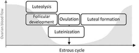
Fig. 2.1
Changes in ovarian blood flow (modified based on [15]) and ovarian processes during the estrous cycle in cattle
Recently, intraovarian blood flow was monitored using color Doppler ultrasonography [16–18]. The blood flow area and velocity (time-averaged maximum velocity, TAMXV) in the preovulatory follicular wall were elevated temporally (a few hours) just after the LH surge and were correlated with an increase in plasma concentration of estradiol-17β and the LH surge [17, 18]. Inducing ovulation by injection of GnRH caused blood flow area and velocity to increase for 120 h, accompanied by an increase in plasma P4 concentration [16, 18]. Similar changes in blood flow have been observed in the mare [19]. The correlation between the increase of blood flow and plasma P4 concentration was similar to the findings of the classical studies that used electromagnetic flow probes [12, 13, 15], so that the blood supply to the ovary and P4 synthesis in the ovary appear to be strongly related. Because the blood supply affects oxygen levels, these changes also strongly suggest a relationship between oxygen supply and steroidogenesis. This relationship is further discussed in Sect. 2.4.
The low levels of ovarian blood flow at the time from luteal regression to luteal formation [12–15], which includes luteinization of follicular granulosa and theca cells before and after ovulation, are thought to be the basis of the decreased oxygen supply to the ovary during the period. The oxygen concentration in bovine arterial blood does not change significantly during the estrous cycle, but tends to be at high levels during the functional luteal phase and then decreases before ovulation [15]. Furthermore, blood supply to the ovary is significantly lower during the several days around ovulation in cows [15]. These findings support the idea that oxygen conditions inside the ovary are low around ovulation, and that the ovarian events in this period, including end-stage maturation of follicles, ovulation, and luteal formation , are considered to progress under low oxygen conditions [6–11].
2.3 Cellular Responses to Hypoxic Conditions
Mammals have cellular mechanisms to adapt to hypoxic conditions. These mechanisms are conserved and expressed in almost every mammalian cell type [20]. The transcription factors activated specifically under hypoxic conditions are called hypoxia-inducible factors (HIFs) , which are heterodimeric transcription factors consisting of two subunits, HIF-α and an aryl hydrocarbon receptor nuclear translocator [ARNT; also called HIF1β (HIF1B)] [21]. Both subunits contain basic helix-loop-helix (bHLH) -Per Arnt-Sim (PAS) domains that mediate heterodimerization and DNA binding [22]. HIF1B is constitutively expressed whereas the activity and expression of HIF1A depends on cellular oxygen concentrations [23–25]. Mammalian cells have three HIF-α genes (HIF1A, 2A, 3A) [23–25]. Each gene contains an oxygen-dependent degradation domain (ODD) [26], which interacts with the von Hippel–Lindau (pVHL) E3 ubiquitin ligase complex [27–31] that targets HIF-α for proteasomal degradation under normoxia [26, 32–35]. HIF1A is expressed ubiquitously, whereas the expression of HIF2A and HIF3A is more restricted [21]. HIF1A and HIF2A dimerize with HIF1B, forming HIF1 and HIF2, both of which activate key transcription factors [21, 22]. HIF3A is found in three isoforms [HIF3A, neonatal and embryonic PAS (NEPAS) , and inhibitory PAS protein (IPAS)] [21, 22]. HIF3A isoforms dimerize with HIF1B, forming HIF3 and HIF3NEPAS [21, 22]. In general, HIFs bind to hypoxia-response elements (HREs) on DNA, which leads to the regulation of some 200 genes, of which 70 have been studied in detail [21, 22]. HIF1 induces the transcription of homeostasis-related genes such as vascular endothelial growth factor (VEGF) and erythropoietin (EPO), whereas HIF2 and HIF3 have more specialized and tissue-specific regulatory roles [21, 22, 36, 37]. HIF1 initiates the defense against hypoxia by a variety of mechanisms. In kidney and liver, hypoxia induces the synthesis of EPO [38, 39], which stimulates erythropoiesis, thereby increasing the O2 capacity of the blood [40]. In virtually all tissues, hypoxia induces the synthesis of proteins controlling local blood flow, such as VEGF [41, 42], endothelial nitric oxide synthase (eNOS) [43], and heme oxygenase-1 (HOX-1) [44]. VEGF stimulates angiogenesis and increases the permeability of blood vessels [45]. eNOS and HOX-1 generate NO and carbon monoxide, which are potent vasodilatory substances that augment perfusion of the hypoxic tissue. At the cellular level, hypoxia induces the expression of virtually all glycolytic enzymes, including phosphoglycerate kinase-1 (PGK-1) , enolase 1, and lactate dehydrogenase-1 [46, 47]. Furthermore, the expression of membranous glucose transporters (primarily GLUT-1) is increased under hypoxic conditions, thereby increasing glucose uptake for glycolysis [48, 49]. To promote gene expression of all these proteins , HIF1 binds to HREs present in the promoter and enhancer regions [24]. HIF1 also induces transcription of an apoptosis regulatory gene, 19-kDa interacting protein-3 (BNIP3) [50]. Apoptosis is important in ovarian physiology, especially during follicular atresia [51] and luteal regression [52]. In cultured bovine luteal cells, the expression of HIF1 and BNIP3 increases under hypoxic conditions [53, 54], suggesting that hypoxia-induced BNIP3 is related to apoptosis during luteolysis. Recently, BNIP3 has been reported to function in mitochondrial autophagy, resulting in suppression of superoxide generation and protection of cells under hypoxic conditions [55, 56]. We found that the expressions of HIF1A [57] and BNIP3 proteins (unpublished data) in bovine CL are significantly higher at the early luteal stage than at other stages. This finding suggests that (1) the HIF1-BNIP3 signal is more active in luteal formation than in luteal regression during the estrous cycle, and (2) apoptosis or mitochondrial autophagy controlled by active BNIP3 under hypoxia has some part in luteal formation. Hypoxic signals mainly controlled by HIFs are now becoming known to regulate ovarian function. These topics are discussed in the following sections.
2.4 Hypoxia Before Ovulation
The involvement of hypoxia as well as HIF1 in the regulation of steroidogenic gene expression has been previously suggested by a number of studies . Hypoxic conditions (1–3 % O2) significantly reduced P4 production in rat [58] and porcine [5] granulosa cells and in bovine luteal cells [53, 59] Culturing bovine mid-luteal cells, culture under 3 %, 5 %, and 10 % O2 for 24 h decreased P4 synthesis and 3 % O2 decreased CYP11A1 (also known as P450scc) mRNA expression and activity [53]. Exposure to cobalt chloride (CoCl2, a chemical inducer of HIF1) decreases CYP11A1 mRNA expression and hence P4 production in testicular Leydig cells [60]. HIF1 is also suggested to bind to and activate the promoter of the HSD3B gene in Leydig cells [61] and the gene for CYP19A1 (also known as aromatase) in breast adenocarcinoma cells [62]. In other cell types, hypoxia has been found to decrease aromatase expression and activity [60, 63]. In human trophoblastic cells, HIF1 decreases aromatase expression via estrogen-related receptor-alpha (EERa), which is an orphan receptor (a receptor whose function is not known) known as an oxygen-dependent transcription factor [60]. HIF1 also inhibits CYP19A1 mRNA expression by activating a micro-RNA (miR-98) in H295R cells [63]. However, it is unclear whether hypoxia also inhibits aromatase activity in ovarian granulosa cells.
The lower pO2 in the FF in the large follicles than in the small follicles has been suggested to promote VEGF production via HIF1 in granulosa cells [5]. On the other hand, in the primate ovary, nuclear immunostaining of HIF1A is mostly absent in growing preantral and antral follicles and is upregulated in the granulosa cells at ovulation [64]. In the periovulatory period, follicles rapidly change their functions, and the CL is formed after ovulation with active angiogenesis [3]. The HIF1-VEGF-induced angiogenesis system may be involved in the later period of follicular development and in the beginning of luteal formation immediately after ovulation.
Regulation of HIF1A expression has been found by the interaction between cAMP and hypoxia in bovine luteinized granulosa cells and human granulosa cells [65]. HIF1A protein expression was increased by chemical hypoxia, and the increased expression was further augmented by LH and cAMP. These results suggest that HIF1A is induced transcriptionally and post-transcriptionally, the first enhanced transcription by LH, which cannot be manifested in higher HIF1A protein levels unless the protein is stabilized under hypoxic conditions.
During the later period of follicular development before ovulation, oxygen levels seem to participate in follicular function by regulating of multiple phenomena, such as steroidogenesis and angiogenesis [66]. This regulation is only a part of the whole system of luteinization and ovulation , and the roles of hypoxia in ovarian function during this period are still largely unknown. Although the levels of oxygen concentration in newly forming luteal tissue immediately after ovulation are not known, the signal generated by hypoxic conditions has been suggested to be a key in luteinization and luteal formation [67, 68].
2.5 Hypoxia in Ovulation
At the time of ovulation, mammalian ovaries express high levels of chemokines, such as interleukin 8 (IL-8), monocyte chemoattractant protein-1 (MCP-1) , growth-regulated oncogene-α (GROα), chemokine CCL5 (also known as RANTES: regulated on activation normal T cell expressed and secreted), and thymus-expressed chemokine (TECK) [69–71]. One of the CXC chemokine families, stromal cell-derived factor-1 (SDF-1) and its receptor CXCR4, have been recently suggested to regulate follicular function before ovulation [72, 73]. SDF-1, first isolated from bone marrow-derived stromal cells, is a natural ligand for CXCR4 and was found to be expressed in several tissues and organs [74]. CXCR4 mRNA is also expressed in bovine ovarian granulosa cells, and the levels are high in the preovulatory follicles [73]. Because CXCR4 mRNA expressions have been found to increase under hypoxic conditions in vitro, the hypoxia-SDF-1/CXCR4 system has been suggested to be involved in ovulation [73].
P4 is required for ovulation and acts via its nuclear receptor progesterone receptor (PGR) [75]. PGR also regulates the genes for HIF1A, HIF2A, and HIF1B in mice [76]. In addition, the expressions of HIF1A, HIF2A, and HIF1B in pgr-null mice after induction of superovulation by gonadotropin were lower than those in wild-type mice, and inhibiting the transcription of HIF1 by echinomycin reduced the expressions of three ovulation-related genes (ADAMTS1, VEGFA, EDN2) [76]. These findings support the idea that P4, PGR, HIF1, and HIF2 signals are operating during ovulation.
Although the importance of hypoxia and the signals that it generates in ovulation have been getting more attention recently, further studies are needed to understand the crosstalk between hypoxic signals and ovulatory signals to clarify the function of hypoxia in ovulation. After ovulation, the tissues of the ruptured follicle immediately form a CL with functional and structural changes. These changes are supported by rapid angiogenesis [1–4], which has been found to be induced by hypoxia [57, 73–75]. These topics are described in the following section.
2.6 Hypoxia After Ovulation
After ovulation, the ruptured follicle is thought to be under hypoxic state because of bleeding, immature vasculature, and cell proliferation without matching blood supply [41] (Fig. 2.2). HIF1A protein expression, a well-known indicator of hypoxic conditions, is high in the newly forming CL 2 days after ovulation in the primate ovary [64], and is also significantly higher at the early and developing luteal tissue (2–6 days after ovulation) than at other stages of the estrous cycle in the bovine ovary [57, 59]. Although the oxygen concentrations in CL tissues have not been determined in any species, these findings about the protein expression of HIF1A [57, 64] strongly support the idea that newly forming CL tissue is under hypoxic conditions.

Fig. 2.2
Schematic of an ovary and its microenvironment after ovulation. The ruptured follicle is thought to be under hypoxic conditions because of bleeding and because the vasculature is immature
Angiogenesis during luteal formation was first investigated in the early 1990s [1] and has been the subject of several reviews [2–4, 77, 78]. VEGF, a potent angiogenic factor, was first identified in 1989 by Ferrarra and Henzel [79], and was found to promote angiogenesis during luteal formation in cows [80] and in women [81]. HIF1, soon after its discovery in 1995 [82], was found to be the most potent transcription factor for VEGF [41]. The early luteal tissue just after ovulation is thought to be under hypoxic conditions because of the destruction of the vasculature by ovulation as an explanation to the hypoxic condition and because the intensive cell proliferation in early CL is not matched initially by number of blood vessels . In bovine luteal endothelial cells, the mRNA expressions of HIF1A and VEGF were not significantly different in normoxic (20 % O2) and hypoxic (1 % O2) culture [83]. On the other hand, the mRNA expression of HIF1A in porcine CL was found to be high at the early luteal stage, which suggested that HIF1 assists in luteal formation [84]. To confirm the participation of HIF1A in luteal formation, analyses of HIF1A protein are crucial as HIF1 is mainly regulated by protein hydroxylation, as detailed in Sect. 2.3 [32]. Under normoxic conditions, the HIF1A subunit is rapidly degraded by the ubiquitin-proteasome pathway, whereas under hypoxic conditions, it becomes concentrated through downregulation of its degradation, and becomes functional after it dimerizes with the other subunit HIF1B [32]. In primates, immunostaining showed that the nuclear localization of HIF1A protein is found in the early CL [64]. In addition, HIF1A protein expression in the bovine CL is higher at the early and developing luteal stages than at the other luteal stages [57, 59]. Hypoxia also induced the expression of HIF1α protein, VEGF mRNA, and protein in cultured developing bovine luteal cells [57]. In luteinizing bovine granulosa cells and human granulosa cells, chemical hypoxia (cobalt chloride) induced HIF1α protein and VEGF mRNA expressions, and LH augmented both of these [65].
In ovarian steroidogenic cells, hypoxia also increased the expressions of several other proteins, including EG-VEGF (prokineticin-1, another type of VEGF) and its receptor PK-R2 [77, 85–87], a vasoactive peptide endothelin-2 (EDN2) [77, 88] and fibroblast growth factor 2 (FGF2) [77], suggesting that these factors also have roles in the hypoxic signals required for luteal formation. EDN2, for instance, induced changes that characterize the developing CL: cell proliferation as well as upregulation of VEGF and cyclooxygenase-2 [88]. The chemokine IL-8, which is a proangiogenic factor [88–94], was also found to be increased by hypoxia in human granulosa cells [95]. Furthermore, expression of IL-8 is higher at the early luteal stage than at other stages of the estrous cycle in the bovine ovary [96]. Thus, the angiogenesis induced by the hypoxia IL-8 system seems to assist luteal formation. Together, these findings suggest that hypoxic conditions generate several signals mainly via HIFs, which are essential for angiogenesis in luteal formation. A schema illustrating these ideas is shown in Fig. 2.3.

Fig. 2.3
Possible hypoxia-related signaling during luteal formation . HIF1 hypoxia-inducible factor-1, LHCGR luteinizing hormone/chorionic gonadotropin receptor, STAR steroidogenic acute regulatory protein, P4 progesterone, VEGFA vascular endothelial growth factor A, PROKR2 prokineticin receptor 2, FGF2 fibroblast growth factor 2, IL-8 interleukin 8
Hypoxia is considered to stimulate the proliferation of luteal endothelial cells during luteal formation [97]. Luteal steroidogenic cells also proliferate during luteal development in cattle as shown by the co-expression of proliferation marker Ki-67 and steroidogenic marker HSD3B [98]. However, rapid growth of CL after ovulation is believed to be mainly the result of an increase in size of steroidogenic cells (hypertrophy) rather than an increase in their number [99–101]. Luteal steroidogenic cells have been suggested to express VEGF in response to the stimulation of hypoxia, resulting in the proliferation of endothelial cells for angiogenesis during luteal formation [57, 64, 77, 88]. Recently, porcine luteal endothelial cells have also been shown to proliferate in response to hypoxic conditions [97]. However, how hypoxia induces steroidogenic cell proliferation remains unclear. In cultured bovine luteinized granulosa cells, severe hypoxia (1 % O2) decreases a proliferation marker, proliferating cell nuclear antigen (PCNA), whereas chemical hypoxia (cobalt chloride) increases the marker as well as HIF1A protein expression [59], implying that chemically induced HIF1 promotes steroidogenic cell proliferation, although the reason why severe hypoxia inhibits luteal cell proliferation is also unclear. To clarify how hypoxia regulates cell proliferation in CL, further studies are needed to determine how hypoxia-induced signals differ, and how their relationships differ, among different cell types, such as endothelial cells, luteinizing granulosa cells, and luteal steroidogenic cells.
2.7 Summary and Future Aspects
The discovery of HIF1 [82] elucidated responses to hypoxia in numerous cell types: these responses are related to pathological (cancer progression) as well as to physiological (female reproductive system) tissue growth [20–25, 102, 103]. HIFs are expressed in a variety of organs, and some of their functions have been determined, which suggests that the cells in such organs have the ability to respond to hypoxic conditions. Hypoxia is an important signal in reproductive physiology [20–22, 24, 25, 102]: hypoxia-generated responses have crucial functions in luteal formation in all species examined thus far [57, 64, 67, 68, 77, 78, 85–88, 97, 104]. Yet, some regulatory mechanisms of hypoxic effects on luteal formation remain unclear. HIF1A is regulated by hormones such as human chorionic gonadotropin (hCG) [104, 105] and LH [65] under both hypoxic and normoxic conditions. Understanding of the crosstalk between hypoxia-generated signals and hormone-induced signals could help to clarify the roles of hypoxia in luteal formation as well as ovarian physiology.
References
1.
Reynolds LP, Killilea SD, Redmer DA. Angiogenesis in the female reproductive system. FASEB J. 1992;6:886–92.PubMed
2.
Reynolds LP, Grazul-Bilska AT, Killilea SD, Redmer DA. Mitogenic factors of corpora lutea. Prog Growth Factor Res. 1994;5:159–75.PubMed
3.
Redmer DA, Reynolds LP. Angiogenesis in the ovary. Rev Reprod. 1996;1:182–92.PubMed
4.
Reynolds LP, Grazul-Bilska AT, Redmer DA. Angiogenesis in the corpus luteum. Endocrine. 2000;12:1–9.PubMed
5.
Basini G, Bianco F, Grasselli F, Tirelli M, Bussolati S, Tamanini C. The effects of reduced oxygen tension on swine granulosa cell. Regul Pept. 2004;120:69–75.PubMed
6.
Fischer B, Künzel W, Kleinstein J, Gips H. Oxygen tension in follicular fluid falls with follicle maturation. Eur J Obstet Gynecol Reprod Biol. 1992;43:39–43.PubMed
7.
Shalgi R, Kraicer PF, Soferman N. Gases and electrolytes of human follicular fluid. J Reprod Fertil. 1972;28:335–40.PubMed
8.
Fraser IS, Baird DT, Cockburn F. Ovarian venous blood PO2, PCO2 and pH in women. J Reprod Fertil. 1973;33:11–7.PubMed
9.
Imoedemhe DA, Chan RC, Ramadan IA, Sigue AB. Changes in follicular fluid gas and pH during carbon dioxide pneumoperitoneum for laparoscopic aspiration and their effect on human oocyte fertilizability. Fertil Steril. 1993;59:177–82.PubMed
10.
Huey S, Abuhamad A, Barroso G, Hsu MI, Kolm P, Mayer J, Oehninger S. Perifollicular blood flow Doppler indices, but not follicular pO2, pCO2, or pH, predict oocyte developmental competence in in vitro fertilization. Fertil Steril. 1999;72:707–12.PubMed
11.
Redding GP, Bronlund JE, Hart AL. Theoretical investigation into the dissolved oxygen levels in follicular fluid of the developing human follicle using mathematical modelling. Reprod Fertil Dev. 2008;20:408–17.PubMed
12.
Niswender GD, Reimers TJ, Diekman MA, Nett TM. Blood flow: a mediator of ovarian function. Biol Reprod. 1976;14:64–81.PubMed
13.
Nett TM, McClellan MC, Niswender GD. Effects of prostaglandins on the ovine corpus luteum: blood flow, secretion of progesterone and morphology. Biol Reprod. 1976;15:66–78.PubMed
14.
Ford SP, Chenault JR. Blood flow to the corpus luteum-bearing ovary and ipsilateral uterine horn of cows during the oestrous cycle and early pregnancy. J Reprod Fertil. 1981;62:555–62.PubMed
15.
Wise TH, Caton D, Thatcher WW, Barron DH, Fields MJ. Ovarian function during the estrous cycle of the cow: ovarian blood flow and progesterone release rate. J Anim Sci. 1982;55:627–37.PubMed
16.
Acosta TJ, Yoshizawa N, Ohtani M, Miyamoto A. Local changes in blood flow within the early and midcycle corpus luteum after prostaglandin F2α injection in the cow. Biol Reprod. 2002;66:651–8.PubMed
17.
Acosta TJ, Hayshi KG, Ohtani M, Miyamoto A. Local changes in blood flow within the preovulatory follicle wall and early corpus luteum in cows. Reproduction. 2003;125:759–67.PubMed
18.
Acosta TJ, Miyamoto A. Vascular control of ovarian function: ovulation, corpus luteum formation and regression. Anim Reprod Sci. 2004;82-83:127–40.PubMed
19.
Ginther OJ, Gastal EL, Gastal MO, Utt MD, Beg MA. Luteal blood flow and progesterone production in mares. Anim Reprod Sci. 2007;99:213–20.PubMed
20.
Lee JW, Bae SH, Jeong JW, Kim SH, Kim KW. Hypoxia-inducible factor (HIF-1)alpha: its protein stability and biological functions. Exp Mol Med. 2004;36:1–12.PubMed
21.
Chen L, Endler A, Shibasaki F. Hypoxia and angiogenesis: regulation of hypoxia-inducible factors via novel binding factors. Exp Mol Med. 2009;41:849–57.PubMedPubMedCentral
22.
Dunwoodie SL. The role of hypoxia in development of the mammalian embryo. Dev Cell. 2009;17:755–73.PubMed
23.
Jiang BH, Semenza GL, Bauer C, Marti HH (1966) Hypoxia-inducible factor 1 levels vary exponentially over a physiologically relevant range of O2 tension. Am J Physiol 271(4 pt 1):C1172–C1180.
24.
Wenger RH. Cellular adaptation to hypoxia: O2-sensing protein hydroxylases, hypoxia-inducible transcription factors, and O2-regulated gene expression. FASEB J. 2002;16:1151–62.PubMed
25.
Bruick RK. Oxygen sensing in the hypoxic response pathway: regulation of the hypoxia-inducible transcription factor. Genes Dev. 2003;17:2614–23.PubMed
26.
Huang LE, Gu J, Schau M, Bunn HF. Regulation of hypoxia-inducible factor 1alpha is mediated by an O2-dependent degradation domain via the ubiquitin-proteasome pathway. Proc Natl Acad Sci USA. 1998;95:7987–92.PubMedPubMedCentral
27.
Maxwell PH, Wiesener MS, Chang GW, Clifford SC, Vaux EC, Cockman ME, Wykoff CC, Pugh CW, Maher ER, Ratcliffe PJ. The tumour suppressor protein VHL targets hypoxia-inducible factors for oxygen-dependent proteolysis. Nature (Lond). 1999;399:271–5.
28.
Cockman ME, Masson N, Mole DR, Jaakkola P, Chang GW, Clifford SC, Maher ER, Pugh CW, Ratcliffe PJ, Maxwell PH. Hypoxia inducible factor-alpha binding and ubiquitylation by the von Hippel-Lindau tumor suppressor protein. J Biol Chem. 2000;275:25733–41.PubMed
29.
Kamura T, Sato S, Iwai K, Czyzyk-Krzeska M, Conaway RC, Conaway JW. Activation of HIF1alpha ubiquitination by a reconstituted von Hippel-Lindau (VHL) tumor suppressor complex. Proc Natl Acad Sci USA. 2000;97:10430–5.PubMedPubMedCentral
30.
Ohh M, Park CW, Ivan M, Hoffman MA, Kim TY, Huang LE, Pavletich N, Chau V, Kaelin WG. Ubiquitination of hypoxia-inducible factor requires direct binding to the beta-domain of the von Hippel-Lindau protein. Nat Cell Biol. 2000;2:423–7.PubMed
31.
Tanimoto K, Makino Y, Pereira T, Poellinger L. Mechanism of regulation of the hypoxia-inducible factor-1 alpha by the von Hippel-Lindau tumor suppressor protein. EMBO J. 2000;19:4298–309.PubMedPubMedCentral
32.
Salceda S, Caro J. Hypoxia-inducible factor 1alpha (HIF-1alpha) protein is rapidly degraded by the ubiquitin-proteasome system under normoxic conditions. Its stabilization by hypoxia depends on redox-induced changes. J Biol Chem. 1997;272:22642–7.PubMed
33.
Kallio PJ, Wilson WJ, O’Brien S, Makino Y, Poellinger L. Regulation of the hypoxia-inducible transcription factor 1alpha by the ubiquitin-proteasome pathway. J Biol Chem. 1999;274:6519–25.PubMed
34.
Ivan M, Kondo K, Yang H, Kim W, Valiando J, Ohh M, Salic A, Asara JM, Lane WS, Kaelin Jr WG. HIFalpha targeted for VHL-mediated destruction by proline hydroxylation: implications for O2 sensing. Science. 2001;292:464–8.PubMed
35.
Jaakkola P, Mole DR, Tian YM, Wilson MI, Gielbert J, Gaskell SJ, Kriegsheim AV, Hebestreit HF, Mukherji M, Schofield CJ, Maxwell PH, Pugh CW, Ratcliffe PJ. Targeting of HIF-alpha to the von Hippel-Lindau ubiquitylation complex by O2-regulated prolyl hydroxylation. Science. 2001;292:468–72.PubMed
36.
Carroll VA, Ashcroft M. Targeting the molecular basis for tumour hypoxia. Expert Rev Mol Med. 2005;7:1–16.PubMed
37.
Ietta F, Wu Y, Winter J, Xu J, Wang J, Post M, Caniggia I. Dynamic HIF1A regulation during human placental development. Biol Reprod. 2006;75:112–21.PubMed
38.
Semenza GL, Wang GL. A nuclear factor induced by hypoxia via de novo protein synthesis binds to the human erythropoietin gene enhancer at a site required for transcriptional activation. Mol Cell Biol. 1992;12:5447–54.PubMedPubMedCentral
39.
Beck I, Weinmann R, Caro J. Characterization of hypoxia-responsive enhancer in the human erythropoietin gene shows presence of hypoxia-inducible 120-Kd nuclear DNA-binding protein in erythropoietin-producing and nonproducing cells. Blood. 1993;82:704–11.PubMed
40.
Jelkmann W. Molecular biology of erythropoietin. Intern Med. 2004;43:649–59.PubMed
41.
Forsythe JA, Jiang BH, Iyer NV, Agani F, Leung SW, Koos RD, Semenza GL. Activation of vascular endothelial growth factor gene transcription by hypoxia-inducible factor 1. Mol Cell Biol. 1996;16:4604–13.PubMedPubMedCentral
42.
Kimura H, Weisz A, Ogura T, Hitomi Y, Kurashima Y, Hashimoto K, D’Acquisto F, Makuuchi M, Esumi H. Identification of hypoxia-inducible factor 1 ancillary sequence and its function in vascular endothelial growth factor gene induction by hypoxia and nitric oxide. J Biol Chem. 2001;276:2292–8.PubMed
43.
Coulet F, Nadaud S, Agrapart M, Soubrier F. Identification of hypoxia-response element in the human endothelial nitric-oxide synthase gene promoter. J Biol Chem. 2003;278:46230–40.PubMed
44.
Lee PJ, Jiang BH, Chin BY, Iyer NV, Alam J, Semenza GL, Choi A. Hypoxia-inducible factor-1 mediates transcriptional activation of the heme oxygenase-1 gene in response to hypoxia. J Biol Chem. 1997;272:5375–81.PubMed
45.
Ferrara N, Gerber HP, LeCouter J. The biology of VEGF and its receptors. Nat Med. 2003;9:669–76.PubMed
46.
Firth JD, Ebert BL, Pugh CW, Ratcliffe PJ (1994) Oxygen-regulated control elements in the phosphoglycerate kinase 1 and lactate dehydrogenase A genes: similarities with the erythropoietin 3′ enhancer. Proc Natl Acad Sci USA 91:6496–6500.
47.
Semenza GL, Jiang BH, Leung SW, Passantino R, Concordet JP, Maire P, Giallongo A. Hypoxia response elements in the aldolase A, enolase 1, and lactate dehydrogenase A gene promoters contain essential binding sites for hypoxia-inducible factor 1. J Biol Chem. 1996;271:32529–37.PubMed
48.
Behrooz A, Ismail-Beigi F. Dual control of glut1 glucose transporter gene expression by hypoxia and by inhibition of oxidative phosphorylation. J Biol Chem. 1997;272:5555–62.PubMed
49.
Gleadle JM, Ratcliffe PJ. Induction of hypoxia-inducible factor-1, erythropoietin, vascular endothelial growth factor, and glucose transporter-1 by hypoxia: evidence against a regulatory role for Src kinase. Blood. 1997;89:503–9.PubMed
50.
Bruick RK. Expression of the gene encoding the proapoptotic Nip3 protein is induced by hypoxia. Proc Natl Acad Sci USA. 2000;97:9082–7.PubMedPubMedCentral
51.
Matsuda F, Inoue N, Manabe N, Ohkura S. Follicular growth and atresia in mammalian ovaries: regulation by survival and death of granulosa cells. J Reprod Dev. 2012;58:44–50.PubMed
52.
McCracken JA, Custer EE, Lamsa JC. Luteolysis: a neuroendocrine-mediated event. Physiol Rev. 1999;79:263–323.PubMed
53.
Nishimura R, Sakumoto R, Tatsukawa Y, Acosta TJ, Okuda K. Oxygen concentration is an important factor for modulating progesterone synthesis in bovine corpus luteum. Endocrinology. 2006;147:4273–80.PubMed
54.
Nishimura R, Komiyama J, Tasaki Y, Acosta TJ, Okuda K. Hypoxia promotes luteal cell death in bovine corpus luteum. Biol Reprod. 2008;78:529–36.PubMed
55.
Zhang H, Bosch-Marce M, Shimoda LA, Tan YS, Baek JH, Wesley JB, Gonzalez FJ, Semenza GL. Mitochondrial autophagy is an HIF-1-dependent adaptive metabolic response to hypoxia. J Biol Chem. 2008;283:10892–903.PubMedPubMedCentral
56.
Semenza GL. Mitochondrial autophagy: life and breath of the cell. Autophagy. 2008;4:534–6.PubMedPubMedCentral
57.
Nishimura R, Okuda K. Hypoxia is important for establishing vascularization during corpus luteum formation in cattle. J Reprod Dev. 2010;56:110–6.PubMed
58.
Koos RD, Feiertag MA. The effect of reduced oxygen tension on progesterone accumulation in rat granulosa cell cultures. Steroids. 1989;54:553–62.PubMed
59.
Jiang YF, Tsui KH, Wang PH, Lin CW, Wang JY, Hsu MC, Chen YC, Chiu CH. Hypoxia regulates cell proliferation and steroidogenesis through protein kinase A signaling in bovine corpus luteum. Anim Reprod Sci. 2011;129:152–61.PubMed
60.
Kumar A, Rani L, Dhole B. Role of oxygen in the regulation of Leydig tumor derived MA-10 cell steroid production: the effect of cobalt chloride. Syst Biol Reprod Med. 2014;60:112–8.PubMed
61.
Lysiak JJ, Kirby JL, Tremblay JJ, Woodson RI, Reardon MA, Palmer LA, Turner TT. Hypoxia-inducible factor-1alpha is constitutively expressed in murine Leydig cells and regulates 3beta-hydroxysteroid dehydrogenase type 1 promoter activity. J Androl. 2009;30:146–56.PubMed
62.
Samarajeewa NU, Yang F, Docanto MM, Sakurai M, McNamara KM, Sasano H, Fox SB, Simpson ER, Brown KA. HIF-1α stimulates aromatase expression driven by prostaglandin E2 in breast adipose stroma. Breast Cancer Res. 2013;15:R30.PubMedPubMedCentral
63.
Yu RM, Chaturvedi G, Tong SK, Nusrin S, Giesy JP, Wu RS, Kong RY. Evidence for microRNA-mediated regulation of steroidogenesis by hypoxia. Environ Sci Technol. 2015;49:1138–47.PubMed
64.
Duncan WC, van den Driesche S, Fraser HM. Inhibition of vascular endothelial growth factor in the primate ovary up-regulates hypoxia-inducible factor-1alpha in the follicle and corpus luteum. Endocrinology. 2008;149:3313–20.PubMed
65.
Yalu R, Oyesiji AE, Eisenberg I, Imbar T, Meidan R. HIF1A-dependent increase in endothelin 2 levels in granulosa cells: role of hypoxia, LH/cAMP, and reactive oxygen species. Reproduction. 2015;149:11–20.PubMed
66.
Thompson JG, Brown HM, Kind KL, Russell DL. The ovarian antral follicle: living on the edge of hypoxia or not? Biol Reprod. 2015;92:153.PubMed
67.
Fadhillah, Yoshioka S, Nishimura R, Okuda K (2014) Hypoxia promotes progesterone synthesis during luteinization in bovine granulosa cells. J Reprod Dev 60:194–201
68.
Kowalewski MP, Gram A, Boos A. The role of hypoxia and HIF1α in the regulation of STAR-mediated steroidogenesis in granulosa cells. Mol Cell Endocrinol. 2015;401:35–44.PubMed
69.
Ujioka T, Matsukawa A, Tanaka N, Matsuura K, Yoshinaga M, Okamura H. Interleukin-8 as an essential factor in the human chorionic gonadotropin-induced rabbit ovulatory process: interleukin-8 induces neutrophil accumulation and activation in ovulation. Biol Reprod. 1998;58:526–30.PubMed
70.
Brännström M, Enskog A. Leukocyte networks and ovulation. J Reprod Immunol. 2002;57:47–60.PubMed
71.
Zhou C, Wu J, Borillo J, Torres L, McMahon J, Bao Y, Lou YH. Transient expression of CC chemokine TECK in the ovary during ovulation: its potential role in ovulation. Am J Reprod Immunol. 2005;53:238–48.PubMed
72.
Nishigaki A, Okada H, Okamoto R, Shimoi K, Miyashiro H, Yasuda K, Kanzaki H. The concentration of human follicular fluid stromal cell-derived factor-1 is correlated with luteinization in follicles. Gynecol Endocrinol. 2013;29:230–4.PubMed
73.
Sayasith K, Sirois J. Expression and regulation of stromal cell-derived factor-1 (SDF1) and chemokine CXC motif receptor 4 (CXCR4) in equine and bovine preovulatory follicles. Mol Cell Endocrinol. 2014;391:10–21.PubMed
74.
Teicher BA, Fricker SP. CXCL12 (SDF-1)/CXCR4 pathway in cancer. Clin Cancer Res. 2010;16:2927–31.PubMed
75.
Park OK, Mayo KE. Transient expression of progesterone receptor messenger RNA in ovarian granulosa cells after the preovulatory luteinizing hormone surge. Mol Endocrinol. 1991;5:967–78.PubMed
76.
Kim J, Bagchi IC, Bagchi MK. Signaling by hypoxia-inducible factors is critical for ovulation in mice. Endocrinology. 2009;150:3392–400.PubMedPubMedCentral
77.
Meidan R, Klipper E, Zalman Y, Yalu R. The role of hypoxia-induced genes in ovarian angiogenesis. Reprod Fertil Dev. 2013;25:343–50.PubMed
78.
Nishimura R, Okuda K. Multiple roles of hypoxia in ovarian function: roles of hypoxia-inducible factor-related and -unrelated signals during the luteal phase. Reprod Fertil Dev. 2015;28:1479–86.
79.
Ferrara N, Henzel WJ. Pituitary follicular cells secrete a novel heparin-binding growth factor specific for vascular endothelial cells. Biochem Biophys Res Commun. 1989;161:851–8.PubMed
80.
Grazul-Bilska AT, Redmer DA, Killilea SD, Zheng J, Reynolds LP. Initial characterization of endothelial mitogens produced by bovine corpora lutea from the estrous cycle. Biochem Cell Biol. 1993;71:270–7.PubMed
81.
Kamat BR, Brown LF, Manseau EJ, Senger DR, Dvorak HF. Expression of vascular permeability factor/vascular endothelial growth factor by human granulosa and theca lutein cells. Role in corpus luteum development. Am J Pathol. 1995;146:157–65.PubMedPubMedCentral
82.
Wang GL, Semenza GL. Purification and characterization of hypoxia-inducible factor 1. J Biol Chem. 1995;270:1230–7.PubMed
83.
Tscheudschilsuren G, Aust G, Nieber K, Schilling N, Spanel-Borowski K. Microvascular endothelial cells differ in basal and hypoxia-regulated expression of angiogenic factors and their receptors. Microvasc Res. 2002;63:243–51.PubMed
84.
Boonyaprakob U, Gadsby JE, Hedgpeth V, Routh PA, Almond GW. Expression and localization of hypoxia inducible factor-1alpha mRNA in the porcine ovary. Can J Vet Res. 2005;69:215–22.PubMedPubMedCentral
85.
Kisliouk T, Levy N, Hurwitz A, Meidan R. Presence and regulation of endocrine gland vascular endothelial growth factor/prokineticin-1 and its receptors in ovarian cells. J Clin Endocrinol Metab. 2003;88:3700–7.PubMed
86.
Kisliouk T, Podlovni H, Spanel-Borowski K, Ovadia O, Zhou QY, Meidan R. Prokineticins (endocrine gland-derived vascular endothelial growth factor and BV8) in the bovine ovary: expression and role as mitogens and survival factors for corpus luteum-derived endothelial cells. Endocrinology. 2005;146:3950–8.PubMed
87.
Kisliouk T, Podlovni H, Meidan R. Unique expression and regulatory mechanisms of EG-VEGF/prokineticin-1 and its receptors in the corpus luteum. Ann Anat. 2005;187:529–37.PubMed
88.
Klipper E, Levit A, Mastich Y, Berisha B, Schams D, Meidan R. Induction of endothelin-2 expression by luteinizing hormone and hypoxia: possible role in bovine corpus luteum formation. Endocrinology. 2010;151:1914–22.PubMed
89.
Ferrer FA, Pantschenko AG, Miller LJ, Anderson K, Grunnet M, McKenna PH, Kreutzer D. Angiogenesis and neuroblastomas: interleukin-8 and interleukin-8 receptor expression in human neuroblastoma. J Urol. 2000;164:1016–20.PubMed
90.
Fujimoto J, Sakaguchi H, Aoki I, Tamaya T. Clinical implications of expression of interleukin 8 related to angiogenesis in uterine cervical cancers. Cancer Res. 2000;60:2632–5.PubMed
91.
Huang S, Robinson JB, Deguzman A, Bucana CD, Fidler IJ. Blockade of nuclear factor-kappaB signaling inhibits angiogenesis and tumorigenicity of human ovarian cancer cells by suppressing expression of vascular endothelial growth factor and interleukin 8 (in process citation). Cancer Res. 2000;60:5334–9.PubMed
92.
Kitadai Y, Takahashi Y, Haruma K, Naka K, Sumii K, Yokozaki H, Yasui W, Mukaida N, Ohmoto Y, Kajiyama G, Fidler IJ, Tahara E. Transfection of interleukin-8 increases angiogenesis and tumorigenesis of human gastric carcinoma cells in nude mice. Br J Cancer. 1999;81:647–53.PubMedPubMedCentral
93.
Yoshino O, Osuga Y, Koga K, Hirota Y, Yano T, Tsutsumi O, Fujimoto A, Kugu K, Momoeda M, Fujiwara T, Taketani Y. Upregulation of interleukin-8 by hypoxia in human ovaries. Am J Reprod Immunol. 2003;50:286–90.PubMed
94.
Koch AE, Polverini PJ, Kunkel SL, Harlow LA, DiPietro LA, Elner VM, Elner SG, Strieter RM. Interleukin-8 as a macrophage-derived mediator of angiogenesis. Science. 1992;258:1798–801.PubMed
95.
Rofstad EK, Halsor EF. Vascular endothelial growth factor, interleukin 8, platelet-derived endothelial cell growth factor, and basic fibroblast growth factor promote angiogenesis and metastasis in human melanoma xenografts. Cancer Res. 2000;60:4932–8.PubMed
96.
Jiemtaweeboon S, Shirasuna K, Nitta A, Kobayashi A, Schuberth HJ, Shimizu T, Miyamoto A. Evidence that polymorphonuclear neutrophils infiltrate into the developing corpus luteum and promote angiogenesis with interleukin-8 in the cow. Reprod Biol Endocrinol. 2011;9:79.PubMedPubMedCentral
97.
Basini G, Falasconi I, Bussolati S, Grolli S, Ramoni R, Grasselli F. Isolation of endothelial cells and pericytes from swine corpus luteum. Domest Anim Endocrinol. 2014;48:100–9.PubMed
98.
Yoshioka S, Abe H, Sakumoto R, Okuda K. Proliferation of luteal steroidogenic cells in cattle. PLoS One. 2013;8, e84186.PubMedPubMedCentral
99.
Baird DT. The ovary. New York: Cambridge University Press; 1984.
100.
Meyer GT, Bruce NW. The cellular pattern of corpus luteal growth during pregnancy in the rat. Anat Rec. 1979;193:823–30.PubMed
101.
Tamura H, Greenwald GS. Angiogenesis and its hormonal control in the corpus luteum of the pregnant rat. Biol Reprod. 1987;36:1149–54.PubMed
102.
Wiesener MS, Maxwell PH. HIF and oxygen sensing; as important to life as the air we breathe? Ann Med. 2003;35:183–90.PubMed
103.
Hellwig-Burgel T, Stiehl DP, Wagner AE, Metzen E, Jelkmann W. Review: hypoxia-inducible factor-1 (HIF-1): a novel transcription factor in immune reactions. J Interferon Cytokine Res. 2005;25:297–310.PubMed
104.
van den Driesche S, Myers M, Gay E, Thong KJ, Duncan WC. HCG up-regulates hypoxia inducible factor-1 alpha in luteinized granulosa cells: implications for the hormonal regulation of vascular endothelial growth factor A in the human corpus luteum. Mol Hum Reprod. 2008;14:455–64.PubMed
105.
Zhang Z, Yu D, Yin D, Wang Z. Activation of PI3K/mTOR signaling pathway contributes to induction of vascular endothelial growth factor by hCG in bovine developing luteal cells. Anim Reprod Sci. 2011;125:42–8.PubMed