
Koumei Shirasuna1 and Akio Miyamoto2
(1)
Faculty of Agriculture, Department of Animal Science, Tokyo University of Agriculture, Atsugi Kanagawa, 243-0034, Japan
(2)
Graduate School of Animal and Food Hygiene, Obihiro University of Agriculture and Veterinary Medicine, Obihiro Hokkaido, 080-8555, Japan
Koumei Shirasuna
Email: ks205312@nodai.ac.jp
Akio Miyamoto (Corresponding author)
Email: akiomiya@obihiro.ac.jp
Abstract
In the past two decades, accumulating evidence has indicated that various types of immune cells (T cells, macrophages, neutrophils, eosinophils, and dendritic cells) exist within the CL and regulate luteal function. These immune cells accumulate during luteal development and support angiogenesis and progesterone production. PGF2α stimulates the production of inflammatory cytokines and chemokines in the mature CL; these factors recruit immune cells into the CL to enhance luteolytic cascades through inflammatory responses. When pregnancy is established, the embryo secretes interferon-tau (IFNT) as a pregnancy recognition signal; this indirectly maintains the CL by inhibiting luteolysis. In addition to its uterine function, IFNT regulates immune cell function and is associated with the transformation of the cyclic CL into the pregnancy CL. This review describes the current state of research on the effect of immune cells on the bovine CL, which is essential for a better understanding of reproductive physiology.
Keywords
Corpus luteum developmentLuteolysisImmune cells
6.1 Introduction
Lobel and Levy [1] first described the presence of white blood cells in the bovine corpus luteum (CL). Later on, experimentally induced lymphopenia caused luteal dysfunction in cattle [2]. During the past two decades, accumulating evidence indicated that various types of immune cells such as T lymphocytes, macrophages, neutrophils, eosinophils, and dendritic cells exist within the CL, and plays key roles to regulate luteal function throughout the lifespan of the CL.
6.2 Immune Cells in the CL : Where Do They Come From?
The immune system is known to contribute to the regulation of ovarian function including ovulation, the development of the CL and luteolysis [3]. Leukocytes present within the ovary are potential regulators of ovarian function by local secretion of modulating cytokines.
T lymphocytes that mature in the thymus are central immune cells to regulate cell-mediated immunity. Within the bovine CL, several types of T lymphocytes include T-helper cells (Th cells, CD4+ T cells), cytotoxic T cells (CD8+ T cells), regulatory T cells (suppressor T cells, Foxp3+ T cells), and γδ+ T cells [4, 5]. All T cells originate from hematopoietic stem cells in the bone marrow. Hematopoietic progenitor cells populate the thymus; these expand by cell division to generate immature thymocytes. Mature thymocytes are released from the thymus to peripheral tissues. In mice, intact thymic function has been found to be necessary in the maintenance of normal ovarian function; congenitally athymic or postnatally thymectomized animals are infertile and exhibit abnormal ovarian function [6].
Cells of the innate immune system , such as macrophages, neutrophils, eosinophils, and dendritic cells, originate from stem cells in the bone marrow. These cells are recruited into the ovary, where they are critical in ovulation and luteal function. Kizuka et al. [7] used a mouse bone marrow transplantation model to demonstrate that transplanted green fluorescent protein (GFP)+ bone marrow cells were recruited into the developing CL; these GFP+ cells were F4/80 (marker of macrophages)+, indicating that macrophages are recruited to the ovary from the bone marrow during CL formation. The spleen is another immune cell reservoir for the ovary. Swirski et al. [8] demonstrated that the spleen is a site for the storage and rapid deployment of monocytes and identified splenic monocytes as a resource that the body exploits to regulate inflammation. Importantly, Oakley et al. [9] showed that a strong inverse relationship exists between the quantity of leukocytes in the ovary and that in the spleen. They also showed a significant reduction in the leukocyte infiltration of ovaries of splenectomized rats, indicating that the spleen may serve as an immediate reservoir of leukocytes for the ovary [9]. Furthermore, the number of leukocytes as well as progesterone concentration significantly decreased in splenectomized pseudo-pregnant rabbits [10].
6.3 Corpus Luteum Development and Maintenance
6.3.1 Role of T Lymphocytes Within the Functional CL
The phenotypes of T lymphocytes present in the bovine CL and the number of CD4+ and CD8+ T cells does not vary from the early to late luteal phase [11]. Poole and Pate [12] observed the proportion of T lymphocytes within the bovine CL to be that 25 % of the T lymphocytes were T-helper cells (CD4+), 45 % were cytotoxic T cells (CD8+), and 30 % were γδ+ T cells. It is well understood that peripheral blood mononuclear cells (PBMC) , including T lymphocytes and monocytes, clearly have a function for steroidogenic cells. Indeed, Hashi et al. [13] reported that PBMC from women could stimulate progesterone production from luteal cells in vitro and that Th-2 cytokines such as IL-10 and IL-4 are candidates for these positive effects of PBMC . Walusimbi and Pate [5] indicated that luteal cells from the mid-luteal phase predominantly induced proliferation of γδ+WC1− T cells together with increased IL-10 expression , but not IFNG expression, on its cell surface. Therefore, it is suggested that a functional CL has an immunosuppressive role to suppress proliferation of resident T lymphocytes by the production of immunosuppressive cytokine IL-10 (Fig. 6.1) [14]. High concentrations of progesterone from a functional CL may be one of the candidates as a communication tool between luteal cells and T lymphocytes. Indeed, progesterone induces IL-10 synthesis and other Th2-associated cytokines in murine γδ+ T cells via progesterone-induced blocking factor [15]. Interestingly, luteal cells regulate proliferation of T lymphocytes devoid of the nuclear progesterone receptor [16, 17]. Alternatively, bovine T lymphocytes express membrane progesterone receptor; therefore, progesterone can induce specific and rapid functional effects on T lymphocytes [18].
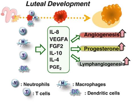
Fig. 6.1
Proposed model of inflammation-like luteal development . The mechanism of luteal development is considered as an inflammation-like response as many types of immune cells such as neutrophils, macrophage, T cells, and dendritic cells are recruited within the CL. They participate in luteal development via increases in angiogenesis, progesterone production, and lymphangiogenesis
6.3.2 Role of Monocytes and Macrophages Within the Functional CL
Macrophages exhibit a high level of phenotypic plasticity and participate in diverse physiological processes, including the innate immune system, host defense together with lymphocytes against external pathogens, removal of apoptotic cells, and angiogenesis. Monocytes originate from bone marrow progenitor cells and travel via the circulation to specific tissues where they differentiate to macrophages depending on the tissue microenvironment.
The number of macrophages increases in the early developing CL in cows [11] and humans [19, 20], as well as in the newly luteinized CL in pigs [21]. Turner et al. [22] reported the importance of macrophages in maintaining vascular integrity in the CL. Progressive macrophage elimination was associated with ovarian hemorrhage, which affected luteal tissue as a result of significant endothelial cell depletion and increased erythrocytes [22]. Additionally, Care et al. [23] convincingly demonstrated that macrophage depletion after conception caused embryo implantation arrest associated with decreased plasma progesterone because of disruption of the luteal microvascular network. In fact, peripheral blood macrophages co-cultured with granulosa cells exerted a luteotropic effect [24–26]. In humans, co-culture of monocytes and granulosa-lutein cells increased interleukin-8 (IL-8) release [27]. Additionally, monocytes have interleukin-8 (IL-8) receptors including CXCR1 and CXCR2 , and a granulosa-lutein cell-conditioned medium stimulated monocyte migration via IL-8 [28]. Thus, an initial interaction between granulosa-lutein cells and monocytes may contribute to increased chemokine release and leukocyte recruitment to the forming CL [27]. Therefore, macrophages play a central role in maintaining the integrity of the ovarian vasculature (Fig. 6.1).
6.3.3 Role of Polymorphonuclear Cells Within the Functional CL
Polymorphonuclear leukocytes (PMNs) such as neutrophils and eosinophils are detected in the CL during the estrous cycle in certain species [29–31]. Neutrophils are important in the primary, unspecific stages of acute inflammatory reaction. In humans, neutrophils account for 60 % of circulating leukocytes, and 90 % of these PMNs are neutrophils.
A considerable number of neutrophils and a high concentration of IL-8 (a neutrophil-specific chemoattractant) are present in the bovine CL during the early luteal phase [31]. Similarly, neutrophils have been detected in large numbers in the human CL [32], and found at relatively high density in the rat CL in the early phases of pregnancy [33]. The formation of the early CL induced PMN migration in vitro using IL-8 and the supernatant of activated PMNs, and IL-8 stimulated the formation of capillary-like structures of CL-derived endothelial cells [31]. Importantly, IL-8 effectively stimulates progesterone production in bovine luteinizing granulosa cells [34] as much as vascular epithelial growth factor (VEGF) A and fibroblast growth factor (FGF)2 in vitro. These findings indicate that IL-8 and neutrophils may function to stimulate the developing CL (Fig. 6.1). However, Talbott et al. [35] reported that in co-culture of neutrophils with luteal cells, neither IL-8 nor activated neutrophils altered luteal cell progesterone synthesis, suggesting the multiple functions of neutrophils depending on its condition or activation.
Neutrophils are thought to be important in physiological and pathophysiological angiogenesis [36]. PMNs and IL-8 could induce angiogenesis in vivo [37, 38] and in vitro [39, 40], indicating that PMNs and IL-8 may function not only in the induction of tissue inflammation and wound healing, but also in the regulation of angiogenesis in the developing CL. In mice, neutrophils expressing VEGFA were detected in the microvessels of the endometrium [41]. Moreover, the proliferation of endothelial cells was significantly reduced in neutrophil-depleted mice compared with control mice [42]. Interestingly, similar to IL-8, FGF2 and VEGFA (10 and 100 ng/ml) stimulated PMN migration in vitro (Shirasuna et al., unpublished observations), and FGF2 and VEGFA are also expressed at high levels within the developing CL. Ancelin et al. [43] demonstrated that VEGFA was chemotactic for neutrophils in humans and that neutralization with anti-VEGFA antibody blocked this effect. Indeed, FGF2 also enhanced recruitment of neutrophils in rats [44]. Therefore, it is speculated that IL-8, VEGFA, and FGF2 act synergistically as stimulators of PMN recruitment in the early CL in cows.
6.3.4 Role of Dendritic Cells Within the Functional CL
Dendritic cells (DCs) are specialized antigen-presenting cells that prime T cells and are thus essential to both innate and adaptive immunity. Increasing evidence suggests that DCs are involved in both the classical induction of immunity against infectious agents and immune tolerance to innocuous antigens. Indeed, a transient ablation of DCs on embryonic day 4.5 leads to complete embryo resorption, which suggests that DCs are pivotal in fetal immune tolerance [45]. Although the presence of DCs within the bovine CL has not been confirmed, Spanel-Borowski [46] suggests that cytokeratin-positive cells isolated from the bovine CL are promising candidates to mature into DCs. Recently, Cohen-Fredarow et al. [47] reported that, in mice, DCs are present in the ovary and accumulate in the newly formed CL. In addition, the conditional depletion of CD11c+ DCs blocks hCG-induced ovulation, interferes with the development of lymphatic vessels, and significantly inhibits progesterone secretion. Hence, they conclude that in the early luteal phase, the DCs localized in the newly formed CL facilitate progesterone production as well as lymphangiogenesis (Fig. 6.1).
6.3.5 Role of Other Blood Cells Within the Functional CL
Platelets are blood cells that have a pivotal role in coagulant systems. They have been reported to produce chemoattractants that are capable of inducing endothelial cell migration and have a role in wound healing and tissue remodeling. Furukawa et al. [48] clearly reported that platelets localize within the human CL in the course of neovascularization. Furthermore, platelets are involved in the development of the CL to upregulate progesterone production [48].
Eosinophils are known to be involved in the immune response against parasitic infection, asthma, and allergic conditions. Eosinophils infiltrate into the CL shortly after ovulation in cows [49], sheep [50], and humans [30], and they appear to be recruited into the developing CL by the expression of P-selectin on endothelial cells [30]. Human granulosa cells express RANTES (eosinophil-attracting chemokines) at the very early stage of development, indicative of their importance in the angiogenic and steroidogenic regulation of CL growth [30]. In cattle, a decrease in the quantity of eosinophils induced by the administration of dexamethasone results in lower progesterone concentrations [51]. However, in pigs, the quantity of eosinophils decreases shortly after ovulation [21].
6.4 Corpus Luteum Regression
6.4.1 Role of T Cells and Macrophages During Luteolysis
Uterine-derived or exogenous prostaglandin F2α (PGF2α) initiates luteolysis and rapidly reduces progesterone secretion by the CL [52]. During luteolysis, leukocytes, particularly macrophages and T lymphocytes, increase significantly in number in cows [53], humans [19], pigs [21], mares [54], and mice [55, 56], suggesting an active role of both cell types in luteolysis. Moreover, inflammatory cytokines such as tumor necrosis factor (TNF)α, IL1β, and IFNG and chemokines such as monocyte chemoattractant protein 1 (CCL2; recruitment of macrophages) are involved in luteal regression [6, 57–61]. These cytokines and chemokines may recruit macrophages and T lymphocytes within the CL to enhance luteolytic cascades through inflammatory and immune responses.
In cows and mares , large numbers of CD4+ and CD8+ T cells were found in the regressing CL [53, 54], suggesting that both cell types play an active role in luteolysis. In vitro, activated PBMC (mainly T cells) clearly inhibit LH-stimulated progesterone secretion from luteal cells [35]. Cannon et al. [17] reported that the stimulation of T cell proliferation was greater in luteal cells isolated after PGF2α administration than in luteal cells isolated before PGF2α administration. Furthermore, exogenous progesterone inhibits T cell proliferation [17]. γδ+ T cells represent a major proportion of circulating T cells in ruminants; γδ+ T cells account for 35 % of the T cells in the bovine CL [12]. Recently, Walusimbi and Pate [14] observed that luteal cells from the regressing CL predominantly induced proliferation of γδ+ WC1+ T cells, whereas luteal cells from the functional CL preferentially induced proliferation of γδ+ WC1− T cells . Interestingly, luteal cells from the functional CL increased the proportion of γδ+ T cells expressing IL-10, an antiinflammatory cytokine, and decreased the proportion of γδ+ T cells expressing IFNG, a major luteolytic factor. Therefore, the environment in a functional CL suppresses the activity of resident T cells, whereas the environment in a regressing CL restores T-cell activity to promote the structural demise of luteal tissue [14]. Regulatory T cells (CD4+ CD25+Foxp3+ T cells) are known to be essential in the regulation of immune tolerance during implantation [62]. Although CD4+ Foxp3+ T cells are more prevalent in the bovine CL than in peripheral blood, PGF2α administration drastically decreases the quantity of Foxp3+ T cells within the regressing CL [12]. These findings suggest that Foxp3+ T cells regulate the function of resident T cells to prevent the initiation of luteolysis; therefore, a decline in the quantity of Foxp3+ T cells is directly associated with luteal regression.
In the regressing CL in the cow, 70 % of proliferating cells are CD14+ macrophages [53]. Macrophages are essential in functionally healthy CL, whereas abundant macrophages exist in the CL during functional regression (early stage of regression) [56]. In rabbits, functional and structural luteolysis correlated with an increased number of X4+ and CD68+ macrophages [63]. Moreover, inflammatory cytokines , such as TNFα, IL1β, and IFNG, and chemokines, such as CCL2 are involved in luteal regression [6, 57–61, 64–67]. These cytokines and chemokines may induce the accumulation of macrophages and T lymphocytes within the CL to support luteolytic cascades such as inflammatory and immune response (Fig. 6.2). Thereafter, the CL regresses primarily through the loss of cells by apoptosis [68, 69], and apoptotic luteal cells are phagocytosed by macrophages in rats [70].
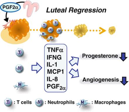
Fig. 6.2
Proposed model of the inflammation-like luteolytic response. The luteolytic cascade is similar to general acute inflammation with respect to the infiltration of immune cells (neutrophils, macrophages, and T cells). The luteolytic cascade is signaled by PGF2α secreted by the uterus and is a type of acute inflammatory immune response that promotes CL regression
In general, macrophages and T lymphocytes have the potential to produce multiple cytokines such as IL-1s, IL-2, IL-4, IL-6, TNFα, IFNA, IFNG, and prostaglandins to increase the immune inflammatory response and to communicate with peripheral resident cells depending on the stimulation conditions. There have been numerous studies on the relationships between luteal regression, leukocytes, particularly macrophages and T lymphocytes, and these cytokines . For example, TNFα protein exists in large and small luteal cells, and endothelial cells, as well as immune cells in the bovine CL [71]. TNFα inhibits progesterone secretion and induces IFNG and Fas-mediated apoptotic cell death in bovine luteal and endothelial cells by increasing caspase-3 activity [72, 73]. IFNG inhibits LH-stimulated progesterone production, increases prostaglandin synthesis , and induces cell death [74, 75].
6.4.2 Role of Neutrophils During Luteolysis
Neutrophils are the first cells recruited to inflammatory sites , providing cytokines and proteolytic enzymes [76]. During luteolysis, PGF2α rapidly induces the accumulation of neutrophils within the bovine CL at 5 min after administration [77]. Also, neutrophils accumulate in the equine CL after PGF2α administration [54]. To occur this rapid response, PGF2α directly stimulates P-selectin expression and enhanced neutrophil adhesion in luteal endothelial cells via P-selectin [77]. Generally, an acute inflammation is characterized by the infiltration of neutrophils within a few minutes and continuous occurrence of T lymphocyte and macrophage migration. Neutrophils can produce various types of inflammatory cytokines recruiting T lymphocytes and macrophages such as IL-8, TNFα, and IFNG [40, 76, 78, 79]. Also, a large number of T lymphocytes and macrophages were observed within the bovine CL at 6–24 h after PGF2α administration, and these immune cells are considered to be essential for a rapid demise of the CL tissue [11, 61, 65, 80]. Therefore, it is suggested that luteolytic cascade by PGF2α involves an acute inflammatory-like response in response to acute migrated neutrophils in cows, and these neutrophils may have a potential to recruit other immune cells in the regressing CL (Fig. 6.2). Indeed, a pretreatment with antibody against CD18 (leukocyte integrin) significantly inhibited not only PGF2α-induced neutrophil accumulation but also the decrease in serum progesterone concentrations [81]. In rats, co-incubation of luteal cells with activated neutrophils by N-formyl-methionyl-leucyl-phenylalanine (fMLP) reduced LH-stimulated cAMP accumulation and progesterone secretion, which was dependent upon the number of neutrophils [82]. However, Talbott et al. [35] reported that treatment of neutrophils with IL-8 and PMA to activate neutrophils did not reduce progesterone secretion under co-culture condition with the bovine luteal cells. Therefore, neutrophils infiltrated within the bovine CL may also play a role as an initiating factor, not directly inducing factor of functional luteolysis.
6.5 Neutrophil and Macrophage Polarization: A New Concept for Luteal Function
6.5.1 Polarization of Neutrophils: “N1” Versus “N2” Neutrophils
As described previously, the CL closely resembles “transitory tumors” because development of the CL is associated with angiogenesis and infiltration of leukocytes. Interestingly, Fridlender et al. [83] demonstrated N1 (antitumoral) and N2 (protumoral) tumor-associated neutrophils. The antitumor activities of N1 neutrophils include increased expression of immune-activating cytokines and chemokines and enhanced killing of tumor. Blockade of TGFβ signaling favors the accumulation of N1 neutrophils, suggesting that TGFβ is a major proximal cytokine within tumors that defines the neutrophil phenotype and inclines differentiation toward the N2 protumorigenic neutrophil phenotype [83]. Indeed, TGFβ can inhibit neutrophil activity and cytotoxicity [84]. N2 neutrophils do not produce high levels of pro-inflammatory cytokines (TNFα, IL-12, and GM-CSF) , whereas tumor-associated N2 neutrophils express higher levels of MMP-9, VEGFA, and CCL2 to stimulate tumor angiogenesis [85]. On the basis of this novel concept of neutrophil polarization, the developing CL may differentiate infiltrated neutrophils to “N2 neutrophil phenotype” through the action of TGFβ, VEGFA, and MMP-9 (Fig. 6.3). On the other hand, N2 neutrophil depletion increased the activation status of CD8+ T cells, whereas N1 neutrophil depletion decreased the activation status of intratumoral CD8+ T cells, suggesting that N2 neutrophils act in an immunosuppressive fashion and N1 neutrophils are an immunostimulatory type [83]. This new concept for neutrophils raises a number of important intellectual prospects when considering luteal development and regression. Therefore, the existence and properties of “N1” versus “N2” neutrophils in the bovine CL should be carefully investigated.
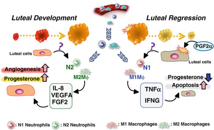
Fig. 6.3
Proposed model of the polarization mechanism of neutrophils (N1 vs. N2) and macrophages (M1 vs. M2), depending on the microenvironment of CL. The polarization of neutrophils and macrophages may be regulated by the luteal microenvironment. Luteotropic and angiogenic factors such as IL-8, VEGFA, and FGF2 facilitate differentiation into N2-neutrophils and M2-macrophages whereas luteolytic factors such as TNFα and IFNG facilitate differentiation into N1-neutrophils and M1-macrophages
6.5.2 Multiple Roles of Macrophages: “M1” Versus “M2” Macrophages
Polarization of macrophages is well understood compared with that of neutrophils [86, 87]. Macrophages activated by pro-inflammatory cytokines (TNFα and IFNG) and microbial products (LPS) are termed M1-type macrophages. M1 macrophages are characterized by high production of pro-inflammatory cytokines including TNFα, IFNG, and IL-12. M1 macrophages promote the differentiation of naïve CD4+ T cells into Th1 effector cells and Th17 cells and secrete high levels of nitric oxide, and thus are a key cell type in the progression of inflammation [86]. In contrast, M2-macrophage polarization is activated by Th2-type cytokines, such as IL-4, IL-10, and IL-13, and stimulates differentiation to CD4+ Th2 cells and regulatory T cells, indicating involvement in regulation of the inflammatory response, Th2 immunity , and tissue remodeling and repair [88].
Development of the bovine CL (angiogenesis and tissue remodeling) contrasts markedly with regression of the CL (angiolysis and tissue disruption); regardless, high numbers of macrophages are observed in both the developing and regressing CL [11]. Therefore, we postulate that the characteristics of these macrophages to differ depending on luteal environment. We investigated the mRNA expression of CD40 (as a marker of the M1 type) and CD163 (as a marker of the M2 type) in the bovine CL. M2-type macrophages were predominant in the developing CL, whereas M1-type macrophages were predominant in the regressing CL (Shirasuna et al., unpublished observations). Thus, the bovine CL has the potential to recruit and regulate macrophages by secretion of cytokines and chemokines, and macrophage function may be closely regulated by the luteal microenvironment (Fig. 6.3).
6.6 The Corpus Luteum in Early Pregnancy
6.6.1 Lymphatic Systems of the CL of Early Pregnancy
The lymphatic vascular system is considered the body’s second circulation system for maintaining interstitial fluid pressure equilibrium and transporting tissue fluid, proteins, and cells [89]. The lymphatic system is also crucial during the immune response to infectious agents, as lymphatic vessels are the route by which dendritic cells, macrophages, and neutrophils migrate to the lymph nodes and lymphoid organs to present antigens to T cells. Two VEGF family members, VEGFC and VEGFD, regulate the lymphatic endothelial cells via their receptor VEGFR-3 [90–92]. Xu and Stouffer [93] have reported that the VEGFC/VEGFD-VEGFR3 system regulates lymphangiogenesis as well as luteal structure and function in the primate CL. Importantly, an injection of soluble VEGFR3 (which acts as an anti-VEGFR3 antibody) into the preovulatory follicle inhibited follicle rupture and ovulation and suppressed progesterone production in the monkey CL [93]. Interestingly, it has been reported that expressions of lymphatic vessel endothelial hyaluronan receptor (LYVE1, a marker of lymphatic vessel) and VEGFC were increased within the bovine CL of pregnancy [94]. In addition, interferon-τ (IFNT), a well-known pregnancy recognition signal for maintenance of CL in ruminants [95] secreted by embryonic trophoblast cells, stimulates lymphatic endothelial cell proliferation and formation of capillary-like tubes in vitro [94]. Hein et al. [96] reported that the concentration of progesterone was higher in ovarian lymph vessels than in uterine lymph or ovarian vein plasma during all stages of pregnancy in cows. These findings suggest that the lymphatic system of the bovine CL may function during early pregnancy [94] (Fig. 6.4).
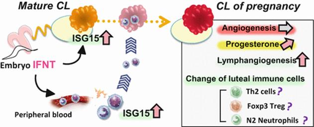
Fig. 6.4
Proposed model of the conversion of the cyclic CL into the pregnancy CL, and the possible role of IFNT as an immune regulator. IFNT produced by the embryo is released from the uterus into peripheral blood, and directly affects immune cells and luteal tissue; this results in the recruitment of immune cells to the CL. IFNT and the recruited immune cells stimulate progesterone secretion and lymphangiogenesis to convert the cyclic CL into the pregnancy CL
6.6.2 Immune System of the CL in Early Pregnancy
In addition to its intrauterine function, IFNT produced by the conceptus passes through the uterine lumen and enters the uterine vein [97]. Compared to other cycling animals, IFNT upregulates the expression of IFN-stimulated gene 15 (ISG15) mRNA in both the endometrium and the CL as well as peripheral immune cells of pregnant ewes and cows [98–100]. These findings indicate that IFNT has a crucial role in transformation of the cyclic CL into the pregnancy CL using the systemic immune system in ruminants (Fig. 6.4). Moreover, ISG15 mRNA levels in bovine PBMC and neutrophils were higher in pregnant cows than in nonpregnant cows after artificial insemination [98, 99, 101]; this suggests the transmission of IFNT signals from the uterus to peripheral immune cells. In addition, IFNT treatment regulates the in vitro expression of ISG15 and IL10 mRNA in PBMCs and PMNs [102]. Therefore, IFNT might cause these changes via ISG responses and the regulation of Th1/Th2 cytokines in cows. We recently reported that compared to the CL of nonpregnant animals, the CL of pregnant animals (day 16 after insemination) had a higher number of neutrophils and a greater expression of IL-8 and ISG15 [103]. Interestingly, IFNT stimulated IL-8 expression in luteal cells; this resulted in the increased migration of IFNT-activated neutrophils [103]. These results suggest that IFNT causes an increase in the number of neutrophils and upregulates their function via IL-8 expression in luteal cells in the early pregnancy CL, and that both neutrophils and IL-8, when stimulated by IFNT, are associated with the maternal recognition in cows (Fig. 6.4).
Fujiwara [104] proposed a new hypothesis that in humans, PBMCs contribute to embryonic–maternal cross-talk through the systemic circulation to transmit pregnancy status to the CL [13]. Indeed, PBMCs from pregnant woman stimulated the progesterone secretion of luteal cells from both pregnant and nonpregnant women [13]. The production levels of Th-2 cytokines such as IL-4 and IL-10 were increased in the co-culture of PBMCs and luteal cells derived from pregnant women, and these cytokines promoted progesterone production in vitro [13]. This hypothesis could be the basis for “systemic maternal recognition of pregnancy in ruminants” in addition to the local interaction between the uterus and embryo.
6.7 Conclusion
The physiological function of the CL is to produce a large amount of progesterone, thereby playing a vital role in the fate of the embryo. After ovulation, immune cells including neutrophils, macrophages, T cells, and dendritic cells are recruited into the developing CL by chemokines and cytokines produced by the luteal cells (Fig. 6.1). During the developing luteal phase, these immune cells can potentially increase progesterone secretion by releasing IL-8, PGE2, VEGFA, and FGF2 within the early CL (Fig. 6.1). In addition, these cytokines released from recruited immune cells and luteal cells stimulate angiogenesis and lymphangiogenesis. We hypothesize that the recruited neutrophils and macrophages may differentiate into “N2-type neutrophils” and “M2-type macrophages,” respectively, in the microenvironment in the developing CL and produce potent promoters of angiogenesis (Fig. 6.3).
Uterine PGF2α, which functions as a start signal of luteolysis, drastically increases the recruitment of various immune cells including macrophages, T lymphocytes, eosinophils, and neutrophils into the CL (Fig. 6.2). Moreover, PGF2α stimulates the production of various types of inflammatory cytokines such as TNFα, IL-1, and IFNG, and chemokines such as CCL2 and IL-8. These cytokines and chemokines recruit macrophages, neutrophils, and T cells into the CL to enhance luteolytic cascades through inflammatory and immune responses (Fig. 6.2). In addition, the inhibition of angiogenesis and induction of vasoconstriction induce an inflammatory immune response, which leads to decreased progesterone production, cell apoptosis, and phagocytosis within the regressing CL. We hypothesize that, in contrast to those in the developing CL, recruited neutrophils and macrophages in the microenvironment of the regressing CL may differentiate into “N1-type neutrophils ” and “M1-type macrophages,” respectively, and produce potent promoters of luteolytic cascades, such as TNFα and IFNG (Fig. 6.3).
In cows, maternal progesterone concentrations have a marked influence on the development of the embryo and on its ability to produce IFNT. The maternal immune system should be able to accept the conceptus as a semi-allograft not only in the local interaction between the embryo and the uterus but also in the peripheral interactions between the conceptus and the maternal organ systems, including immune cells and the CL (Fig. 6.4). A proposed hypothesis is that in response to embryo recognition, peripheral immune cells, especially T cells and neutrophils, transmit signals (detected by ISG15 expression) to various organs in the whole body, including the uterus and maternal vessels, to prepare for and maintain embryo implantation. The immune cells that are activated by IFNT may accumulate within the CL and participate in the establishment of the pregnancy CL, which includes an increase in progesterone concentration and lymphangiogenesis (Fig. 6.4). Furthermore, we speculate that IFNT (and/or ISGs stimulated by IFNT) in peripheral blood regulates immune tolerance to stimulate the differentiation of Th2 cells, forkhead/winged helix transcription factor (Foxp3)+ Treg cells, and N2-type neutrophils, which support the acceptance of the conceptus as a semi-allograft.
Further investigations of the regulatory mechanisms of luteal function involving angiology and immunology are essential for better understanding of reproductive physiology.
Acknowledgments
This study was supported by the Grant-in-Aid for Scientific Research of the Japan Society for the Promotion of Science (JSPS) and the Global COE Program, Ministry of Education, Culture, Science and Technology, Japan.
References
1.
Lobel BL, Levy E. Enzymic correlates of development, secretory function and regression of follicles and corpora lutea in the bovine ovary. Acta Endocrinol (Copenh). 1968;(Suppl 132):5–63.
2.
Alila HW, Hansel W. Induction of lymphopenia causes luteal dysfunction in cattle. Biol Reprod. 1984;31(4):671–8.PubMed
3.
Bukovsky A, Presl J, Krabec Z, Bednarik T. Ovarian function in adult rats treated with antithymocyte serum. Experientia (Basel). 1977;33(2):280–1.
4.
Pate JL, Toyokawa K, Walusimbi S, Brzezicka E. The interface of the immune and reproductive systems in the ovary: lessons learned from the corpus luteum of domestic animal models. Am J Reprod Immunol. 2010;64(4):275–86.PubMed
5.
Walusimbi SS, Pate JL. Physiology and Endocrinology Symposium: role of immune cells in the corpus luteum. J Anim Sci. 2013;91:1650–9.PubMed
6.
Bukulmez O, Arici A. Leukocytes in ovarian function. Hum Reprod Update. 2000;6:1–15.PubMed
7.
Kizuka F, Tokuda N, Takagi K, Adachi Y, Lee L, Tamura I, Maekawa R, Taketani T, Tamura H, Suzuki T, Owada Y, Sugino N. Involvement of bone marrow-derived vascular progenitor cells in neovascularization during formation of the corpus luteum in mice. Biol Reprod. 2012;87:1–7.
8.
Swirski FK, Nahrendorf M, Etzrodt M, Wildgruber M, Cortez-Retamozo V, Panizzi P, Figueiredo JL, Kohler RH, Chudnovskiy A, Waterman P, Aikawa E, Mempel TR, Libby P, Weissleder R, Pittet MJ. Identification of splenic reservoir monocytes and their deployment to inflammatory sites. Science. 2009;325:612–6.PubMedPubMedCentral
9.
Oakley OR, Kim H, El-Amouri I, Lin PC, Cho J, Bani-Ahmad M, Ko C. Periovulatory leukocyte infiltration in the rat ovary. Endocrinology. 2010;151(9):4551–9.PubMedPubMedCentral
10.
Endo T, Kanayama K. Effects of splenectomy on luteal function in pseudopregnant rabbits. J Int Med Res. 1998;26:93–7.PubMed
11.
Penny LA, Armstrong D, Bramley TA, Webb R, Collins RA, Watson ED. Immune cells and cytokine production in the bovine corpus luteum throughout the oestrous cycle and after induced luteolysis. J Reprod Fertil. 1999;115:87–96.PubMed
12.
Poole DH, Pate JL. Luteal microenvironment directs resident T lymphocyte function in cows. Biol Reprod. 2012;86(2):29.PubMed
13.
Hashii K, Fujiwara H, Yoshioka S, Kataoka N, Yamada S, Hirano T, Mori T, Fujii S, Maeda M. Peripheral blood mononuclear cells stimulate progesterone production by luteal cells derived from pregnant and non-pregnant women: possible involvement of interleukin-4 and interleukin-10 in corpus luteum function and differentiation. Hum Reprod. 1998;13:2738–44.PubMed
14.
Walusimbi SS, Pate JL. Luteal cells from functional and regressing bovine corpora lutea differentially alter the function of gamma delta T cells. Biol Reprod. 2014;90:1–7.
15.
Szekeres-Bartho J, Wegmann TG. A progesterone-dependent immunomodulatory protein alters the Th1/Th2 balance. J Reprod Immunol. 1996;31:81–95.PubMed
16.
Petroff M, Coggeshall KM, Jones LS, Pate JL. Bovine luteal cells elicit major histocompatibility complex class II-dependent T-cell proliferation. Biol Reprod. 1997;57:887–93.PubMed
17.
Cannon MJ, Pate JL. The role of major histocompatibility complex molecules in luteal function. Reprod Biol Endocrinol. 2003;1:93.PubMedPubMedCentral
18.
Ndiaye K, Poole DH, Walusimbi S, Cannon MJ, Toyokawa K, Maalouf SW, Dong J, Thomas P, Pate JL. Progesterone effects on lymphocytes may be mediated by membrane progesterone receptors. J Reprod Immunol. 2012;95:15–26.PubMed
19.
Best CL, Pudney J, Welch WR, Burger N, Hill JA. Localization and characterization of white blood cell populations within the human ovary throughout the menstrual cycle and menopause. Hum Reprod. 1996;11:790–7.PubMed
20.
Gaytan F, Morales C, Garcia-Pardo L, Reymundo C, Bellido C, Sanchez-Criado JE. Macrophages, cell proliferation, and cell death in the human menstrual corpus luteum. Biol Reprod. 1998;59:417–25.PubMed
21.
Standaert FE, Zamora CS, Chew BP. Quantitative and qualitative changes in blood leukocytes in the porcine ovary. Am J Reprod Immunol. 1991;25:163–8.PubMed
22.
Turner EC, Hughes J, Wilson H, Clay M, Mylonas KJ, Kipari T, Duncan WC, Fraser HM. Conditional ablation of macrophages disrupts ovarian vasculature. Reproduction. 2011;141:821–31.PubMedPubMedCentral
23.
Care AS, Diener KR, Jasper MJ, Brown HM, Ingman WV, Robertson SA. Macrophages regulate corpus luteum development during embryo implantation in mice. J Clin Invest. 2013;123:3472–87.PubMedPubMedCentral
24.
Emi N, Kanzaki H, Yoshida M, Takakura K, Kariya M, Okamoto N, Imai K, Mori T. Lymphocytes stimulate progesterone production by cultured human granulosa luteal cells. Am J Obstet Gynecol. 1991;165:1469–74.PubMed
25.
Halme J, Hammond MG, Syrop CH, Talbert LM. Peritoneal macrophages modulate human granulosa-luteal cell progesterone production. J Clin Endocrinol Metab. 1985;61:912–6.PubMed
26.
Adashi EY. The potential relevance of cytokines to ovarian physiology: the emerging role of resident ovarian cells of the white blood cell series. Endocr Rev. 1990;11:454–64.PubMed
27.
Polec A, Tanbo T, Fedorcsak P. Cellular interaction regulates interleukin-8 secretion by granulosa-lutein cells and monocytes/macrophages. Am J Reprod Immunol. 2009;61:85–94.PubMed
28.
Polec A, Raki M, Abyholm T, Tanbo TG, Fedorcsak P. Interaction between granulosa-lutein cells and monocytes regulates secretion of angiogenic factors in vitro. Hum Reprod. 2011;26:2819–29.PubMed
29.
Murdoch WJ. Treatment of sheep with prostaglandin F2 alpha enhances production of a luteal chemoattractant for eosinophils. Am J Reprod Immunol Microbiol. 1987;15:52–6.PubMed
30.
Aust G, Simchen C, Heider U, Hmeidan FA, Blumenauer V, Spanel-Borowski K. Eosinophils in the human corpus luteum: the role of RANTES and eotaxin in eosinophil attraction into periovulatory structures. Mol Hum Reprod. 2000;6:1085–91.PubMed
31.
Jiemtaweeboon S, Shirasuna K, Nitta A, Kobayashi A, Schuberth HJ, Shimizu T, Miyamoto A. Evidence that polymorphonuclear neutrophils infiltrate into the developing corpus luteum and promote angiogenesis with interleukin-8 in the cow. Reprod Biol Endocrinol. 2011;9:79.PubMedPubMedCentral
32.
Brannstrom M, Pascoe V, Norman RJ, McClure N. Localization of leukocyte subsets in the follicle wall and in the corpus luteum throughout the human menstrual cycle. Fertil Steril. 1994;61:488–95.PubMed
33.
Brannstrom M, Giesecke L, Moore IC, van den Heuvel CJ, Robertson SA. Leukocyte subpopulations in the rat corpus luteum during pregnancy and pseudopregnancy. Biol Reprod. 1994;50:1161–7.PubMed
34.
Shimizu T, Kaji A, Murayama C, Magata F, Shirasuna K, Wakamiya K, Okuda K, Miyamoto A. Effects of interleukin-8 on estradiol and progesterone production by bovine granulosa cells from large follicles and progesterone production by luteinizing granulosa cells in culture. Cytokine. 2012;57:175–81.PubMed
35.
Talbott H, Delaney A, Zhang P, Yu Y, Cushman RA, Cupp AS, Hou X, Davis JS. Effects of IL8 and immune cells on the regulation of luteal progesterone secretion. Reproduction. 2014;148:21–31.PubMedPubMedCentral
36.
Nozawa H, Chiu C, Hanahan D. Infiltrating neutrophils mediate the initial angiogenic switch in a mouse model of multistage carcinogenesis. Proc Natl Acad Sci USA. 2006;103:12493–8.PubMedPubMedCentral
37.
Koch AE, Polverini PJ, Kunkel SL, Harlow LA, DiPietro LA, Elner VM, Elner SG, Strieter RM. Interleukin-8 as a macrophage-derived mediator of angiogenesis. Science. 1992;258:1798–801.PubMed
38.
Goto J, Suganuma N, Takata K, Kitamura K, Asahina T, Kobayashi H, Muranaka Y, Furuhashi M, Kanayama N. Morphological analyses of interleukin-8 effects on rat ovarian follicles at ovulation and luteinization in vivo. Cytokine. 2002;20:168–73.PubMed
39.
Yasuda M, Shimizu S, Tokuyama S, Watanabe T, Kiuchi Y, Yamamoto T. A novel effect of polymorphonuclear leukocytes in the facilitation of angiogenesis. Life Sci. 2000;66:2113–21.PubMed
40.
Schruefer R, Lutze N, Schymeinsky J, Walzog B. Human neutrophils promote angiogenesis by a paracrine feedforward mechanism involving endothelial interleukin-8. Am J Physiol Heart Circ Physiol. 2005;288(3):H1186–92.PubMed
41.
Mueller MD, Lebovic DI, Garrett E, Taylor RN. Neutrophils infiltrating the endometrium express vascular endothelial growth factor: potential role in endometrial angiogenesis. Fertil Steril. 2000;74:107–12.PubMed
42.
Heryanto B, Girling JE, Rogers PA. Intravascular neutrophils partially mediate the endometrial endothelial cell proliferative response to oestrogen in ovariectomised mice. Reproduction. 2004;127:613–20.PubMed
43.
Ancelin M, Chollet-Martin S, Herve MA, Legrand C, El Benna J, Perrot-Applanat M. Vascular endothelial growth factor VEGF189 induces human neutrophil chemotaxis in extravascular tissue via an autocrine amplification mechanism. Lab Invest. 2004;84:502–12.PubMed
44.
Zittermann SI, Issekutz AC. Endothelial growth factors VEGF and bFGF differentially enhance monocyte and neutrophil recruitment to inflammation. J Leukoc Biol. 2006;80:247–57.PubMed
45.
Freitag N, Tirado-Gonzalez I, Barrientos G, Herse F, Thijssen VL, Weedon-Fekjaer SM, Schulz H, Wallukat G, Klapp BF, Nevers T, Sharma S, Staff AC, Dechend R, Blois SM. Interfering with Gal-1-mediated angiogenesis contributes to the pathogenesis of preeclampsia. Proc Natl Acad Sci USA. 2013;110:11451–6.PubMedPubMedCentral
46.
Spanel-Borowski K. Ovulation as danger signaling event of innate immunity. Mol Cell Endocrinol. 2011;333:1–7.PubMed
47.
Cohen-Fredarow A, Tadmor A, Raz T, Meterani N, Addadi Y, Nevo N, Solomonov I, Sagi I, Mor G, Neeman M, Dekel N. Ovarian dendritic cells act as a double-edged pro-ovulatory and anti-inflammatory sword. Mol Endocrinol. 2014;28:1039–54.PubMed
48.
Furukawa K, Fujiwara H, Sato Y, Zeng BX, Fujii H, Yoshioka S, Nishi E, Nishio T. Platelets are novel regulators of neovascularization and luteinization during human corpus luteum formation. Endocrinology. 2007;148:3056–64.PubMed
49.
Reibiger I, Spanel-Borowski K. Difference in localization of eosinophils and mast cells in the bovine ovary. J Reprod Fertil. 2000;118:243–9.PubMed
50.
Murdoch WJ, Van Kirk EA. Aetiology of attenuated luteal development in prednisolone-induced eosinopenic ewes. Reprod Fertil Dev. 2000;12:127–32.PubMed
51.
Kliem H, Rodler D, Ulbrich SE, Sinowatz F, Berisha B, Meyer HH, Schams D. Dexamethasone-induced eosinopenia is associated with lower progesterone production in cattle. Reprod Domest Anim. 2013;48:137–48.PubMed
52.
McCracken JA, Schramm W, Barcikowski B, Wilson Jr L. The identification of prostaglandin F2 alpha as a uterine luteolytic hormone and the hormonal control of its synthesis. Acta Vet Scand Suppl. 1981;77:71–88.PubMed
53.
Bauer M, Reibiger I, Spanel-Borowski K. Leucocyte proliferation in the bovine corpus luteum. Reproduction. 2001;121:297–305.PubMed
54.
Al-Zi'abi MO, Fraser HM, Watson ED. Cell death during natural and induced luteal regression in mares. Reproduction. 2002;123:67–77.PubMed
55.
Petrovska M, Dimitrov DG, Michael SD. Quantitative changes in macrophage distribution in normal mouse ovary over the course of the estrous cycle examined with an image analysis system. Am J Reprod Immunol. 1996;36:175–83.PubMed
56.
Komatsu K, Manabe N, Kiso M, Shimabe M, Miyamoto H. Changes in localization of immune cells and cytokines in corpora lutea during luteolysis in murine ovaries. J Exp Zool A Comp Exp Biol. 2003;296:152–9.PubMed
57.
Brannstrom M, Friden B. Immune regulation of corpus luteum function. Semin Reprod Endocrinol. 1997;15:363–70.PubMed
58.
Townson DH, Liptak AR. Chemokines in the corpus luteum: implications of leukocyte chemotaxis. Reprod Biol Endocrinol. 2003;1:94.PubMedPubMedCentral
59.
Penny LA. Monocyte chemoattractant protein 1 in luteolysis. Rev Reprod. 2000;5:63–6.PubMed
60.
Okuda K, Sakumoto R. Multiple roles of TNF super family members in corpus luteum function. Reprod Biol Endocrinol. 2003;95:1–10.
61.
Pate JL, Landis KP. Immune cells in the corpus luteum: friends or foes? Reproduction. 2001;122:665–76.PubMed
62.
Shima T, Sasaki Y, Itoh M, Nakashima A, Ishii N, Sugamura K, Saito S. Regulatory T cells are necessary for implantation and maintenance of early pregnancy but not late pregnancy in allogeneic mice. J Reprod Immunol. 2010;85:121–9.PubMed
63.
Krusche CA, Vloet TD, Herrler A, Black S, Beier HM. Functional and structural regression of the rabbit corpus luteum is associated with altered luteal immune cell phenotypes and cytokine expression patterns. Histochem Cell Biol. 2002;118:479–89.PubMed
64.
Pate JL. Involvement of immune cells in regulation of ovarian function. J Reprod Fertil Suppl. 1995;49:365–77.PubMed
65.
Bowen JM, Towns R, Warren JS, Landis KP. Luteal regression in the normally cycling rat: apoptosis, monocyte chemoattractant protein-1, and inflammatory cell involvement. Biol Reprod. 1999;60:740–6.PubMed
66.
Townson DH, O’Connor CL, Pru JK. Expression of monocyte chemoattractant protein-1 and distribution of immune cell populations in the bovine corpus luteum throughout the estrous cycle. Biol Reprod. 2002;66:361–6.PubMed
67.
Neuvians TP, Schams D, Berisha B, Pfaffl MW. Involvement of pro-inflammatory cytokines, mediators of inflammation, and basic fibroblast growth factor in prostaglandin F2alpha-induced luteolysis in bovine corpus luteum. Biol Reprod. 2004;70:473–80.PubMed
68.
Sawyer HR, Niswender KD, Braden TD, Niswender GD. Nuclear changes in ovine luteal cells in response to PGF2 alpha. Domest Anim Endocrinol. 1990;7:229–37.PubMed
69.
Juengel JL, Garverick HA, Johnson AL, Youngquist RS, Smith MF. Apoptosis during luteal regression in cattle. Endocrinology. 1993;132:249–54.PubMed
70.
Kato S, Shiratsuchi A, Nagaosa K, Nakanishi Y. Phosphatidylserine- and integrin-mediated phagocytosis of apoptotic luteal cells by macrophages of the rat. Dev Growth Differ. 2005;47:153–61.PubMed
71.
Sakumoto R, Vermehren M, Kenngott RA, Okuda K, Sinowatz F. Localization of gene and protein expressions of tumor necrosis factor-{alpha} (TNF), and TNF receptor types I and II in the bovine corpus luteum during the estrous cycle. J Anim Sci. 2011;89(10):3040–7.PubMed
72.
Taniguchi H, Yokomizo Y, Okuda K. Fas-Fas ligand system mediates luteal cell death in bovine corpus luteum. Biol Reprod. 2002;66:754–9.PubMed
73.
Pru JK, Lynch MP, Davis JS, Rueda BR. Signaling mechanisms in tumor necrosis factor alpha-induced death of microvascular endothelial cells of the corpus luteum. Reprod Biol Endocrinol. 2003;1:17.PubMedPubMedCentral
74.
Fairchild DL, Pate JL. Interferon-gamma induction of major histocompatibility complex antigens on cultured bovine luteal cells. Biol Reprod. 1989;40:453–7.PubMed
75.
Fairchild DL, Pate JL. Modulation of bovine luteal cell synthetic capacity by interferon-gamma. Biol Reprod. 1991;44:357–63.PubMed
76.
Paape MJ, Bannerman DD, Zhao X, Lee JW. The bovine neutrophil: structure and function in blood and milk. Vet Res. 2003;34:597–627.PubMed
77.
Shirasuna K, Jiemtaweeboon S, Raddatz S, Nitta A, Schuberth HJ, Bollwein H, Shimizu T, Miyamoto A. Rapid accumulation of polymorphonuclear neutrophils in the corpus luteum during prostaglandin F(2alpha)-induced luteolysis in the cow. PLoS One. 2012;7, e29054.PubMedPubMedCentral
78.
Diez-Fraile A, Meyer E, Duchateau L, Paape MJ, Burvenich C. In vitro regulation of Mac-1 expression on bovine polymorphonuclear leukocytes by endotoxin and tumor necrosis factor-alpha at different stages of lactation. Can J Vet Res. 2004;68:232–5.PubMedPubMedCentral
79.
Sohn EJ, Paape MJ, Connor EE, Bannerman DD, Fetterer RH, Peters RR. Bacterial lipopolysaccharide stimulates bovine neutrophil production of TNF-alpha, IL-1beta, IL-12 and IFN-gamma. Vet Res. 2007;38:809–18.PubMed
80.
Benyo DF, Haibel GK, Laufman HB, Pate JL. Expression of major histocompatibility complex antigens on the bovine corpus luteum during the estrous cycle, luteolysis, and early pregnancy. Biol Reprod. 1991;45:229–34.PubMed
81.
Minegishi K, Tanaka M, Nishimura O, Tanigaki S, Miyakoshi K, Ishimoto H, Yoshimura Y. Reactive oxygen species mediate leukocyte-endothelium interactions in prostaglandin F2alpha -induced luteolysis in rats. Am J Physiol Endocrinol Metab. 2002;283:E1308–15.PubMed
82.
Pepperell JR, Wolcott K, Behrman HR. Effects of neutrophils in rat luteal cells. Endocrinology. 1992;130:1001–8.PubMed
83.
Fridlender ZG, Sun J, Kim S, Kapoor V, Cheng G, Ling L, Worthen GS, Albelda SM. Polarization of tumor-associated neutrophil phenotype by TGF-beta: “N1” versus “N2” TAN. Cancer Cell. 2009;16:183–94.PubMedPubMedCentral
84.
Shen L, Smith JM, Shen Z, Eriksson M, Sentman C, Wira CR. Inhibition of human neutrophil degranulation by transforming growth factor-beta1. Clin Exp Immunol. 2007;149:155–61.PubMedPubMedCentral
85.
Piccard H, Muschel RJ, Opdenakker G. On the dual roles and polarized phenotypes of neutrophils in tumor development and progression. Crit Rev Oncol Hematol. 2012;82(3):296–309.PubMed
86.
Coffelt SB, Hughes R, Lewis CE. Tumor-associated macrophages: effectors of angiogenesis and tumor progression. Biochim Biophys Acta. 1796;2009:11–8.
87.
Nagamatsu T, Schust DJ. The contribution of macrophages to normal and pathological pregnancies. Am J Reprod Immunol. 2010;63:460–71.PubMed
88.
Gordon S. Alternative activation of macrophages. Nat Rev Immunol. 2003;3:23–35.PubMed
89.
Wang Y, Oliver G. Current views on the function of the lymphatic vasculature in health and disease. Genes Dev. 2010;24:2115–26.PubMedPubMedCentral
90.
Yamada Y, Nezu J, Shimane M, Hirata Y. Molecular cloning of a novel vascular endothelial growth factor VEGF-D. Genomics. 1997;42:483–8.PubMed
91.
Joukov V, Sorsa T, Kumar V, Jeltsch M, Claesson-Welsh L, Cao Y, Saksela O, Kalkkinen N, Alitalo K. Proteolytic processing regulates receptor specificity and activity of VEGF-C. EMBO J. 1997;16:3898–911.PubMedPubMedCentral
92.
Karkkainen MJ, Haiko P, Sainio K, Partanen J, Taipale J, Petrova TV, Jeltsch M, Jackson DG, Talikka M, Rauvala H, Betsholtz C, Alitalo K. Vascular endothelial growth factor C is required for sprouting of the first lymphatic vessels from embryonic veins. Nat Immunol. 2004;5:74–80.PubMed
93.
Xu F, Stouffer RL. Existence of the lymphatic system in the primate corpus luteum. Lymphat Res Biol. 2009;7:159–68.PubMedPubMedCentral
94.
Nitta A, Shirasuna K, Haneda S, Matsui M, Shimizu T, Matsuyama S, Kimura K, Bollwein H, Miyamoto A. Possible involvement of IFNT in lymphangiogenesis in the corpus luteum during the maternal recognition period in the cow. Reproduction. 2011;142:879–92.PubMed
95.
Imakawa K, Anthony RV, Kazemi M, Marotti KR, Polites HG, Roberts RM. Interferon-like sequence of ovine trophoblast protein secreted by embryonic trophectoderm. Nature (Lond). 1987;330:377–9.
96.
Hein WR, Shelton JN, Simpson-Morgan MW, Seamark RF, Morris B. Flow and composition of lymph from the ovary and uterus of cows during pregnancy. J Reprod Fertil. 1988;83:309–23.PubMed
97.
Oliveira JF, Henkes LE, Ashley RL, Purcell SH, Smirnova NP, Veeramachaneni DN, Anthony RV, Hansen TR. Expression of interferon (IFN)-stimulated genes in extrauterine tissues during early pregnancy in sheep is the consequence of endocrine IFN-tau release from the uterine vein. Endocrinology. 2008;149:1252–9.PubMed
98.
Han H, Austin KJ, Rempel LA, Hansen TR. Low blood ISG15 mRNA and progesterone levels are predictive of non-pregnant dairy cows. J Endocrinol. 2006;191:505–12.PubMed
99.
Gifford CA, Racicot K, Clark DS, Austin KJ, Hansen TR, Lucy MC, Davies CJ, Ott TL. Regulation of interferon-stimulated genes in peripheral blood leukocytes in pregnant and bred, nonpregnant dairy cows. J Dairy Sci. 2007;90:274–80.PubMed
100.
Yang L, Wang XL, Wan PC, Zhang LY, Wu Y, Tang DW, Zeng SM. Up-regulation of expression of interferon-stimulated gene 15 in the bovine corpus luteum during early pregnancy. J Dairy Sci. 2010;93:1000–11.PubMed
101.
Green JC, Okamura CS, Poock SE, Lucy MC. Measurement of interferon-tau (IFN-tau) stimulated gene expression in blood leukocytes for pregnancy diagnosis within 18-20 d after insemination in dairy cattle. Anim Reprod Sci. 2010;121:24–33.PubMed
102.
Shirasuna K, Matsumoto H, Kobayashi E, Nitta A, Haneda S, Matsui M, Kawashima C, Kida K, Shimizu T, Miyamoto A. Upregulation of interferon-stimulated genes and interleukin-10 in peripheral blood immune cells during early pregnancy in dairy cows. J Reprod Dev. 2012;58:84–90.PubMed
103.
Shirasuna K, Matsumoto H, Matsuyama S, Kimura K, Bollwein H, Miyamoto A. Possible role of IFNT on the bovine corpus luteum and neutrophils during the early pregnancy. Reproduction. 2015;150(3):217–25.PubMed
104.
Fujiwara H. Do circulating blood cells contribute to maternal tissue remodeling and embryo-maternal cross-talk around the implantation period? Mol Hum Reprod. 2009;15:335–43.PubMed