
Robert Rekawiecki1 , Magdalena K. Kowalik1 and Jan Kotwica1
(1)
Department of Physiology and Toxicology of Reproduction, Institute of Animal Reproduction and Food Research, The Polish Academy of Sciences, Olsztyn, 10-748, Poland
Robert Rekawiecki
Email: r.rekawiecki@pan.olsztyn.pl
Magdalena K. Kowalik
Email: m.kowalik@pan.olsztyn.pl
Jan Kotwica (Corresponding author)
Email: j.kotwica@pan.olsztyn.pl
Abstract
The function of the corpus luteum (CL) is to produce progesterone (P4), which is the main regulator of estrous cycle duration and creates suitable conditions for embryo implantation and development. The CL also synthesizes moderate amounts of estradiol (E2). The action of these steroid hormones on target cells are evoked by specific nuclear receptors that belong to the family of receptor-dependent transcription factors. The physiological effect of P4 upon target cells is mediated through interaction of this hormone with nuclear progesterone receptor (PGR) isoforms A (PGRA) and B (PGRB) and that of E2 through the alpha (ERα) and beta (ERβ) receptors. Steroids may also affect cells through a nongenomic mechanism, which involves the membrane steroid-binding proteins such as the progesterone receptor membrane component (PGRMC) 1 and 2 and the membrane progestin receptors (mPR) alpha (mPRα), beta (mPRβ), and gamma (mPRγ), and the G protein-coupled estrogen receptor (GPR30). These proteins rapidly activate the appropriate intracellular signal transduction pathways, and subsequently they can initiate specific cell responses or modulate genomic cell responses. The diversity of nuclear and membrane steroid hormone receptors enhances their regulatory influence on the CL function.
Keywords
Corpus luteumProgesterone receptorEstradiol receptorSteroid receptor isoforms
5.1 Introduction
The corpus luteum (CL) is a transient endocrine gland formed from the secretory cells of the ovarian follicle following ovulation. The main function of the CL is the production of progesterone (P4), which has a key role in many processes that regulate female fertility; however, the CL also synthesizes a moderate amount of estradiol (E2) . The action of these steroid hormones is carried out by specific nuclear receptors that belong to the family of receptor-dependent transcription factors, which affect the regulation of specific target gene expression after their activation. It was also found that P4 and E2 affect cells by a nongenomic mechanism because the effect of hormone action occurs in a few minutes or even seconds after application and the effect is not inhibited by inhibitors of transcription and translation.
5.2 Structure of the Progesterone and Estradiol Receptors
The classical, well-studied mechanism by which steroids influence cells is via nuclear receptors. Progesterone works mainly by two distinct isoforms of the receptor: A (PGRA ) and B (PGRB ), which are encoded by the same gene but are transcribed under the influence of two different promoters. The bovine PGR gene consists of eight exons and is located on chromosome 15 [1]. The specific element that differentiates PGRB from PGRA is an additional section located at the N-terminal end of the protein. The length of this section ranges from 128 amino acids in chickens [2] to approximately 164 amino acids in humans [3]. The receptor protein is composed of a number of different regions, which are responsible for different functions of the receptor. Starting from the N-terminus part of the PGR, there are two domains: AF-1 and AF-3 (Fig. 5.1), which bind transcriptional factors that are responsible for the activation of the appropriate promoter and turn on transcription of the isoforms. The AF-1 domain is present in both isoforms of PGR, but AF-3 is only found in isoform B. The AF-1 domain is located upstream in the inhibitor domain (IF), which includes approximately 140 amino acids. The antagonist receptor is connected to this domain and thereby inhibits receptor activity. AF-3 contributes to PGRB transcriptional activity by suppression of the IF domain activity, which is contained within the sequence common to PGRA and PGRB [4]. The most conserved part of the receptor isoforms is the DNA-binding domain (DBD) , adjacent to the AF-1 domain. It contains approximately 66 to 68 amino acids that form two zinc fingers; these are responsible for the interaction of a hormone–receptor complex with the appropriate regulatory sequences within the promoter of the target gene and therefore regulate transcription [4]. A ligand-binding domain (LBD) is located on the C-terminal domain of the DBD. An additional AF-2 domain is found in this part of the receptor, which is responsible for the activation of the receptor by connecting transcription factors. Moreover, the AF-2 domain binds an inactive receptor with heat shock proteins (HSPs) , and it is also responsible for receptor dimerization [3]. PGRA and PGRB affect the target genes in a different manner. PGRB is a potent activator of progesterone-dependent genes in different cells. When both PGR isoforms are activated in the cell, PGRA acts as a potent inhibitor of PGRB and decreases the effect of P4 on target cells [5].

Fig. 5.1
Schematic representation of the human progesterone receptor gene and protein domains of progesterone receptor (PGR) B (PGRB), progesterone receptor A (PGRA), and progesterone receptor C (PGRC) isoforms. In humans, the progesterone receptor gene consists of eight exons. All receptor isoforms are transcribed from the same gene but are under the influence of different promoters. DBDDNA-binding domain, LBD ligand-binding domain, AF1–AF3 activation domains, ID inhibitory domain
Moreover, in human breast cancer cell lines, but not in the CL, isoform C (PGRC) was found, which does not have one of the zinc fingers in the DBD domain [6] and therefore shows no transcriptional activity. The sequence of PGRC is limited to the full ligand-binding domain (LBD), and the sequence responsible for the dimerization and receptor localization is in the nucleus. PGRC exhibits the ability to bind P4 and its antagonists with the same affinity as PGRA and PGRB. The action of PGRC is not yet fully understood, but it has been claimed it can form heterodimers with the isoforms PGRA and PGRB, thus controlling their transcription properties [7].
In the CL, there are two types of estradiol receptors (ERs): alpha (ERα) and beta (ERβ) [8, 9], which are encoded by two separate genes (ESR1 and ESR2) [10]. Both receptors have a modular structure and contain all the domains typical for the construction of nuclear receptors.
5.3 Activation of the Steroid Receptor
Receptor activation involves the conversion of the biologically inactive form of the receptor to the active form that is capable of binding to genes and regulating their transcription . The inactive form of the receptor is associated with a complex of chaperone proteins including HSP 90, HSP 70, p23, and immunophilins [11]. Formation of this intermediate complex requires energy released from ATP breakdown, which suggests involvement of phosphorylation processes . P4 binding initiates activation of the receptor, which entails a change in the conformation of the receptor and disconnection of the chaperone proteins, leading to unveiling of the DBD and nuclear translocation (Fig. 5.2). This process also requires energy from ATP breakdown [12]. Phosphorylation of the receptor causes a change in electric charge, which causes further changes that enable receptor dimerization. Both PGR isoforms, PGRA and PGRB, can bind as a homodimer A:A, a homodimer B:B, and a heterodimer A:B. Dimerization consequently modulates the transcriptional activities of PGR and determines the diversity of physiological responses associated with P4 action [3]. After translocation to the nucleus, receptors bind (as a dimer) to a hormone response element (HRE) , which is located in the promoter of a target gene. The next step is connection of coregulators to the receptor dimer, and then, the transcription process of the target gene is initiated or inhibited [13]. The activation of ERs is also followed by a classical pattern of nuclear receptor activation [10].

Fig. 5.2
Schematic illustration of PGR receptor action. The inactive form of the receptor is located in the cytoplasm and is associated with a complex of chaperone proteins. Progesterone penetrates the cell membrane and connects to the LBD of the receptor. Aggregation of the hormone causes disconnection of the associated chaperone protein complex and dislocation of the receptor to the nucleus where it undergoes dimerization. Receptor dimers connect to the hormone response element (HRE) located within the specific gene promoter. After activation of the receptor dimer by the receptor coactivators, the transcription process begins. P4 progesterone, IF immunophilins, HSP heat shock protein
5.4 Regulation of Steroid Receptor Transcriptional Activity
Coregulators are a large group of transcription factors that regulate gene transcription activated by P4. They interact with the AF-2 domain of the receptor without binding to the DNA of the target gene sequence [14]. There are two groups of coregulators: coactivators that enhance the transcription of genes, and corepressors, which are proteins that inhibit the transcription of genes. The main PGR coactivators are representative of steroid receptor coactivators, which contain SRC-1, SRC-2, SRC-3, and the CREB-binding protein (also described as CREBBP or CBP), which includes CBP and p300 protein [15] and also the P300/CBP-associated factor (p/CAF) (also described as K(lysine) acetyltransferase 2B; KAT2B) [16]. Moreover, there are proteins that do not belong to any of these groups of coactivators, including L7/SPA, RIP140, TIF1, ARA70, HMG-1/2E6-AP, and RPF-1 [17]. The coactivators interact with PGR through the highly conserved motif known as the “NR box,” which consists of three leucine amino acids and two unspecified amino acids (Leu-X-X-Leu-Leu motif) [18, 19]. Coactivators also have the activity of histone acetyltransferase (HAT), which transfers an acetyl group from acetyl CoA to lysine amino acids on histone proteins, leading to their acetylation and causing loosening of chromatin and, consequently, greater availability of transcription factors and polymerase to the appropriate gene sequence. This process is also referred to as transformation of heterochromatin to euchromatin [20]. Another group of coregulators are corepressors, which include two main proteins: nuclear receptor corepressor (N-CoR) and silencing mediator for retinoid and thyroid hormone receptor (SMRT) [19]. Corepressors also have a conservative sequence containing additional amino acids compared to the “NR box” forming the following sequence: Leu-Leu-X-X-X-Ile-X-X-X-Leu. This motif is defined as a CoRNR (‘corner’) box and is responsible for the interaction of the corepressor with the PGR receptor [21]. Corepressor proteins are connected with histone deacetylases (HDACs), which, in contrast to the HAT, remove an acetyl group from the lysine amino acid on a histone; this results in an increase in chromatin condensation and transcription of the target genes not being initialized [22]. We recently found that mRNA expression of the coactivator P300/CBP-associated factor (PCAF) and nuclear receptor corepressor (NCoR) are positively correlated with luteal level of P4 and negatively correlated with mRNA expression for both PGR isoforms in the CL during the estrous cycle in cows (unpublished data). This finding indicated that coregulator involvement is an important step in the regulation of PGR isoform action in the CL. Transcriptional activity of ERs is also regulated by the same groups of coregulators [10, 23].
Other factors involved in the regulation of PGR activity are their antagonists. These compounds negatively regulate receptor interaction with HRE and weakly bind or prevent proper binding of agonists to the receptor, which impairs the activation of the receptor. One of the PGR antagonists is mifepristone (RU 486), which competes with greater affinity than P4 for the LBD [24]. The removal of 42 amino acids from the C-terminus of the receptor abolished P4 binding to the LBD but had no effect on RU 486 binding [25]. However, a single substitution of Gly-Cys amino acids at position 722 of the LBD inhibited binding of the antagonist to this domain and did not affect the binding of P4 to the LBD [26, 27]. The inhibition of PGR may occur in different ways. Antagonists modify the C-terminus segment of the receptor, which is followed by the blockade of coactivators binding to the AF-2 domain, leading to a lack of receptor activation [28]. Full activity of PGR requires interactions between the C- and N-terminus parts of the receptor. RU 486 causes conformational changes in PGR that inhibit this interaction, and as a result, none of the coactivators can be attached to the receptor [29]. Receptor antagonists may also act indirectly, by interaction of PGR with another transcriptional factor, as this happens when HRE of the receptor is partially overlapping with the transcription factor-binding site. For example, RU486 induced inhibition of nuclear factor kappa-light-chain-enhancer of activated B cells (NF-kB) activity associated with blocking of PGR receptor activity. Moreover, Rothchild [30] suggested that RU486 after binding to PGRA may act as an inhibitor of the receptor; however, after connection to the PGRB isoform, it may be a highly active agonist of the receptor, as was found earlier [31, 32]. A similar effect of PGR antagonists was observed in bovine endometrial cells. Both ZK299 and RU486 appeared to affect the mRNA and protein expression levels of the PGRA and PGRB isoforms . Thus, the final physiological effect evoked by an antagonist depends on the PGR isoform that is bound to it [33].
5.5 Progesterone and Estradiol Receptor Isoforms in the CL
The variable expression of isoforms PGRA and PGRB during the estrous cycle has been observed in the CL. The profile of their expression is similar; however, PGRB mRNA levels are minor than those of PGRA mRNA in human (100–1000 times) [34] and bovine CL (500–2000 times) [35]. The highest level of PGRA and PGRB mRNA in the human [36] and bovine [35] CL is at the beginning of the ovarian cycle, and thereafter it is gradually decreased (Fig. 5.3). Sakumoto et al. [37] suggested that, in the newly formed CL, high PGR mRNA and protein expression appeared to be related to the increase in the number of blood vessels that occur in the CL. Additionally, an increase in PGR mRNA expression may also be induced by the LH surge in granulosa cells of preovulatory follicles in cattle [38], and in this way PGR signaling pathways may help mediate the effects of the preovulatory LH surge on follicle rupture in cattle. Moreover, the LH surge increases the oxytocin (OT) level and mRNA expression for the oxytocin receptor in the newly formed CL. Because OT is involved in the regulation of P4 production in the bovine luteal cells, it thus forms a positive feedback loop with P4 [38, 39].

Fig. 5.3
Graphic demonstration of the influence of P4 on the mRNA level of PGRA and PGRB isoforms. A high concentration of P4 within luteal cells induces the expression of PGRA mRNA expression, which results in the repression of PGRB mRNA transcription and finally reduces the cell response. A low level of P4 may decrease the expression of PGRA mRNA followed by an increase in PGRB mRNA transcription. This will induce PGR action and increase the cell response to P4. White arrows impact of high P4 concentrations; black arrowsimpact of low P4 concentrations [36]
Earlier studies by Misao et al. [36] on human CL suggested that a high concentration of P4 within luteal cells induces the expression of PGRA mRNA expression, which results in repression of the transcription of PGRB mRNA, and the effects of P4 in the luteal gland are suppressed [36]. On the other hand, a low level of P4 may decrease the expression of PGRA mRNA, which is followed by an increase in PGRB mRNA transcription: this will induce PGR action and increase the effects of P4 in target cells (Fig. 5.3). However our recent studies in cows revealed the expression of mRNA for PGRA and PGRB decreases from day 6 of the estrous cycle in the CL [35] (Fig. 5.4) along with an increasing concentration of P4 in the CL. So, it is possible there are essential differences between species in regulation of PGR isoform expression. Progesterone was also found to enhance its own effect by increasing the activity of 3-beta-hydroxysteroid dehydrogenase (3β-HSD) on days 6–10 of the estrous cycle in cows [40] and to stimulate gene expression of 3β-HSD, steroidogenic acute regulatory protein (StAR), and cytochrome P450scc [41] without affecting the level of mRNA for total PGR in the CL of the cycling cows [42, 43]. Furthermore, luteotrophic factors (e.g., LH, E2, and PGE2) affect the expression level of mRNA in contrast to the NO donor (NONate) and inhibitor of cytochrome P450scc (aminoglutethimide), and in this way, they modify the action of P4, as shown in the endometrial cells from cycling cows [44].

Fig. 5.4
Progesterone receptor isoform mRNA (mean ± SEM; n = 4 per stage) levels in bovine corpora lutea collected on days 1–5, 6–10, 11–16, and 17–20 of the estrous cycle and 3–5, 6–8, and 9–12 weeks of early pregnancy. The probe and primers for the PGRA isoform were designed against the sequence common to both isoforms; therefore, the mRNA expression determined was the total mRNA expression for both isoforms A and B (PGRAB). The mRNA level for PGRA was obtained after subtracting the mRNA level of PGRB from the mRNA level of PGRAB. (a) PGRB mRNA levels; (b) PGRAB progesterone receptor mRNA levels; (c) PGRA mRNA. Values with different superscripts are significantly different (P< 0.05). Reproduced with permission from [35]
Toward the end of the estrous cycle, P4 production is markedly reduced, which entails a decrease in the mRNA and protein concentration of PGRA and PBRB to their lowest levels [35]: this is the effect of luteolysis initiation and intensification of luteal cells apoptosis, characterized by changes in the nucleus structure, chromatin condensation, and the DNA cutting by endonucleases [45]. Thus, it is possible that the decrease in the level of mRNA and protein expression of both PGR isoforms is a part of the luteolytic events.
During the initial period of pregnancy, the expression of mRNA and protein for both receptor isoforms is low, but it increases as pregnancy progresses [35] (Fig. 5.4). At that time, relatively intense secretion of P4 also occurs, which is followed by a decrease in the transcription of both PGR isoforms. At approximately 45 days of pregnancy, the placenta becomes an additional source of P4 [46]. Thus, the local countercurrent transfer of P4 from the uterus to the ovary [47] may increase the impact of P4 on the CL. As a result, the increased impact of P4 on luteal cells can lead to higher mRNA and protein expression levels for each isoform. Additionally, the ratio of PGRA to PGRB may indicate predominance of isoform B over isoform A at the beginning of pregnancy, which is a crucial period for embryo development. However, secretion of P4 from the placenta may account for the alteration of the PGRA:PGRB ratio and may lead to a modification of cell responsiveness to P4 [48].
A different response of P4 action during the estrous cycle in the CL of monkeys has been reported. Although the P4 profile is similar to that of humans [49], the level of PGRB protein expression in the luteal tissue predominates, and this persists for the duration of the estrous cycle. However, the protein expression of PGRA decreases from its highest level at the early phase of the estrous cycle to its lowest level at the end of the cycle [50]. These data indicated that PGRA is not the dominant isoform in all species and suggests different regulation of PGR isoform expression in luteal cells of different species.
The highest mRNA expression for ERα was detected in the bovine CL during the early luteal phase, followed by a significant decrease to the end of the estrous cycle. In contrast, ERβ mRNA expression is relatively high during the early stage, decreases during the mid-stage, and increases significantly again during the late luteal phase and after CL regression [8]. These data suggested that both isoforms of ERs are involved in CL formation, but that ERβ may also take part in luteolysis.
5.5.1 Nongenomic Effects of Steroid Action Hormones in Cells
Steroids can also affect cells by a nongenomic mechanism in which the effects of the hormone are observed after a very short time following its application (i.e., several seconds or minutes) and are not diminished by inhibitors of transcription and translation [51–53]. These nongenomic actions of P4 and E2 have been demonstrated in several tissues from the female reproductive tract in different species [54–58], including that of cows [59–62]. The mechanism of this steroid action is not fully understood. It has been proposed that the cytoplasmic fractions of nuclear PGR, mainly isoform B, may participate in the nongenomic signaling pathway of P4 [63]. However, this rapid action of P4 has also been observed in cells lacking PGRs, and several studies have shown that it is initiated at the cell membrane [52, 56–58]. Hence, the following putative mechanisms have been suggested: (a) P4 modulates other membrane receptors or impairs the binding of these receptors with their ligands, as has been demonstrated for the OT receptor [51, 59]; (b) P4, as a lipophilic substance, may modify the fluidity of the cell membrane and thus alter the affinity of other membrane receptors for their ligands [64]; and (c) P4 could interact with specific proteins that function as a membrane progesterone receptor [52, 56, 57]. This group consists of membrane progesterone-binding proteins such as the progesterone receptor membrane component (PGRMC) 1 and 2 and the membrane progestin receptors (mPR) alpha (mPRα), beta (mPRβ), and gamma (mPRγ) (Fig. 5.5). Similarly, estradiol may also affect the cell in a nongenomic manner via ERα and ERβ and the G protein-coupled estrogen receptor (GPR30 or GPER1) localized in the cell membranes [65, 66].

Fig. 5.5
Hypothetic model of progesterone (P4) action in the cell. In the genomic pathway, P4 binds to the nuclear progesterone receptor (PGR) and activates PGR gene expression, which stimulates or inhibits the cellular synthesis of proteins. This pathway requires a long time from hormonal activation to induction of a biological effect. In the nongenomic pathway (1), P4 can bind to the membrane progestin receptor (mPR) and activate the mitogen-activated protein (MAP) kinase pathway by decreasing cyclic AMP (cAMP). Moreover, mPR can stimulate protein kinase C (PKC) and phospholipase C (PLC), leading to an increase in the mobilization of Ca2+ in the cell. (2) P4 can also activate the progesterone receptor membrane component (PGRMC), which may form a membrane complex with the serpine mRNA-binding protein 1 (SERBP1) and activate protein kinase G (PKG) to decrease the levels of Ca2+ in the cell. Stimulation of second messengers allows for the target cells to quickly respond to the changes in the hormonal milieu but can also modulate the genomic pathway, leading to the synthesis of new proteins
The presence of membrane proteins that bind steroids ensures the generation of a more rapid cellular response compared with the genomic responses to the steroid. This mechanism allows the target cells to respond quickly to changes in the hormonal milieu and modulate the cell response elicited by the signals that activate the genomic mode of action. Therefore, it is possible that P4 and other steroids can activate the synthesis of new proteins within cells, and at the same time, they can initiate a series of changes at the level of the cell membrane. This effect of steroid hormones can essentially affect cell sensitivity to P4 and to other hormonal factors.
5.5.2 PGRMC1 and PGRMC2 Structure, Expression, and Function
PGRMC1 and PGRMC2 belong to a family of membrane-associated progesterone receptor (MAPR) proteins [53, 56, 57]. PGRMC1 protein was isolated for the first time from porcine vascular smooth muscle cells [54]. This protein is composed of 194 amino acids [54] with a molecular weight of approximately 25–28 kDa in different species [52, 57, 67]. It contains a short N-terminal extracellular domain, a single transmembrane domain, and a cytoplasmic domain with a sequence that binds cytochrome b5 and steroids, and it also contains three Src homology domains that are involved in ligand-dependent signal transduction [52, 57, 67]. PGRMC1 protein is localized mainly to the cell membrane [52, 68–70] but is also found in the endoplasmic reticulum and the Golgi apparatus of rodents and humans [52, 54]. The expression of PGRMC1 mRNA/protein was detected in human [71], mouse [72], rat [73], and cow [74, 75] granulosa and luteal cells.
It has been found that PGRMC1 is involved in the regulation of cholesterol metabolism, [76], steroidogenesis [77], myometrium contractility [78], oocyte maturation [79], and survival of normal and cancerous ovarian cells in vitro [80, 81]. It was assumed that PGRMC1 can associate with another polypeptide, serpin mRNA-binding protein 1 (SERBP1), and these proteins form a membrane receptor complex that binds P4 [53, 80, 81]. This membrane complex may mediate the antiapoptotic effect of P4 in ovarian cells via activation of protein kinase G and reduction of the calcium levels [53, 80, 82].
There are fewer data available on PGRMC2. This protein has high homology with PGRMC1, but the sequences of these two proteins differ in the N-terminal and transmembrane domains, suggesting that these two receptors can potentially interact with different proteins [83]. The expression of PGRMC2 mRNA or protein was detected in the endometrium of mice [70] and monkeys [84] and in the endometrium, myometrium [85, 86], oviduct [87], and CL [75] of cattle. Moreover, this protein has been proposed to be involved in oviduct function [87] and preterm labor [88].
5.5.3 Membrane Progestin Receptor (mPR) Structure, Expression, and Function
The nongenomic effects of P4 on target cells may also be mediated by its binding to membrane receptors (i.e., mPRs), belonging to the progestin and adipoQ receptor family (PAQR) family of proteins [58, 89]. These receptors were initially isolated from the spotted sea trout ovary [89] and were subsequently identified in other species, including the female reproductive tract tissues in humans [90, 91], pigs [92], mice [93], sheep [94, 95], rats [73], and cows (M.K. Kowalik, unpublished data). Three isoforms of the receptor encoded by different genes, mPRα, mPRβ, and mPRγ, also called PAQR7, PAQR8, and PAQR5, respectively, have been detected in humans and other vertebrates [57, 58]. The mPRa receptor is a protein composed of 352 amino acids with a mass of approximately 40 kDa in humans [89]. This protein and other mPR isoforms contain an extracellular N-terminal domain, seven transmembrane domains, and a cytoplasmic domain [57, 89], and are localized mainly in the cell membrane and endoplasmic reticulum [89, 90].
It was also found that the mPR isoforms were involved in the maturation [92] and transport of oocytes [93], as well as preparation of the uterus for implantation [92, 94, 95], pregnancy [90], and labor [91].
5.5.4 The Hypothetical Role of Membrane Progesterone Receptors in the CL
A number of studies have demonstrated nongenomic effects of P4 in the CL in different species [52, 71, 73, 82] including cows [59, 75, 96]. Data obtained from cattle confirmed the existence of transcription-independent effects of P4 on the luteal cells because the administration of actinomycin D, which is an inhibitor of transcription, did not change the effect evoked by P4 on PGE2 secretion from bovine luteal cells [59, 97]. Moreover, P4 decreased the mobilization of intracellular calcium within seconds in PGE2-stimulated luteal cells in vitro [96, 97]. These findings suggested that the effect of P4 on the secretion or production of PGE2 in luteal cells takes place through nongenomic action, probably by membrane P4 receptors, because the mRNA and protein expression of PGRMC1, PGRMC2, mPRα, mPRβ, and mPRγ were all found in the CL [74, 75, 98] (Fig. 5.6; Kowalik, unpublished data). Moreover, the expression of mRNA or protein for these membrane receptors in the bovine CL changed during the estrous cycle and first trimester of pregnancy, and PGRMC1 and PGRMC2 expression was positively correlated with P4 concentrations in luteal tissue [74, 75, 98]. These findings suggested that membrane progesterone receptors participate in signaling in the bovine CL during the estrous cycle and pregnancy. However, how P4 influences these processes via membrane P4 receptors or which P4 receptors are involved remains unclear. The highest expression of PGRMC1 and PGRMC2 mRNA is observed on days 6–16 and days 11–16, respectively, during the estrous cycle and the first trimester of the pregnancy in cows [75]. Similarly, expression of mPRα and mPRβ mRNA is high during the second half of the estrous cycle, but the mPRγ mRNA level is the highest on days 17–20 of the estrous cycle (Kowalik, unpublished data). Moreover, PGRMC1 and PGRMC2 protein expression occurs mainly in large luteal cells (Fig. 5.6), which are the major source of P4 in the bovine CL , but also in small luteal cells, which produce P4 in response to LH stimulation [45]. However, mPR proteins are mainly present in small luteal cells but also in large luteal cells (Fig. 5.6) (Kowalik, unpublished data). Therefore, it is possible that the membrane P4 receptor genes and their protein products present in middle and late CL stages may participate in luteal steroidogenesis and, in this way, protect the CL before premature luteolysis. This suggestion is further supported by data on the participation of PGRMC1 in steroidogenesis in adrenocortical cells [99] and in rat granulosa cells [52, 80]. Moreover, it was found that PGRMC1 can bind to cytochrome P450 [77, 100] and to form complexes with SCAP (SREBP cleavage activation protein) and Insig1 (insulin-induced gene) proteins [76, 101], which are involved in cholesterol biosynthesis. These results support the notion that the expression and function of PGRMC1 and PGRMC2 may be associated with the synthesis of P4 in the CL.
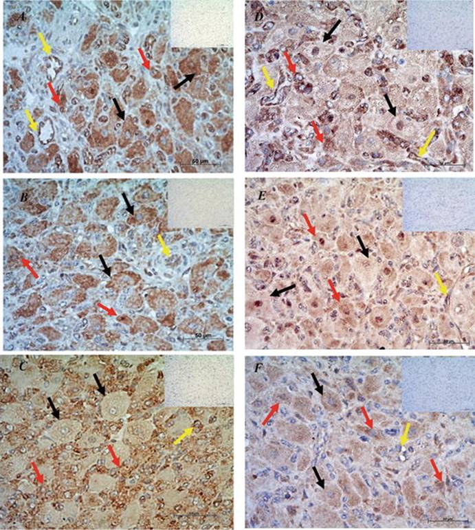
Fig. 5.6
Cellular localization of PGRMC1 (a), PGRMC2 (b), SERBP1 (c), mPRα (d), mPRβ (e), and mPRγ (f) in the bovine CL on days 11–16 of the estrous cycle. Control immunohistochemistry was performed without primary antibodies (inserts). Black arrows large luteal cells; red arrows small luteal cells; yellow arrows endothelial cells of blood vessels. Bars 50 μm
Localization of membrane P4 receptors in the endothelium of blood vessels in the bovine CL (Fig. 5.6) and uterus [75, 85] indicated that these receptors may contribute to the nongenomic effects of P4 in blood vessels in the female reproductive tract and, in this way, participate in the processes promoting the development and maintenance of pregnancy, such as cell differentiation, regulation of cell apoptosis, steroidogenesis, and contractility of the uterus [70, 102] and uterine blood flow. Expression of membrane P4 receptors in the endothelial cells of blood vessels suggested that they can participate in the fast, nongenomic effects of P4 on blood flow and make it an important regulator of reproductive system function.
The presence of P4 membrane receptors , except those of the nuclear receptor isoforms, illustrates the different ways hormones influence cellular processes. Selective blockade of these receptors by means of specific blockers or silencing genes using siRNA on one hand or receptors stimulation with agonists on the other hand may demonstrate the physiological importance of these receptors. This method of selective stimulation or inhibition of P4 receptors can also be a convenient tool to modify intracellular processes and subsequently to obtain a diverse cell response.
Acknowledgments
The authors’ research was supported by the National Science Centre (2012/05/B/NZ4/01810) the Ministry of Science and Higher Education (N311 113638) and the Polish Academy of Sciences.
References
1.
Misrahi M, Venencie PY, Saugier-Veber P, Sar S, Dessen P, Milgrom E. Structure of the human progesterone receptor gene. Biochim Biophys Acta. 1993;1216(2):289–92.CrossRefPubMed
2.
Conneely OM, Kettelberger DM, Tsai MJ, Schrader WT, O’Malley BW. The chicken progesterone receptor A and B isoforms are products of an alternate translation initiation event. J Biol Chem. 1989;264(24):14062–4.PubMed
3.
Mulac-Jericevic B, Conneely OM. Reproductive tissue selective actions of progesterone receptors. Reproduction. 2004;128(2):139–46.CrossRefPubMed
4.
Giangrande PH, Pollio G, McDonnell DP. Mapping and characterization of the functional domains responsible for the differential activity of the A and B isoforms of the human progesterone receptor. J Biol Chem. 1997;272(52):32889–900.CrossRefPubMed
5.
Pieber D, Allport VC, Bennett PR. Progesterone receptor isoform A inhibits isoform B-mediated transactivation in human amnion. Eur J Pharmacol. 2001;427(1):7–11.CrossRefPubMed
6.
Taylor AH, McParland PC, Taylor DJ, Bell SC. The cytoplasmic 60 kDa progesterone receptor isoform predominates in the human amniochorion and placenta at term. Reprod Biol Endocrinol. 2009;7:22.CrossRefPubMedPubMedCentral
7.
Wei LL, Hawkins P, Baker C, Norris B, Sheridan PL, Quinn PG. An amino-terminal truncated progesterone receptor isoform, PRc, enhances progestin-induced transcriptional activity. Mol Endocrinol. 1996;10(11):1379–87.PubMed
8.
Berisha B, Pfaffl MW, Schams D. Expression of estrogen and progesterone receptors in the bovine ovary during estrous cycle and pregnancy. Endocrine. 2002;17(3):207–14.CrossRefPubMed
9.
Shibaya M, Matsuda A, Hojo T, Acosta TJ, Okuda K. Expressions of estrogen receptors in the bovine corpus luteum: cyclic changes and effects of prostaglandin F2alpha and cytokines. J Reprod Dev. 2007;53(5):1059–68.CrossRefPubMed
10.
Ascenzi P, Bocedi A, Marino M. Structure-function relationship of estrogen receptor alpha and beta: impact on human health. Mol Aspects Med. 2006;27(4):299–402.CrossRefPubMed
11.
Cheung J, Smith DF. Molecular chaperone interactions with steroid receptors: an update. Mol Endocrinol. 2000;14(7):939–46.CrossRefPubMed
12.
Smith DF. Chaperones in progesterone receptor complexes. Semin Cell Dev Biol. 2000;11(1):45–52.CrossRefPubMed
13.
Griekspoor A, Zwart W, Neefjes J, Michalides R. Visualizing the action of steroid hormone receptors in living cells. Nucl Recept Signal. 2007;5, e003.PubMedPubMedCentral
14.
Glass CK, Rosenfeld MG. The coregulator exchange in transcriptional functions of nuclear receptors. Genes Dev. 2000;14(2):121–41.PubMed
15.
Chakravarti D, LaMorte VJ, Nelson MC, Nakajima T, Schulman IG, Juguilon H, Montminy M, Evans RM. Role of CBP/P300 in nuclear receptor signalling. Nature. 1996;383(6595):99–103.CrossRefPubMed
16.
Soutoglou E, Viollet B, Vaxillaire M, Yaniv M, Pontoglio M, Talianidis I. Transcription factor-dependent regulation of CBP and P/CAF histone acetyltransferase activity. EMBO J. 2001;20(8):1984–92.CrossRefPubMedPubMedCentral
17.
Rowan BG, O’Malley BW. Progesterone receptor coactivators. Steroids. 2000;65(10-11):545–9.CrossRefPubMed
18.
Heery DM, Kalkhoven E, Hoare S, Parker MG. A signature motif in transcriptional co-activators mediates binding to nuclear receptors. Nature. 1997;387(6634):733–6.CrossRefPubMed
19.
McKenna NJ, Lanz RB, O’Malley BW. Nuclear receptor coregulators: cellular and molecular biology. Endocr Rev. 1999;20(3):321–44.PubMed
20.
Tyler JK, Kadonaga JT. The “dark side” of chromatin remodeling: repressive effects on transcription. Cell. 1999;99(5):443–6.CrossRefPubMed
21.
Hu X, Lazar MA. The CoRNR motif controls the recruitment of corepressors by nuclear hormone receptors. Nature. 1999;402(6757):93–6.CrossRefPubMed
22.
Lazar MA. Nuclear receptor corepressors. Nucl Recept Signal. 2003;1, e001.CrossRefPubMedPubMedCentral
23.
Feng Q, O’Malley BW. Nuclear receptor modulation--role of coregulators in selective estrogen receptor modulator (SERM) actions. Steroids. 2014;90:39–43.CrossRefPubMedPubMedCentral
24.
Cadepond F, Ulmann A, Baulieu EE. RU486 (mifepristone): mechanisms of action and clinical uses. Annu Rev Med. 1997;48:129–56.CrossRefPubMed
25.
Vegeto E, Allan GF, Schrader WT, Tsai MJ, McDonnell DP, O’Malley BW. The mechanism of RU486 antagonism is dependent on the conformation of the carboxy-terminal tail of the human progesterone receptor. Cell. 1992;69(4):703–13.CrossRefPubMed
26.
Benhamou B, Garcia T, Lerouge T, Vergezac A, Gofflo D, Bigogne C, Chambon P, Gronemeyer H. A single amino acid that determines the sensitivity of progesterone receptors to RU486. Science. 1992;255(5041):206–9.CrossRefPubMed
27.
Leonhardt SA, Edwards DP. Mechanism of action of progesterone antagonists. Exp Biol Med (Maywood). 2002;227(11):969–80.
28.
Oñate SA, Tsai SY, Tsai MJ, O’Malley BW. Sequence and characterization of a coactivator for the steroid hormone receptor superfamily. Science. 1995;270(5240):1354–7.CrossRefPubMed
29.
Tetel MJ, Giangrande PH, Leonhardt SA, McDonnell DP, Edwards DP. Hormone-dependent interaction between the amino- and carboxyl-terminal domains of progesterone receptor in vitro and in vivo. Mol Endocrinol. 1999;13(6):910–24.CrossRefPubMed
30.
Rothchild I. The corpus luteum revisited: are the paradoxical effects of RU486 a clue to how progesterone stimulates its own secretion? Biol Reprod. 1996;55(1):1–4.CrossRefPubMed
31.
Meyer ME, Pornon A, Ji JW, Bocquel MT, Chambon P, Gronemeyer H. Agonistic and antagonistic activities of RU486 on the functions of the human progesterone receptor. EMBO J. 1990;9(12):3923–32.PubMedPubMedCentral
32.
Conneely OM, Lydon JP. Progesterone receptors in reproduction: functional impact of the A and B isoforms. Steroids. 2000;65(10-11):571–7.CrossRefPubMed
33.
Rekawiecki R, Kowalik MK, Kotwica J. Onapristone (ZK299) and mifepristone (RU486) regulate the messenger RNA and protein expression levels of the progesterone receptor isoforms A and B in the bovine endometrium. Theriogenology. 2015;84(3):348–57.CrossRefPubMed
34.
Ottander U, Hosokawa K, Liu K, Bergh A, Ny T, Olofsson JI. A putative stimulatory role of progesterone acting via progesterone receptors in the steroidogenic cells of the human corpus luteum. Biol Reprod. 2000;62(3):655–63.CrossRefPubMed
35.
Rekawiecki R, Kowalik MK, Kotwica J. Cloning and expression of progesterone receptor isoforms A and B in bovine corpus luteum. Reprod Fertil Dev. 2014;67:215–25
36.
Misao R, Nakanishi Y, Iwagaki S, Fujimoto J, Tamaya T. Expression of progesterone receptor isoforms in corpora lutea of human subjects: correlation with serum oestrogen and progesterone concentrations. Mol Hum Reprod. 1998;4(11):1045–52.CrossRefPubMed
37.
Sakumoto R, Vermehren M, Kenngott RA-M, Okuda K, Sinowatz F. Changes in the levels of progesterone receptor mRNA and protein in the bovine corpus luteum during the estrous cycle. J Reprod Dev. 2010;56(2):219–22.CrossRefPubMed
38.
Cassar CA, Dow MPD, Pursley JR, Smith GW. Effect of the preovulatory LH surge on bovine follicular progesterone receptor mRNA expression. Domest Anim Endocrinol. 2002;22(3):179–87.CrossRefPubMed
39.
Okuda K, Miyamoto A, Sauerwein H, Schweigert FJ, Schams D. Evidence for oxytocin receptors in cultured bovine luteal cells. Biol Reprod. 1992;46(6):1001–6.CrossRefPubMed
40.
Kotwica J, Rekawiecki R, Duras MA. Stimulatory influence of progesterone on its own synthesis in bovine corpus luteum. Bull Vet Inst Pulawy. 2004;48(2):139–46.
41.
Rekawiecki R, Nowik M, Kotwica J. Stimulatory effect of LH, PGE2 and progesterone on StAR protein, cytochrome P450 cholesterol side chain cleavage and 3beta hydroxysteroid dehydrogenase gene expression in bovine luteal cells. Prostaglandins Other Lipid Mediat. 2005;78(1-4):169–84.CrossRefPubMed
42.
Rekawiecki R, Kotwica J. Molecular regulation of progesterone synthesis in the bovine corpus luteum. Vet Med (Praha). 2007;52(9):405–12.
43.
Rekawiecki R, Kotwica J. Involvement of progesterone, oxytocin, and noradrenaline in the molecular regulation of steroidogenesis in the corpus luteum of the cow. Bull Vet Inst Pulawy. 2008;52:573–80.
44.
Rekawiecki R, Kowalik MK, Kotwica J. Luteotropic and luteolytic factors regulate mRNA and protein expression of progesterone receptor isoforms A and B in the bovine endometrium. Reprod Fertil Dev. 2014;67:215–25
45.
Niswender GD, Juengel JL, Silva PJ, Rollyson MK, McIntush EW. Mechanisms controlling the function and life span of the corpus luteum. Physiol Rev. 2000;80(1):1–29.PubMed
46.
Laven RA, Peters AR. Bovine retained placenta: aetiology, pathogenesis and economic loss. Vet Rec. 1996;139(19):465–71.CrossRefPubMed
47.
Krzymowski T, Stefańczyk-Krzymowska S. The oestrous cycle and early pregnancy--a new concept of local endocrine regulation. Vet J. 2004;168(3):285–96.CrossRefPubMed
48.
Graham JD, Yager ML, Hill HD, Byth K, O’Neill GM, Clarke CL. Altered progesterone receptor isoform expression remodels progestin responsiveness of breast cancer cells. Mol Endocrinol. 2005;19(11):2713–35.CrossRefPubMed
49.
Auletta FJ, Kelm LB, Schofield MJ. Responsiveness of the corpus luteum of the rhesus monkey (Macaca mulatta) to gonadotrophin in vitro during spontaneous and prostaglandin F2 alpha-induced luteolysis. J Reprod Fertil. 1995;103(1):107–13.CrossRefPubMed
50.
Duffy DM, Wells TR, Haluska GJ, Stouffer RL. The ratio of progesterone receptor isoforms changes in the monkey corpus luteum during the luteal phase of the menstrual cycle. Biol Reprod. 1997;57(4):693–9.CrossRefPubMed
51.
Grazzini E, Guillon G, Mouillac B, Zingg HH. Inhibition of oxytocin receptor function by direct binding of progesterone. Nature. 1998;392(6675):509–12.CrossRefPubMed
52.
Peluso JJ. Multiplicity of progesterone actions and receptors in the mammalian ovary. Biol Reprod. 2006;75(1):2–8.CrossRefPubMed
53.
Lösel RM, Besong D, Peluso JJ, Wehling M. Progesterone receptor membrane component 1--many tasks for a versatile protein. Steroids. 2008;73(9-10):929–34.CrossRefPubMed
54.
Falkenstein E, Meyer C, Eisen C, Scriba PC, Wehling M. Full-length cDNA sequence of a progesterone membrane-binding protein from porcine vascular smooth muscle cells. Biochem Biophys Res Commun. 1996;229(1):86–9.CrossRefPubMed
55.
Kelly MJ, Levin ER. Rapid actions of plasma membrane estrogen receptors. Trends Endocrinol Metab. 2001;12(4):152–6.CrossRefPubMed
56.
Cahill MA. Progesterone receptor membrane component 1: an integrative review. J Steroid Biochem Mol Biol. 2007;105(1-5):16–36.CrossRefPubMed
57.
Gellersen B, Fernandes MS, Brosens JJ. Non-genomic progesterone actions in female reproduction. Hum Reprod Update. 2009;15(1):119–38.CrossRefPubMed
58.
Dressing GE, Goldberg JE, Charles NJ, Schwertfeger KL, Lange CA. Membrane progesterone receptor expression in mammalian tissues: a review of regulation and physiological implications. Steroids. 2011;76(1-2):11–7.CrossRefPubMed
59.
Bogacki M, Silvia WJ, Rekawiecki R, Kotwica J. Direct inhibitory effect of progesterone on oxytocin-induced secretion of prostaglandin F(2alpha) from bovine endometrial tissue. Biol Reprod. 2002;67(1):184–8.CrossRefPubMed
60.
Duras M, Mlynarczuk J, Kotwica J. Non-genomic effect of steroids on oxytocin-stimulated intracellular mobilization of calcium and on prostaglandin F2alpha and E2 secretion from bovine endometrial cells. Prostaglandins Other Lipid Mediat. 2005;76(1-4):105–16.CrossRefPubMed
61.
Kowalik MK, Slonina D, Kotwica J. Genomic and non-genomic effects of progesterone and pregnenolone on the function of bovine endometrial cells. Vet Med. 2009;54(5):205–14.
62.
Slonina D, Kowalik MK, Subocz M, Kotwica J. The effect of ovarian steroids on oxytocin-stimulated secretion and synthesis of prostaglandins in bovine myometrial cells. Prostaglandins Other Lipid Mediat. 2009;90(3-4):69–75.CrossRefPubMed
63.
Boonyaratanakornkit V, McGowan E, Sherman L, Mancini MA, Cheskis BJ, Edwards DP. The role of extranuclear signaling actions of progesterone receptor in mediating progesterone regulation of gene expression and the cell cycle. Mol Endocrinol. 2007;21(2):359–75.CrossRefPubMed
64.
Gimpl G, Fahrenholz F. Cholesterol as stabilizer of the oxytocin receptor. Biochim Biophys Acta. 2002;1564(2):384–92.CrossRefPubMed
65.
Revankar CM, Cimino DF, Sklar LA, Arterburn JB, Prossnitz ER. A transmembrane intracellular estrogen receptor mediates rapid cell signaling. Science. 2005;307(5715):1625–30.CrossRefPubMed
66.
Thomas P, Pang Y, Filardo EJ, Dong J. Identity of an estrogen membrane receptor coupled to a G protein in human breast cancer cells. Endocrinology. 2005;146(2):624–32.CrossRefPubMed
67.
Raza FS, Takemori H, Tojo H, Okamoto M, Vinson GP. Identification of the rat adrenal zona fasciculata/reticularis specific protein, inner zone antigen (IZAg), as the putative membrane progesterone receptor. Eur J Biochem. 2001;268(7):2141–7.CrossRefPubMed
68.
Peluso JJ, Liu X, Gawkowska A, Johnston-MacAnanny E. Progesterone activates a progesterone receptor membrane component 1-dependent mechanism that promotes human granulosa/luteal cell survival but not progesterone secretion. J Clin Endocrinol Metab. 2009;94(7):2644–9.CrossRefPubMedPubMedCentral
69.
Krebs CJ, Jarvis ED, Chan J, Lydon JP, Ogawa S, Pfaff DW. A membrane-associated progesterone-binding protein, 25-Dx, is regulated by progesterone in brain regions involved in female reproductive behaviors. Proc Natl Acad Sci USA. 2000;97(23):12816–21.CrossRefPubMedPubMedCentral
70.
Zhang L, Kanda Y, Roberts DJ, Ecker JL, Losel R, Wehling M, Peluso JJ, Pru JK. Expression of progesterone receptor membrane component 1 and its partner serpine 1 mRNA binding protein in uterine and placental tissues of the mouse and human. Mol Cell Endocrinol. 2008;287(1-2):81–9.CrossRefPubMed
71.
Sasson R, Rimon E, Dantes A, Cohen T, Shinder V, Land-Bracha A, Amsterdam A. Gonadotrophin-induced gene regulation in human granulosa cells obtained from IVF patients. Modulation of steroidogenic genes, cytoskeletal genes and genes coding for apoptotic signalling and protein kinases. Mol Hum Reprod. 2004;10(5):299–311.CrossRefPubMed
72.
McRae RS, Johnston HM, Mihm M, O’Shaughnessy PJ. Changes in mouse granulosa cell gene expression during early luteinization. Endocrinology. 2005;146(1):309–17.CrossRefPubMed
73.
Cai Z, Stocco C. Expression and regulation of progestin membrane receptors in the rat corpus luteum. Endocrinology. 2005;146(12):5522–32.CrossRefPubMed
74.
Luciano AM, Corbani D, Lodde V, Tessaro I, Franciosi F, Peluso JJ, Modina S. Expression of progesterone receptor membrane component-1 in bovine reproductive system during estrous cycle. Eur J Histochem. 2011;55(3), e27.CrossRefPubMedPubMedCentral
75.
Kowalik MK, Rekawiecki R, Kotwica J. Expression and localization of progesterone receptor membrane component 1 and 2 and serpine mRNA binding protein 1 in the bovine corpus luteum during the estrous cycle and the first trimester of pregnancy. Theriogenology. 2014;82(8):1086–93.CrossRefPubMed
76.
Rohe HJ, Ahmed IS, Twist KE, Craven RJ. PGRMC1 (progesterone receptor membrane component 1): a targetable protein with multiple functions in steroid signaling, P450 activation and drug binding. Pharmacol Ther. 2009;121(1):14–9.CrossRefPubMed
77.
Hughes AL, Powell DW, Bard M, Eckstein J, Barbuch R, Link AJ, Espenshade PJ. Dap1/PGRMC1 binds and regulates cytochrome P450 enzymes. Cell Metab. 2007;5(2):143–9.CrossRefPubMed
78.
Wu H, Zhang Y. Mechanisms and functions of Tet protein-mediated 5-methylcytosine oxidation. Genes Dev. 2011;25(23):2436–52.CrossRefPubMedPubMedCentral
79.
Luciano AM, Lodde V, Franciosi F, Ceciliani F, Peluso JJ. Progesterone receptor membrane component 1 expression and putative function in bovine oocyte maturation, fertilization, and early embryonic development. Reproduction. 2010;140(5):663–72.CrossRefPubMed
80.
Peluso JJ, Romak J, Liu X. Progesterone receptor membrane component-1 (PGRMC1) is the mediator of progesterone’s antiapoptotic action in spontaneously immortalized granulosa cells as revealed by PGRMC1 small interfering ribonucleic acid treatment and functional analysis of PGRMC1 mutations. Endocrinology. 2008;149(2):534–43.CrossRefPubMed
81.
Peluso JJ, Liu X, Gawkowska A, Lodde V, Wu CA. Progesterone inhibits apoptosis in part by PGRMC1-regulated gene expression. Mol Cell Endocrinol. 2010;320(1-2):153–61.CrossRefPubMedPubMedCentral
82.
Engmann L, Losel R, Wehling M, Peluso JJ. Progesterone regulation of human granulosa/luteal cell viability by an RU486-independent mechanism. J Clin Endocrinol Metab. 2006;91(12):4962–8.CrossRefPubMed
83.
Wendler A, Wehling M. PGRMC2, a yet uncharacterized protein with potential as tumor suppressor, migration inhibitor, and regulator of cytochrome P450 enzyme activity. Steroids. 2013;78(6):555–8.CrossRefPubMed
84.
Keator CS, Mah K, Slayden OD. Alterations in progesterone receptor membrane component 2 (PGRMC2) in the endometrium of macaques afflicted with advanced endometriosis. Mol Hum Reprod. 2012;18(6):308–19.CrossRefPubMedPubMedCentral
85.
Slonina D, Kowalik MK, Kotwica J. Expression of progesterone receptor membrane component 1, serpine mRNA binding protein 1 and nuclear progesterone receptor isoforms A and B in the bovine myometrium during the estrous cycle and early pregnancy. J Reprod Dev. 2012;58(3):288–94.CrossRefPubMed
86.
Kowalik MK, Slonina D, Rekawiecki R, Kotwica J. Expression of progesterone receptor membrane component (PGRMC) 1 and 2, serpine mRNA binding protein 1 (SERBP1) and nuclear progesterone receptor (PGR) in the bovine endometrium during the estrous cycle and the first trimester of pregnancy. Reprod Biol. 2013;13(1):15–23.CrossRefPubMed
87.
Saint-Dizier M, Sandra O, Ployart S, Chebrout M, Constant F. Expression of nuclear progesterone receptor and progesterone receptor membrane components 1 and 2 in the oviduct of cyclic and pregnant cows during the post-ovulation period. Reprod Biol Endocrinol. 2012;10:76.CrossRefPubMedPubMedCentral
88.
Shankar R, Johnson MP, Williamson NA, Cullinane F, Purcell AW, Moses EK, Brennecke SP. Molecular markers of preterm labor in the choriodecidua. Reprod Sci. 2010;17(3):297–310.CrossRefPubMed
89.
Zhu Y, Bond J, Thomas P. Identification, classification, and partial characterization of genes in humans and other vertebrates homologous to a fish membrane progestin receptor. Proc Natl Acad Sci USA. 2003;100(5):2237–42.CrossRefPubMedPubMedCentral
90.
Fernandes MS, Pierron V, Michalovich D, Astle S, Thornton S, Peltoketo H, et al. Regulated expression of putative membrane progestin receptor homologues in human endometrium and gestational tissues. J Endocrinol. 2005;187(1):89–101.CrossRefPubMed
91.
Karteris E, Zervou S, Pang Y, Dong J, Hillhouse EW, Randeva HS, Thomas P. Progesterone signaling in human myometrium through two novel membrane G protein-coupled receptors: potential role in functional progesterone withdrawal at term. Mol Endocrinol. 2006;20(7):1519–34.CrossRefPubMed
92.
Qiu HB, Lu SS, Ji KL, Song XM, Lu YQ, Zhang M, Lu KH. Membrane progestin receptor beta (mPR-beta): a protein related to cumulus expansion that is involved in in vitro maturation of pig cumulus-oocyte complexes. Steroids. 2008;73(14):1416–23.CrossRefPubMed
93.
Nutu M, Weijdegård B, Thomas P, Thurin-Kjellberg A, Billig H, Larsson DJ. Distribution and hormonal regulation of membrane progesterone receptors β and γ in ciliated epithelial cells of mouse and human fallopian tubes. Reprod Biol Endocrinol. 2009;7(1):89.CrossRefPubMedPubMedCentral
94.
Ashley RL, Clay CM, Farmerie TA, Niswender GD, Nett TM. Cloning and characterization of an ovine intracellular seven transmembrane receptor for progesterone that mediates calcium mobilization. Endocrinology. 2006;147(9):4151–9.CrossRefPubMed
95.
Ashley RL, Arreguin-Arevalo JA, Nett TM. Binding characteristics of the ovine membrane progesterone receptor alpha and expression of the receptor during the estrous cycle. Reprod Biol Endocrinol. 2009;7:42.CrossRefPubMedPubMedCentral
96.
Duras M, Brzósko E, Kotwica J. Influence of progesterone, pregnenolone and 17-beta-hydroxyprogesterone on the function of bovine luteal cells treated with luteinizing hormone, noradrenaline and prostaglandin E2. Pol J Vet Sci. 2005;8(2):113–9.PubMed
97.
Mlynarczuk J, Sasiadek J, Kotwica J. Non-genomic action of progesterone in cultured bovine luteal and endometrial epithelial cells. Bull Vet Inst Pulawy. 2005;49(2):193–8.
98.
Kowalik MK, Kotwica J. Progesterone receptor membrane component 1 (PGRMC1) gene expression in corpus luteum during the estrous cycle in cows. Reprod Biol. 2008;8(3):291–7.CrossRefPubMed
99.
Min L, Takemori H, Nonaka Y, Katoh Y, Doi J, Horike N, Osamu H, Raza FS, Vinson GP, Okamoto M. Characterization of the adrenal-specific antigen IZA (inner zone antigen) and its role in the steroidogenesis. Mol Cell Endocrinol. 2004;215(1-2):143–8.CrossRefPubMed
100.
Albrecht C, Huck V, Wehling M, Wendler A. In vitro inhibition of SKOV-3 cell migration as a distinctive feature of progesterone receptor membrane component type 2 versus type 1. Steroids. 2012;77(14):1543–50.CrossRefPubMed
101.
Suchanek M, Radzikowska A, Thiele C. Photo-leucine and photo-methionine allow identification of protein-protein interactions in living cells. Nat Methods. 2005;2(4):261–7.CrossRefPubMed
102.
Wu W, Shi S-Q, Huang H-J, Balducci J, Garfield RE. Changes in PGRMC1, a potential progesterone receptor, in human myometrium during pregnancy and labour at term and preterm. Mol Hum Reprod. 2011;17(4):233–42.CrossRefPubMed