Gab Kovacs1 and Paula Briggs2
(1)
Department of Obstetrics and Gynaecology, Monash University, Clayton, Victoria, Australia
(2)
Sexual and Reproductive Health, Southport and Ormskirk Hospital, Southport, UK
With the joining of the oocyte and the sperm an embryo is created (Fig. 2.1). As both the oocyte and the sperm (gametes) contribute 23 chromosomes (haploid), the embryo now is made up of 23 pairs of chromosomes (46). The developing embryo inherits half its genetic material from each of its parents, thus it is diploid, and its genetic makeup is determined for life. As the cells continue to divide rapidly, each nucleus contains an identical chromosomal template. By the time it reaches the uterine cavity, the embryo has developed to the blastocyst stage (Fig. 2.2). The blastocyst differentiates into outer cells, the trophoectoderm or trophoblast, which will form the placenta as it combines with the uterine endometrium, and inner cells which form the inner cell mass (Fig. 2.3). These will form the embryo, as well as the amnion and yolk sac. This is the stage at which the process of implantation commences. By the eighth day, the cells of the inner cell mass proliferate into a rounded bilaminar structure. The embryo will develop from, this and a small slit like space forms to become the amniotic cavity. The ectoderm develops from the floor of this cavity and makes up one of the layers of the bilaminar embryonic disc, the other layer being the endoderm. The mesoderm develops as a further layer between the ectoderm and endoderm. As it grows outwards, in combination with the trophoblast, the chorion is formed. The cells continuous with the endoderm extend along the inner aspect of the blastocyst producing another fluid filled sac – the primary yolk sac. Ultimately the ectoderm will form the skin, nervous system and parts of the eyes, ears and nose. The endoderm is the origin of the linings of the gut and respiratory system, whereas the mesoderm is the origin of muscle, bone, blood tissues and connective tissue.
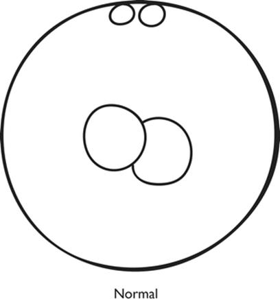
Fig. 2.1
The fertilised oocyte
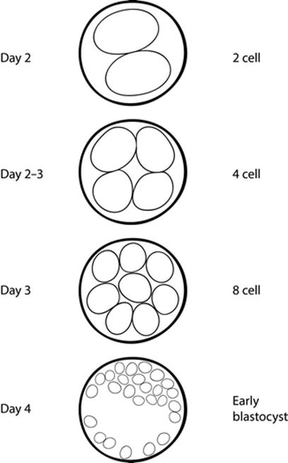
Fig. 2.2
Early embryonic development
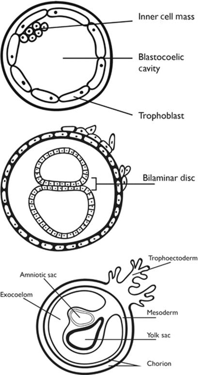
Fig. 2.3
Development of the embryo after implantation
Between the yolk sac and the trophoblast, tissue is present, which is called the extra embryonic mesoderm. Within the extra embryonic mesoderm, cavities appear which become confluent, and as the embryo folds, it is surrounded by what is now called the extra embryonic coelom. The part of the extra embryonic mesoderm joining the early embryo to the trophoblast forms the body stalk, which will subsequently form the umbilical cord of the fetus. By about 15 days of age, the embryo becomes oriented in a longitudinal axis with the development of a keel-like thickening of the ectoderm known as the primitive streak. The anterior end of the primitive streak forms a clump of cells, which will give rise to the brain of the fetus. The embryo then rapidly elongates with the head process growing quickly and becoming larger than the streak itself. As the cells proliferate, a third layer develops between the ectoderm and the endoderm, the intra-embryonic mesoderm. A longitudinal in-pouching of the ectoderm then takes place and this gives rise to the neural tube which will develop into the central nervous system. This neural tube will develop three dilations at its cephalic end, which give rise to the ventricles of the brain.
By about 21 days after fertilisation (5 weeks of age) the blood vessel plexuses within the embryo and the mesoderm form the primitive heart, with circulation developing to supply the brain (future carotid arteries). The vitelline vessels to the gut, the blood supply to the nervous system, and umbilical vessels to the placenta all develop from these plexuses.
By week 6 the heart is formed, and can be seen beating using ultrasound, an important sign to confirm that the embryo is alive and well. The limbs and genital system develop by week 8, the liver and kidney with its drainage system by week 12. The eyes and spinal cord develop by 20 weeks, with the gastrointestinal system and respiratory system developing by week 24–28.
As all the organs are now formed, the last 3 months of intrauterine development will consist of maturation and growth of the systems.