Maureen P. Malee
Hematologic Disease
Anemias
Anemia is defined as a hemoglobin (Hb) concentration of less than 12 g/dL in nonpregnant women. Anemia can be acquired or inherited. During pregnancy, plasma volume expands proportionately more than Hb or red blood cell volume, resulting in Hb dilution, such that anemia is defined as a Hb concentration of less than 10 g/dL. In addition to blood loss, anemia can result from decreased production or increased destruction of red blood cells. The initial workup consists of a history and physical examination, as well as an examination of the red blood cell indices and a peripheral smear, with additional tests as indicated (Fig. 17.1).
|
|
|
FIG. 17.1. Workup of anemia in pregnancy. |
Acquired Anemias
Iron Deficiency Anemia
Iron deficiency is an acquired anemia and is the most common cause of anemia in gravid women, occurring in 15% to 25% of all pregnancies. Iron deficiency is suspected when the mean corpuscular volume (MCV) is less than ã80/µm3 and is confirmed by demonstrating an elevated total iron-binding capacity (TIBC), a low serum iron level, a serum iron-to-TIBC ratio less than 20%, or a low ferritin level. Effects of iron deficiency on the fetus are usually minimal, although neonatal anemia is increased. Iron is transported actively across the placenta, and fetal iron and ferritin levels are 3 times higher than maternal levels. However, iron deficiency anemia has been weakly associated with preterm birth, and when maternal anemia is severe (Hb less than 6 g/dL), intrauterine growth restriction (IUGR) may occur. In pregnant women, iron deficiency can cause symptoms including fatigue, headache, lightheadedness, and reduced exercise tolerance. Blood loss at delivery may be tolerated poorly in anemic patients, and postpartum tissue healing may be compromised. For these reasons, treatment during pregnancy is recommended.
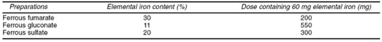
The total iron requirement of pregnancy is 1,000 mg: 500 mg increases the maternal red blood cell mass, 300 mg is transported to the fetus and placenta, and 200 mg compensates for blood loss at delivery. The iron requirements of pregnancy increase steadily toward term but average 3.5 mg per day. Even though iron absorption efficiency increases during pregnancy, excess iron must be ingested to ensure sufficient dosage. Recommended supplementation for nonanemic gravidas is 300 mg of ferrous sulfate per day, which contains 60 mg of elemental iron. Anemic gravidas (Hb of 8 or 9 g/dL) should take 300 mg ferrous sulfate 2 or 3 times a day. Patients who cannot tolerate iron tablets may take an enteric-coated tablet or a liquid suspension (Table 17.1). Vitamin C facilitates iron absorption. Therapeutic results can be expected after 3 weeks of therapy.
|
|
|
TABLE 17.1. Iron preparations and dosages |
The severely anemic patient (Hb less than 8 g/dL) may require parenteral therapy in the form of intramuscular or intravenous iron dextran. Because 0.2% to 0.3% of patients have an anaphylactic response to iron dextran, all patients should receive a small test dose 1 hour before the initiation of treatment, and therapy should be provided in an area with ready access to resuscitative medication and equipment. The total dose of iron required can be calculated using this formula:
Total dose of iron dextran (mL) = (0.0476 × body weight in kg × [desired Hb concentration - observed Hb concentration]) + 1 mL/5 kg of body weight up to a maximum of 14 mL
This dose can be given intramuscularly or intravenously (by slow push), 2 mg per day, until the total dose has been given, or the entire dose can be given diluted in 500 to 1,000 mL of 0.9% saline and administered intravenously over 1 to 6 hours. Adequate parenteral therapy should result in a marked increase in the reticulocyte count within 7 to 14 days.
Megaloblastic Anemia
Megaloblastic anemia is characterized by red blood cells with increased MCV and white blood cells with altered morphology (hypersegmented neutrophils, anisocytosis, and poikilocytosis). It complicates up to 1% of pregnancies and usually is caused by folate deficiency, although it can occur after exposure to sulfa drugs or hydroxyurea or, rarely, because of vitamin B12 deficiency.
Folate deficiency can develop over a relatively short time, because liver stores of folate are sufficient to meet the body's needs for only 1 to 2 months. Malnutrition (e.g., alcoholism), malabsorption, anticonvulsant therapy, oral contraceptive use, or pregnancy can rapidly deplete the body's folate stores. Hypersegmented neutrophils (more than 5% of neutrophils having five or more lobes) appear after 7 weeks of deficiency, red blood cell folate is reduced after 18 weeks, and anemia occurs after 20 weeks. The daily folate requirement for a nonpregnant individual is 50 to 100 µg; a pregnant woman needs 300 to 400 µg. This dosage may be difficult to achieve through dietary manipulation, because folate is found primarily in fresh fruits and vegetables and is destroyed by cooking. As a separate issue, it now seems apparent that some women require excess folate to overcome a relative enzyme deficiency leading to high blood and amniotic fluid levels of homocysteine and an increased risk for fetal neural tube defects. For these reasons, women contemplating pregnancy should be advised to ingest a daily folic acid supplement (0.4 mg per day if there is no family history of neural tube defects; 4 mg per day if there is a family history) beginning before conception and continuing throughout the first trimester of pregnancy.
In contrast, vitamin B12 deficiency is rare, because very little of the body's stores is used each day. Ingested vitamin B12 is bound to intrinsic factor produced by the parietal cells of the stomach and then absorbed through the mucosa of the distal ileum. Patients who have had a gastrectomy, ileitis, or ileal resection, or who have pernicious anemia, pancreatic insufficiency, or intestinal parasites eventually may become vitamin B12 deficient.
When megaloblastic anemia is suspected, the history should be reviewed for predisposing factors. The peripheral smear should be examined both to confirm altered cell morphology and to rule out a mixed (i.e., folate and iron) deficiency. Serum folate and vitamin B12 levels should be measured. A fasting folate level less than 3 ng/mL or a vitamin B12 level less than 80 pg/mL indicates deficiency. Folate deficiency responds to 0.5 to 1.0 mg folate orally per day, while a B12 deficiency requires vitamin B12, 1 mg intramuscularly, weekly for 6 weeks.
Hereditary Anemias
The most commonly encountered hereditary anemias in pregnancy are the thalassemias and sickle cell variants. Hb is a tetramer composed of two copies each of two different polypeptide chains; the identity of the chains determines the type of Hb produced. During embryonic and fetal life, genes directing production of different types of polypeptide chains and, thus, different types of Hb, are switched on and then off sequentially. At birth, a normal individual produces α and β chains, along with very small quantities of δ and τ chains (Fig. 17.2). Normal adults produce primarily hemoglobin A (HbA), composed of two α and two β polypeptide chains.
|
|
|
FIG. 17.2. Production of hemoglobin polypeptide chains in relationship to gestational age. (From Rucknagel DL, Laros RK. Hemoglobinopathies: genetics and implications for studies of human reproduction. Clin Obstet Gynecol 1969;12:4, with permission.) |
Thalassemias
Thalassemias are characterized by impaired production of one or more of the peptide chains. Thalassemia has a high incidence in certain ethnic groups, especially those originating in the Mediterranean basin, the Middle East, Africa, Asia, and India. Four clinical syndromes are associated with α-thalassemia, and two syndromes are associated with β-thalassemia.
Two genes direct β-chain production, one on each copy of chromosome 11. Over 100 different gene mutations have been identified that prevent or reduce β-chain transcription; if one gene carries such a mutation, β-chain production will be reduced by one half, and abnormally low quantities of Hb will be produced. This results in β-thalassemia minor. The excess α chains combine, instead, with δ chains, producing a molecule called HbA2, or with ν chains, producing fetal hemoglobin (HbF). If β-thalassemia minor is suspected because the patient has microcytic anemia without iron deficiency, Hb electrophoresis should be performed. Levels of HbA2 greater than 3.5% and HbF greater than or equal to 2% confirm the diagnosis (Table 17.2). The gravid patient with β-thalassemia minor generally tolerates pregnancy well. She should receive folic acid supplementation, but not iron supplementation unless iron deficiency is diagnosed, also.
|
|
|
TABLE 17.2. Hemoglobin electrophoresis findings in various hemoglobinopathies |
Patients with mutations preventing transcription of both β-chain genes have β-thalassemia major (β-thalassemia), or Cooley anemia. Erythropoiesis is ineffective because there is no β-chain production, and the α chains precipitate, causing red blood cell destruction. Occasionally, the mutations allow some β-chain production, resulting in a less severe reduction of Hb synthesis (β-thalassemia). Aggressive intervention in infancy using transfusion therapy ultimately leads to iron overload and hemosiderosis, with multiple organ system dysfunction and infertility. Increasing numbers of pregnancies in this population are being reported by virtue of aggressive transfusion and iron chelation therapy. Such pregnancies can be complicated by an increased risk of cardiac arrhythmias and congestive heart failure secondary to severe anemia, chronic hypoxemia, and myocardial hemosiderosis. Folate supplementation is routine. The safety of iron-chelating agents such as deferoxamine has not been established in pregnancy. Prenatal diagnosis is available, and such pregnancies show improved outcome with stable maternal disease.
An entity designated thalassemia intermedia has been described also, in which the clinical course is milder than with homozygous β-thalassemia. Some individuals with this condition produce large quantities of τ chains that combine with α chains to produce fetal HbF; the presence of 17% to 35% HbF defines hereditary persistence of HbF. Alternatively, these individuals may have some degree of α-thalassemia in addition to β-thalassemia, resulting in less β-chain precipitation and hemolysis. Patients with thalassemia intermedia have severe hemolytic anemia but generally are not transfusion dependent.
Before genetic counseling is provided to the patient with β-thalassemia minor, MCV screening, followed by Hb electrophoresis if the MCV is low, should be offered to the father of the fetus. If the father has normal Hb, the fetus has a 50% chance to have β-thalassemia minor and a 50% chance to have normal Hb. If the father has β-thalassemia minor, the fetus has a 25% chance to have β-thalassemia major, which is associated with increased morbidity and mortality, a 50% chance of thalassemia minor, and a 25% chance of having normal hemoglobin. Prenatal testing of the fetus should be offered to high-risk women; at least 20% of β-chain mutations can be detected by chorionic villus sampling (CVS) or amniocentesis.
Alpha-chain production is directed by four genes, two on each copy of chromosome 16. Mutation of only one gene results in no clinical or laboratory abnormalities and is thus referred to as the silent carrier state. Mutations in two of the four genes results in β-thalassemia minor, a condition characterized by mild microcytic hypochromic anemia. Patients with β-thalassemia minor have a low MCV but normal levels of HbA2. The patient with these laboratory results should be referred for genetic evaluation and family studies to confirm the diagnosis, but typically they tolerate pregnancy fairly well.
Mutation of three of the four α genes results in hemoglobin H (HbH) disease. Affected patients have some HbA and a large percentage of HbH (four β chains). The clinical course is characterized by chronic hemolytic anemia that may worsen during pregnancy. Loss of all four β-chain genes causes β-thalassemia major, resulting in fetal hydrops and perinatal death. As with β-thalassemia, testing of the father is crucial for accurate genetic counseling. Consideration of the patient's ethnic background is also important. Asians with β-thalassemia minor usually have the two mutant genes on the same chromosome (cis position) and thus have a 50% risk of passing on both affected genes with each conception. In contrast, patients of other ethnic origins usually carry the mutant genes on opposite chromosomes (trans position), so that only one affected gene can be transmitted with each conception. All forms of β-thalassemia can be detected by CVS or amniocentesis.
Hemoglobinopathies
Hemoglobinopathies involve Hb gene mutations. Over 400 have been identified that alter polypeptide function instead of preventing production. These Hb variants generally have either reduced oxygen transport capabilities or cause hemolytic anemia. Hemoglobin S (HbS) and hemoglobin C (HbC) are the most frequent variants and can occur in association with thalassemia, as well (Table 17.3).
|
|
|
TABLE 17.3. Frequency of sickle hemoglobinopathies in African Americans |
Sickle Disease
A mutation causing a single amino acid substitution of valine for glutamic acid at position 6 on the β chain changes normal Hb to sickle Hb. An individual who is homozygous for this mutation has sickle cell anemia, producing only HbS and a small quantity of HbF, but no HbA. Sickle Hb functions well in the oxygenated state but aggregates, forming rod-shaped polymers, in the deoxygenated state. Polymerized Hb precipitates in the red blood cell, changing the cell from a biconcave disc to an elongated crescent or sickle shape. Sickled red blood cells are not deformable and cannot squeeze through the microcirculation. Microvascular obstruction results in local hypoxia that leads to a vicious cycle of further sickling and obstruction. Localized schemia and infarction cause tissue damage.
Patients with sickle cell anemia usually produce increased quantities of HbF. HbF is not distributed uniformly among all red blood cells but is present at levels of zero to 20% per cell. In cells containing HbF, restoration of normal oxygen tension may reverse the sickling and halt the destructive process. Cells containing little or no HbF become irreversibly sickled and are rapidly cleared from the system in a process leading to hemolytic anemia. Patients with homozygous HbS typically have hematocrits of 20% to 30% and reticulocyte counts of 10% to 25%. Hydroxyurea therapy has been shown to increase both the number of red blood cells containing HbF and the quantity of HbF per cell. Unfortunately, there are few data regarding the safety of hydroxyurea use in pregnancy. It is a category D drug.
Any pathologic state causing acidosis, dehydration, or hypoxemia can precipitate sickling, hemolysis, vasoocclusion, and infarction. Pregnancy often is characterized by an increase in sickle crises and associated problems (e.g., pneumonia, pyelonephritis, pulmonary emboli, congestive heart failure) and by pregnancy complications such as IUGR, preterm birth, and preeclampsia. The goal of pregnancy management should be to maintain adequate hydration and oxygen delivery to the tissues, and to avoid or rapidly control infections or other stressors that could precipitate a crisis.
Patients with sickle cell anemia should ingest 1 mg of folate per day to support increased erythropoiesis in the face of chronic hemolysis and should receive the polyvalent pneumococcal vaccine, because chronic splenic infarction leads to functional asplenia by adulthood. Iron supplementation should not be given prophylactically but should be prescribed if there is laboratory evidence of iron deficiency anemia. All sickle cell patients should undergo a funduscopic examination, with laser therapy as needed, because they are at increased risk for proliferative retinopathy. Asymptomatic bacteriuria (ASB) and other infections should be treated aggressively.
One controversy concerns the possible benefit of antepartum prophylactic exchange transfusions. Available data indicate that although some women with sickle cell anemia may escape prophylactic transfusion because they have no associated organ damage, very few crises, and a high percentage of HbF, many will require antepartum transfusion. Even those patients who avoid transfusion during pregnancy should be transfused prior to delivery, because the stresses of labor, anesthesia, operative delivery, and any associated complications (e.g., preeclampsia, chorioamnionitis) can precipitate a serious crisis. Transfusions should be planned to achieve a hematocrit above 30% and HbA above 50%. Unless complications dictate otherwise, delivery can be at term, with cesarean section for obstetric indications only.
Substitution of lysine for glutamic acid at the sixth position of the α chain results in the production of HbC. HbC is less soluble than HbA and can cause a mild hemolytic anemia, but it is more stable than HbS under hypoxic conditions. Nonpregnant women who are compound homozygotes for HbS and HbC generally have less severe anemia and fewer pain crises than women with HbSS, but under the stress of pregnancy, they experience the same maternal morbidity and pregnancy complications. Additionally, severe bone pain frequently occurs in individuals with HbSC, and acute respiratory compromise as the result of embolization of necrotic bone marrow has been reported. The antenatal management of women with HbSC should be the same as that of women with HbSS.
If one β-chain gene carries the sickle cell mutation and the other gene is functionally deleted, the patient has sickle cell β-thalassemia. Pregnancy-related morbidity in these patients is the same as for sickle cell anemia, and they should be managed similarly. Patients with HbCC or C-β-thalassemia have a very mild anemia and usually do not experience hemoglobinopathy-related pregnancy complications.
Heterozygotes for the sickle Hb mutation have sickle cell trait. Individuals with sickle trait have red blood cells that sickle under conditions of markedly reduced oxygen tension (i.e., the sickle Dex test), but Hb electrophoresis confirms the presence of 55% to 60% HbA, in addition to 35% to 40% HbS. Sickling does not occur in vivo, except under conditions of severe stress and hypoxia. Because the renal medulla is especially sensitive to reduced oxygen tension, patients with sickle trait may have episodes of painless, self-limited hematuria. During pregnancy they exhibit an increased susceptibility to urinary tract infections. There are also reports of increased preeclampsia in these patients. Patients with sickle trait should be offered genetic counseling. The father of the fetus should be tested so that the precise risk to the fetus can be provided. Prenatal diagnosis is possible by CVS or amniocentesis. No special therapy is required generally during labor and delivery.
Congenital Hemolytic Anemias
Hereditary Spherocytosis, Elliptocytosis, and Pyropoikilocytosis
Hemolytic anemia can occur for a variety of reasons. It may result from a hemoglobinopathy, may be autoimmune, drug induced, or pregnancy induced (very rarely), or may occur as the result of inherited red blood cell membrane abnormalities. Hereditary spherocytosis, elliptocytosis, and pyropoikilocytosis result from congenital defects of different red blood cell membrane proteins. All are autosomal dominant disorders occurring at an incidence of 1 in 4,000 to 5,000. All result in variant red blood cell shapes, such that affected red blood cells cannot pass readily through the spleen. While trapped in the spleen, the cell membranes are damaged, leading to red blood cell lysis, hemolytic anemia, jaundice, and splenomegaly. Splenectomy is the treatment of choice and effectively eliminates the anemia. Most women of reproductive age will already have undergone splenectomy. Although the abnormality of red blood cell shape persists, affected women tolerate pregnancy, labor, and delivery well, with few associated problems. The rare patient who has not undergone splenectomy may experience hemolytic anemia sufficient to require red blood cell transfusions. All patients should receive the polyvalent pneumococcal vaccine and should ingest a folic acid supplement throughout pregnancy. Infection should be treated aggressively, because it may cause hemolysis. The offspring of affected individuals have a 50% chance of inheriting the condition. Affected neonates may experience severe neonatal jaundice requiring exchange transfusion or splenectomy.
Glucose-6-phosphate Dehydrogenase Deficiency
Glucose-6-phosphate dehydrogenase (G6PD) deficiency is an inherited defect of an enzyme essential to the hexose monophosphate shunt. Because of this defect, when under oxidant stress, Hb sulfhydryl groups become oxidized and Hb precipitates in the red blood cell, leading to hemolytic anemia. The gene is most prevalent among individuals of African, Asian, Mediterranean, or Middle Eastern origin. Known stressors include viruses, bacteria, toxins, fava beans, and certain drugs such as antimalarial agents, sulfa drugs, and nitrofurantoin. Over 400 different gene mutations leading to G6PD deficiency have been described; the A variant is most common and is present in 1 in 20 black men and 1 in 10 black women in the United States. Although G6PD deficiency is X linked and males are affected preferentially, women with this gene defect can be symptomatic. Some heterozygotes have markedly reduced G6PD levels because unfavorable lyonization can lead to a large proportion of cells expressing the defect, and homozygosity for G6PD deficiency can occur (in at least 1 in 400 black women). Precipitating drugs should be avoided in known carriers.
Platelet Disorders
Thrombocytopenia, defined as a platelet count less than 150,000/mm3, occurs relatively frequently in pregnancy, complicating 7% to 8% of all pregnancies. The diagnosis of benign or essential gestational thrombocytopenia is one of exclusion, however, requiring that other pathologic forms of thrombocytopenia be ruled out. Thrombocytopenia in pregnancy can be caused by defective platelet production (bone marrow pathology such as leukemia, lymphoma, metastatic disease), sequestration (splenomegaly), or accelerated platelet destruction. Accelerated destruction occurs most commonly. Destructive processes may be nonimmunologic and unique to pregnancy (e.g., preeclampsia, placental abruption), may occur as part of sepsis or disseminated intravascular coagulation, or may result from immune dysfunction (e.g., systemic lupus erythematosus, immune thrombocytopenic purpura). These causes of thrombocytopenia are discussed elsewhere.
Thrombotic Thrombocytopenic Purpura
Thrombotic thrombocytopenic purpura (TTP) is a disorder characterized by the pentad of thrombocytopenia, hemolytic anemia, fever, neurologic abnormalities, and renal failure. It is rare and of unknown etiology. TTP affects individuals of all ages, although most commonly young women. The untreated mortality rate exceeds 90%. Patients typically experience bleeding (uterine, gastrointestinal, or other) along with a mild Coombs-negative hemolytic anemia, thrombocytopenia, and mild jaundice. Hypertension and renal failure occur later in the course of the disease. All disease signs and symptoms result from microvascular damage caused by platelet thrombi, fibrin deposition, and microaneurysms in arterioles. Endothelial cell function, including prostaglandin production, is abnormal, although it is not known whether this causes TTP or results from it. Immune dysfunction may play a role.
When TTP manifests in the third trimester, it may be difficult to distinguish from preeclampsia or the syndrome of hemolysis, elevated liver enzymes, and low platelets (HELLP syndrome). One distinguishing feature is that tests of coagulation (prothrombin time, partial thromboplastin time, fibrinogen, fibrin dimers) usually have normal results in TTP. The advent of a fever of unknown origin or transient neurologic symptoms, as well as nonspecific complaints of arthralgias, nausea, or abdominal pain, may aid in the diagnosis of TTP. End-organ damage worsens as the disease persists. Delirium, seizures, hemiparesis, visual field defects, and coma indicate a very poor prognosis and an increased risk of mortality.
Distinguishing TTP from preeclampsia in its various forms is vital, because management is dramatically different. TTP responds only to plasmapheresis or exchange transfusion, although delivery is eventually curative for preeclampsia. Steroids, heparin, splenectomy, and antiplatelet drugs have had only variable success in management of TTP. Plasmapheresis should be initiated as soon as the diagnosis is made, regardless of the clinical severity. If the patient is at or near term, magnesium sulfate therapy and delivery should also be initiated because of the possibility that the true diagnosis is preeclampsia. Cesarean delivery should be for obstetric indications only.
Hemolytic Uremic Syndrome
Hemolytic uremic syndrome (HUS) is similar to TTP, with similar microangiopathy, except that the kidneys are primarily affected in HUS. The patient usually manifests hemolytic anemia, thrombocytopenia, and oliguric renal failure. Laboratory evaluation reveals a normal coagulation profile and hemoglobinuria. Most patients are hypertensive. The pathologic process usually is confined to the kidney, although some patients have mild neurologic symptoms. Postpartum renal failure is probably the same entity, except that the pregnancy has already ended. Treatment in both cases consists of dialysis and red blood cell transfusions to maintain the hematocrit above 20%. Maternal morbidity and mortality are significant, with death frequently resulting from uncontrollable hemorrhage.
Coagulation Defects
Von Willebrand Disease
Von Willebrand disease is an inherited defect of von Willebrand factor (vWF), one of the proteins in the coagulation cascade. vWF is a large glycoprotein synthesized by endothelial cells and megakaryocytes and serves two functions: it is the plasma carrier for factor VIII, and it allows normal platelet aggregation at sites of endothelial injury. These two functions are directed by two different regions of the molecule, and several different mutations in both of these domains have been identified. There are three forms of von Willebrand disease.
Type I (approximately 75% of cases) and type II von Willebrand disease (25%) are inherited as autosomal dominant traits. Affected individuals have one normal vWF gene in addition to the abnormal gene, and some normal vWF will, therefore, be produced. As a result, individuals with type I disease usually are mildly affected, exhibiting easy bruising or bleeding only after dental procedures. Individuals with type II disease usually experience more severe bleeding problems, such as menorrhagia or corpus luteum hemorrhage. Type III disease is autosomal recessive and extremely rare. It usually is associated with severe symptoms, because affected patients have no normal allele and thus produce no vWF. Clinical manifestations in Type III disease are similar to those associated with hemophilia. The many different known mutations and heterozygosity in the majority of cases account for the variability observed in symptoms and in laboratory test results.
If the diagnosis is not made before pregnancy, it may be considered after excessive bleeding from a surgical or episiotomy site. Retrospectively, the patient may describe easy bruising or heavy menses. The pedigree is likely to include other similarly affected family members. The diagnosis is confirmed by all or some combination of the following laboratory tests: a prolonged bleeding time, decreased vWF concentration, reduced ristocetin cofactor activity, and reduced factor VIII activity.
Women with von Willebrand disease usually tolerate pregnancy well, in large part because the production of all coagulation factors is increased and vWF factor levels can reach near-normal levels. Despite this, the bleeding time may still be prolonged, and treatment may be required. If the bleeding time is prolonged at term, levels of vWF must be increased so that postpartum or surgical hemorrhage can be avoided. One way to increase the vWF level is to administer desmopressin acetate (DDAVP) for 48 hours prior to planned delivery. Patients with type I disease have the best response to desmopressin; those with type III disease usually do not respond at all. Thus, a trial of the therapy should be conducted in the second trimester. Alternatively, vWF replacement can be provided. Fresh frozen plasma contains all coagulation factors in equal proportions, cryoprecipitate contains factor VIII, vWF, and fibrinogen, and lyophilized factor VIII contains only that protein. For patients with von Willebrand disease, the recommended therapy is 15 to 20 U of cryoprecipitate given twice daily just prior to delivery and for 2 to 3 days afterward. Factor VIII concentrate can be administered instead. Effective treatment should normalize the bleeding time.
Women with type I or type II von Willebrand disease have a 50% risk of having an affected child; those with type III disease have minimal risk, unless they are related to their spouses. Prenatal diagnosis is possible but, unless termination is a consideration, is unlikely to affect labor and delivery management, because affected neonates experience minimal bleeding difficulties.
Hemophilias A and B
Hemophilias A and B result from X-linked deficiencies of two different coagulation proteins. Female carriers of hemophilia A have a mutation in one factor VIII gene, while carriers of hemophilia B have a mutation in one gene for factor IX; levels of these factors are thus reduced by one half or more. These decreased factor levels are adequate for normal hemostasis, and carrier women usually are clinically unaffected. In rare circumstances, a woman may exhibit all the classic features of hemophilia (i.e., if she is homozygous for the mutation or if she is a carrier and has unfavorable lyonization leading to preferential expression of the X chromosome carrying the mutation); such patients benefit from factor replacement.
Carrier mothers should be offered genetic counseling. One half of their daughters will be carriers, and one half of their sons will have hemophilia. Prenatal diagnosis is available. Knowledge of fetal hemophilia status allows consideration of pregnancy termination. In ongoing pregnancies, knowledge that a male fetus carries a hemophilia gene allows the obstetrician to plan to avoid placing a scalp electrode during labor and to avoid vacuum-assisted or forceps-assisted vaginal delivery. Cesarean delivery should be for obstetric indications only, because atraumatic spontaneous vaginal delivery does not entail additional risk for the affected fetus.
GASTROINTESTINAL DISEASE
Nausea and Vomiting
Mild and self-limited nausea and vomiting in the first trimester of pregnancy occur in 60% to 80% of women. Chronic nausea and vomiting, or hyperemesis gravidarum, complicates 1 in 200 to 300 pregnancies. This disorder is characterized by dehydration, electrolyte imbalance, and nutrition depletion and prompts medical intervention.
The etiology of hyperemesis is unclear. Theories have suggested the influence of human chorionic gonadotropin, the pituitary–adrenal axis, transient hyperthyroidism, and psychogenic factors. Regardless of the cause, intervention is appropriate, ranging from intravenous hydration and antiemetic medications (e.g., droperidol, metoclopramide, and prochlorperazine) to nasogastric enteral feeding and hyperalimentation. Pregnancies complicated by mild or severe hyperemesis are not at increased risk for growth abnormalities, congenital anomalies, or prematurity (Table 17.4).
|
|
|
TABLE 17.4. Gastrointestinal disease in pregnancy |
Gastrointestinal Reflux Disease
One half of all pregnant women complain of gastroesophageal reflux disease (GERD), commonly known as heartburn, sometime during pregnancy and particularly in the third trimester. Complaints include burning substernal discomfort with or without radiation, dysphagia exacerbated by meals, and increased intraabdominal pressure, all worsening in the recumbent position. The differential diagnosis includes angina, achalasia, and structural or functional causes of dysphagia.
Risk factors for gestational GERD include heartburn prior to or in previous pregnancies, multiparity, and advanced gestational age. There is no association between GERD and race, prepregnancy weight, or weight gain during pregnancy. Treatment options are similar to treatment of the nonpregnant population, depend on the severity of symptoms, and are initiated sequentially beginning with lifestyle modifications and antacids. In severe refractory cases, cimetidine and metoclopramide are appropriate therapeutic interventions.
Peptic Ulcer Disease
Gastric secretion and motility are reduced and mucus secretion is increased during gestation. As a result, peptic ulcer disease (PUD) is uncommon in pregnancy, and its complications, such as hemorrhage and perforation, quite rare. Patients with PUD often experience considerable improvement, if not remission, of disease in pregnancy. However, PUD recurs in most women within 2 years of delivery.
Upper Gastrointestinal Bleeding
Hyperemesis can be accompanied by gastrointestinal bleeding. Although gastrointestinal bleeding prompts a concern for PUD with hemorrhage, most pregnant women with hematemesis will prove to have Mallory-Weiss tears. These small, linear mucosal tears near the gastroesophageal junction respond to iced saline lavage, antacids, and intravenous cimetidine. Endoscopy can be performed during pregnancy and will detect esophageal rupture with bleeding (Boerhaave syndrome), a much more serious diagnosis for which surgery and gastroenterology consultations are appropriate.
Cholelithiasis and Biliary Disease
Studies using serial ultrasonographic examinations over the course of pregnancy confirm that the risk of gallstones is increased, to an incidence of 2% to 10%, because pregnancy is characterized by decreased gallbladder motility and increased biliary sludge. Many women with cholelithiasis are relatively asymptomatic during pregnancy and require no intervention. However, acute cholecystitis complicates about 1 in 1,000 to 1,600 gestations. It is heralded by postprandial pain in the right upper quadrant or epigastric area, with radiation to the back or shoulder. This type of pain, with anorexia, nausea, emesis, low-grade fever, and leukocytosis, suggests stone obstruction of a duct. Ultrasonographic examination is very helpful, detecting approximately 95% of stones.
Management is the same as in a nonpregnant individual. Three fourths of patients with acute cholecystitis will respond to medical therapy consisting of bowel rest, nasogastric suction, intravenous hydration, antibiotics, and analgesics. The remainder will require surgical intervention for persistent pain, empyema, gangrene, or perforation. Open laparoscopic cholecystectomy during pregnancy is becoming more widely accepted. Although the second trimester is considered optimal for any surgical procedure, delay in treatment should be avoided regardless of gestational age.
Pancreatitis
Pancreatitis occurs with an incidence of 1 in 1,500 to 4,000 during pregnancy, with the majority of cases due to cholelithiasis. Other far less common etiologies include ethanol abuse, certain medications, trauma, and hypertriglyceridemia. Symptoms include midepigastric pain with back radiation, anorexia, nausea, and emesis. In normal pregnancy, serum amylase and lipase levels tend to increase only slightly with advancing gestation. The upper limits of normal for amylase and lipase in the first two trimesters are 100 U/dL and 200 U/dL, respectively. Significant elevations of these enzymes are, therefore, consistent with pancreatitis, although the degree of elevation does not correlate with disease severity. As in the nonpregnant population, pancreatitis is managed by bowel rest, nasogastric suction, analgesia, and intravenous hydration.
In most patients, inflammation subsides within 2 to 7 days. In the minority, abscess or pseudocyst formation prompts abdominal exploration. In this population, perinatal morbidity ranges from 5% to 15%, and perinatal mortality can be as high as 38%, most likely resulting from accompanying hypovolemia, hypoxia, and acidosis.
Inflammatory Bowel Disease
The term inflammatory bowel disease (IBD) refers to two forms of intestinal inflammation, namely, Crohn disease and ulcerative colitis. These diseases share many features but usually can be differentiated. IBDs are genetic diseases with complex nonmendelian patterns of inheritance. The greatest risk factor for IBD is a family history of IBD. When both parents have IBD, the risk to the offspring is as high as 36% and is unaffected by disease activity in either parent at the time of conception. Figures for healthy offspring, congenital abnormalities, spontaneous abortions, and fetal demise are the same in pregnancies complicated by IBD as in the control population. Some report an increased risk of low birth weight in patients with Crohn disease, particularly if there is ileal disease, a history of bowel resection, or current tobacco abuse.
Ulcerative Colitis
Ulcerative colitis is a mucosal disease, almost always involves the rectum, and extends proximally and continuously for a variable distance. Symptoms include diarrhea, often with bleeding, and some degree of abdominal pain. Affected individuals may also have arthritis, uveitis, or erythema nodosum. Colon cancer occurs in 1% per year. The clinical course is one of exacerbations and remissions. The most serious complication is toxic megacolon, which can necessitate an emergency colectomy. Medical management includes sulfasalazine, 5-aminosalicylic acid, and prednisone. If ulcerative colitis is quiescent at the time of conception, only one third to one half of patients will experience reactivation, often in the first trimester. Active disease at the time of conception has a worse prognosis. When the disease is active, aggressive medical management, including parenteral nutrition, is essential.
Crohn Disease
Crohn disease is a transmural granulomatous inflammatory process, which involves the rectum about 50% of the time. It may involve any part of the gastrointestinal tract but most often involves the terminal ileum and colon. “Skip” areas are common. Diarrhea and hematochezia can occur, and abdominal pain is almost always a problem. Nutritional deficiencies are more common than with ulcerative colitis. Complications include toxic megacolon and fistula formation, which is problematic for vaginal delivery if the perineum is involved. Eighteen percent of patients develop de novo perineal involvement after vaginal delivery, most often if an episiotomy was performed. As in ulcerative colitis, the patient may also have arthritis, and the risk of cancer is increased. Cancer risk correlates with the extent of mucosal pathology (pancolitis confers the highest risk) and the duration of the disease. In patients with long-standing disease, the risk exceeds 1% per year. Quiescent disease at conception carries a good prognosis. Prednisone, sulfasalazine, and immunosuppressant drugs help control disease activity. Surgery is necessary in about 5% of such pregnant patients.
Hepatitis
Acute viral hepatitis in pregnancy is a systemic illness with fever, nausea, emesis, and fatigue. Jaundice is common initially, and liver function tests are elevated markedly. With the exception of hepatitis E viral (HEV) infection, viral hepatitides do not occur more frequently or with greater severity in pregnancy. HEV infection is more dangerous in a pregnant patient, with a mortality of 15% to 20%. It is transmitted by the fecal-oral route and occurs most frequently in countries with poor sanitation (e.g., the Middle East, Africa, and India). Infection in the third trimester often is associated with fulminant hepatitis, as well as preterm delivery, and neonatal and maternal death.
Hepatitis A
Hepatitis A virus (HAV) is an RNA virus, with fecal-oral transmission and an incubation period of 15 to 50 days. This highly contagious disease is self-limited, with resolution over 2 to 3 weeks. Acute HAV infection is confirmed by a positive anti-HAV immunoglobulin M (IgM) antibody test. There are no chronic sequelae, and HAV does not cross the placenta. A single dose of hepatitis immune globulin is recommended as soon as possible after exposure. If the exposed pregnant patient becomes infected, close contacts, including the neonate, should be offered passive immunotherapy.
Hepatitis B
Hepatitis B virus (HBV) is a double-stranded DNA virus with worldwide distribution, transmitted by parenteral and sexual contact. Risk factors include multiple sexual partners, intravenous drug abuse, and receipt of blood products. Its incubation period is 40 to 100 days, and it can be recovered from all body fluids, most importantly, blood, breast milk, and amniotic fluid. HBV surface antigen (HBsAg) and anti-HBc IgM antibody are seen in the early clinical phase of infection, before icteric changes or elevations in liver function tests. They indicate infectivity (Fig. 17.3). The presence of HBe antigen (HBeAg) denotes active viral replication. Although HBeAg usually indicates acute infection, its persistence correlates both with the chronic carrier state and with the ultimate development of hepatocellular carcinoma. The risk of maternal–fetal transmission increases dramatically to 90% when acute infection occurs in the third trimester or in the presence of both HBsAg and HBeAg positivity, and is a consequence of intrapartum exposure to blood and genital secretions. If the mother develops HBV infection remote from delivery and has developed anti-HB antibodies, the risk of fetal or neonatal infection is considerably less. The neonate's risk of active or chronic disease is reduced significantly by HB immune globulin and the HBV vaccine; these should be given at delivery. Breast-feeding does not increase the risk of infection in these infants. The absence of HBsAg excludes active or chronic infection, and there is no risk for neonatal transmission. In the at-risk patient who is HBsAg negative and antibody negative, vaccination should be offered, because it is not contraindicated in pregnancy.
|
|
|
FIG. 17.3. Timing of hepatitis B antigen and antibody production in acute hepatitis B infection. (From Dienstag JL, Isselbacher KJ. Acute hepatitis. In: Isselbacher KJ, Braunwald E, Wilson JD, et al., eds. Harrison's principles of internal medicine, 13th ed. New York: McGraw-Hill, 1994:1458, with permission.) |
Hepatitis C
Hepatitis C virus (HCV) is the agent primarily responsible for non-A, non-B (posttransfusion) hepatitis. HCV is a single-stranded RNA virus. Principal risk factors for HCV transmission are blood product transfusion and intravenous drug use. Acute HCV infection follows an incubation period of 3 to 60 days, and only 25% of infected patients will be symptomatic. The presence of HCV antibody indicates chronic infection and does not confer immunity; approximately one half of those infected develop chronic liver disease. No specific therapy has been shown to be efficacious in decreasing the morbidity of the disease. Coinfection with HCV and human immunodeficiency virus (HIV) is thought to accelerate the progression of hepatic injury.
Seroprevalence studies in pregnant patients in the United States indicate an incidence of HCV of 2% to 4%. Vertical transmission is proportional to the maternal HCV RNA titer, and approximately 8% of patients transmit the disease to their offspring. Coinfection with HIV is associated with an increased rate of perinatal transmission to 23% to 44%. Breast-feeding in the HCV-positive patient is not contraindicated by virtue of the 4% transmission rate in breast and bottle-fed infants.
Hepatitis D
Hepatitis D virus (HDV) is an RNA virus that is dependent on coinfection with HBV for replication. HDV is acquired as a coinfection with HBV or as a superinfection in a chronic HBV carrier. Coinfection rarely leads to chronic disease, whereas superinfection is associated with an 80% likelihood of chronic hepatitis. Perinatal transmission of HDV can be prevented by the immunoprophylaxis used for HBV.
Pregnancy Following Liver Transplantation
Following liver transplantation, most authorities recommend that pregnancy be avoided for at least 12 months, so that graft viability can be assessed and immunosuppression can be achieved and maintained with the lowest possible medication dosages. Thirty-eight percent of liver transplant patients are hypertensive; pregnancy does not increase this incidence or hasten graft rejection. The incidence of spontaneous abortion is similar to that of the general pregnant population, and the incidence of preeclampsia is 13.5%. Anemia complicates 31% of pregnancies in liver transplant patients, and rejection develops or worsens in 9%. Fifty-eight percent deliver at term, and the majority deliver appropriately-grown babies vaginally.
Acute Fatty Liver
Acute fatty liver of pregnancy (AFLP) has an incidence of 1 in 13,000 deliveries. AFLP accounts for a large percentage of severe liver disease in pregnancy and is accompanied by a mortality of up to 25%. Primiparity, male fetal sex, and multiple gestation appear to confer a higher risk. The etiology is unknown, and liver biopsy reveals microvesicular fatty infiltrates.
Symptoms typically appear in the late third trimester and include malaise, persistent nausea, and vomiting. Right upper quadrant or epigastric pain is noted in 50% to 80%. Laboratory abnormalities include elevated liver function tests, increased ammonia and uric acid levels, hemolysis, hypoglycemia, and coagulopathy. Early recognition is essential; if untreated, AFLP progresses to multiorgan system failure and death. Once it is diagnosed, intensive supportive care is provided, and delivery is necessarily accomplished. Under these circumstances, maternal and fetal mortality are less than 20%. Survivors have no long-term sequelae, and recurrence in subsequent pregnancies is a rarity.
CARDIOVASCULAR DISEASE
Physiologic Changes in Pregnancy
Normal pregnancy entails many physiologic changes that can stress the cardiovascular system. Plasma volume increases are measurable by 6 to 8 weeks gestation and 45% greater by 30 to 34 weeks. Red blood cell volume increases about 25%, resulting in a physiologic anemia. Cardiac output increases by 30% to 50% during the first half of pregnancy (as the result of an increase in both stroke volume and heart rate), by a further 30% during active labor, and by 45% during pushing. Systemic vascular resistance decreases during pregnancy, with both systolic and diastolic blood pressures falling during the second trimester and then returning to prepregnancy values in the third trimester. During labor, each uterine contraction results in an autotransfusion of 300 to 500 mL of blood. Cardiac output during this time is influenced by maternal vascular volume, maternal position, pain, and the method of pain relief (epidural anesthesia, spinal anesthesia, or intravenous narcotics). Cardiac output rapidly increases at delivery as the result of autotransfusion and relief of caval compression by the involuting uterus.
Women with cardiovascular disease may tolerate these physiologic changes poorly. Knowledge of the pregnancy-associated risks and complications associated with each type of heart disease allows the physician to choose management that optimizes the chances for a good pregnancy outcome. For each patient, the prepregnancy cardiovascular status should be established and used as a reference in assessing any pregnancy-related cardiac changes. The New York Heart Association (NYHA) classification scheme is useful for quantifying symptomatology:
· Class I: patients are asymptomatic in all situations.
· Class II: patients are symptomatic with greater-than-normal exertion.
· Class III: patients are symptomatic with normal activities.
· Class IV: patients are symptomatic at rest.
Although useful for categorizing symptoms, this classification scheme does not necessarily predict pregnancy outcome. In one large retrospective study, for example, the majority of cases of pulmonary edema and maternal death occurred in women who were functional class I or class II. However, this scheme can be used to assess changes in cardiac function. Any change in cardiac classification during the pregnancy, even if only from class I to class II, can be ominous and should prompt a thorough evaluation and aggressive management. Bed rest or hospitalization often is required.
Rheumatic Heart Disease
Approximately 4% of reproductive-age women have heart disease. Although this number has remained fairly constant, the relative incidence of the various forms of heart disease has changed dramatically during the last few decades. During most of the 20th century, the majority of heart disease resulted from rheumatic fever (group A β-hemolytic Streptococcus); the ratio of rheumatic heart disease to congenital heart disease was 20 to 1. During the last few decades, however, the prevalence of rheumatic heart disease has decreased significantly, while the number of adult survivors with congenital heart disease has increased; the ratio is now 3 to 1 or less. Nevertheless, rheumatic valvular disorders still account for a substantial proportion of heart disease in reproductive-age women.
Mitral Stenosis
Mitral stenosis is the most common form of rheumatic heart disease in women. Rheumatic fever typically occurs at ages 6 to 15 years. If myocarditis is present, mitral insufficiency will develop, followed in approximately 5 years by mitral stenosis. Symptoms usually do not begin for another 15 years after that, with severe complications such as right-sided heart failure occurring in another 5 to 10 years. The mean age for the initiation of symptoms is thus 31, with incapacity occurring at age 38 if the condition is not treated. Initial symptoms include fatigue and dyspnea on exertion, which progress to dyspnea at rest and hemoptysis. Atrial arrhythmias, infection, or pulmonary embolism can lead to heart failure.
The stenotic mitral valve impairs left ventricular filling and thus limits any increase in cardiac output. Pregnancy-mediated cardiovascular changes, especially increased intravascular volume and increased heart rate, can exacerbate the impaired filling and lead to decompensation during pregnancy and especially during labor, delivery, and the puerperium. Left atrial volume and pressure increase, pulmonary venous pressure increases and, eventually, features of pulmonary hypertension and right ventricular hypertrophy and failure can develop. The goals of management are to optimize cardiac output by preventing rapid ventricular rates and avoiding decreases in systemic vascular resistance, and to reduce stress on the right ventricle by minimizing increases in blood volume and avoiding situations in which pulmonary artery pressure is increased (i.e., hypercarbia, hypoxia, or acidosis). Two serious complications associated with mitral stenosis are atrial fibrillation and pulmonary edema. Both have been associated with maternal death.
During pregnancy, tachyarrhythmias should be treated, because a rapid heart rate prevents adequate ventricular filling and decreases cardiac output. Beta blockers should be considered for the patient with a heart rate above 90 beats per minute. Digoxin and heparin may be required for the patient with atrial fibrillation. Rarely, surgery becomes necessary during the pregnancy, including balloon valvuloplasty and surgical commissurotomy. During labor, bedside cardiac monitoring is routine; central hemodynamic monitoring is routine if the patient is in NYHA class III to IV or the valve diameter is less than 2.5 cm2. Pain must be managed effectively. Epidural anesthesia can be used if care is taken not to overload the patient with fluid beforehand and not to decrease systemic vascular resistance during the infusion. Fluid management must be meticulous, with extra attention given to the patient during the immediate postpartum period, when autotransfusion rapidly increases the central blood volume. Pulmonary function must be followed closely for pulmonary edema. A pulmonary artery catheter may assist in the management of patients with severe disease. Because the pulmonary capillary wedge pressure (PCWP) may not accurately reflect left ventricular filling pressure in severe mitral stenosis, the PCWP should be maintained in the high-normal to elevated range. If general anesthesia becomes necessary, agents that produce tachycardia (e.g., atropine, meperidine, ketamine) should be avoided. The high-risk period for severe decompensation continues for 24 to 48 hours postpartum.
Although the American Heart Association recommends antibiotic prophylaxis only for women who have a vaginal delivery in the presence of an infection or who undergo urethral catheterization, many clinicians provide prophylaxis to all cardiac patients. Subacute bacterial endocarditis (SBE) prophylaxis usually includes ampicillin 2 g and gentamicin 1.5 mg/kg intravenously, 30 minutes before delivery, and ampicillin 1 g intravenously or amoxicillin 1 g orally 6 hours after delivery. Penicillin-allergic patients should receive vancomycin 1 g before delivery and again 8 hours later, instead of ampicillin.
Mitral Insufficiency
Mitral insufficiency results in regurgitation of blood from the left ventricle back into the left atrium, with resulting left atrial enlargement. Most patients tolerate mitral insufficiency well and remain asymptomatic for 30 to 40 years. However, because pulmonary edema or embolism, atrial tachycardia, and infective endocarditis can occur during pregnancy, patients with mitral insufficiency should be monitored closely. Anything that stresses or impairs the function of the left ventricle should be avoided. Increases in systemic vascular resistance, atrial fibrillation, bradycardia, or myocardial depressants can all result in left ventricular decompensation. During labor, pain should be treated effectively and fluid management calculated to maintain left ventricular volume without increasing it. Epidural anesthesia can be very effective, as long as preprocedure hydration is conducted cautiously. SBE prophylaxis should be given. Occasionally, surgical valve replacement is necessary during pregnancy.
Aortic Insufficiency
Aortic insufficiency (AI) usually occurs 7 to 10 years after an episode of rheumatic fever myocarditis and the patient remains asymptomatic for another 7 to 10 years. The regurgitant valve causes a chronic increase in left ventricle volume, eventually leading to increased compliance, increased end-diastolic pressure, and pulmonary congestion and edema. Most pregnant women with AI are relatively asymptomatic. This is, in part, because the decreased systemic vascular resistance and increased heart rate typical of pregnancy tend to increase forward flow through the insufficient valve. However, cardiovascular changes occurring during labor and delivery can lead to decompensation, especially if intravascular volume is increased markedly or systemic vascular resistance is increased by pain or other stressors.
Epidural anesthesia is ideal for such patients, because it eliminates pain and decreases systemic vascular resistance. However, care must be taken not to reduce diastolic blood pressure or provoke a bradycardic episode, because left ventricular output will decrease as a result. Myocardial depressants should be avoided, and fluids must be managed carefully to maintain adequate volume but not overload the left side of the heart. Frequent pulmonary examinations to rule out pulmonary congestion may be helpful. SBE prophylaxis should be given.
Aortic Stenosis
Aortic stenosis (AS) resulting from rheumatic fever rarely complicates pregnancy, because the time lag between the rheumatic fever episode and the occurrence of stenosis is usually 35 to 40 years. However, AS can occur in reproductive-age women, and those who are symptomatic (e.g., angina, syncope, shortness of breath) have a risk of sudden death out of proportion to the severity of their symptoms; left ventricular failure and infective endocarditis are other serious complications.
The normal cross-sectional area of the aortic valve is 2.6 to 3.5 cm2; an orifice less than 2.6 cm2 usually is heralded by a loud systolic murmur, while an orifice less than 1 cm2 produces symptoms of dyspnea, chest pain, and syncope. AS results in a relatively fixed stroke volume that is dependent on both adequate diastolic filling and heart rate. Although some increase in heart rate helps to maintain an adequate cardiac output, tachycardia greater than 140 beats per minute, bradycardia, and decreased systemic vascular resistance are poorly tolerated.
For these reasons, epidural anesthesia may be a poor choice for pain relief during labor, and the patient could instead be managed with parenteral narcotics and pudendal block. Fluid management must be meticulous, taking care to maintain an adequate intravascular and thus end-diastolic volume. A pulmonary artery catheter may be very helpful in directing fluid management. Because hypovolemia is a far greater threat to this patient than is pulmonary edema, the pulmonary artery wedge pressure should be maintained in the range of 14 to 16 mm Hg to provide a margin of safety against unexpected peripartum blood loss.
Congenital Heart Disease
Congenital heart disease accounts for the majority of all heart disease in reproductive-age women. Many women now reach adulthood without surgical correction of their lesions, while for others, early surgery has been lifesaving. Women who have undergone surgical correction, have normal hemodynamics, and are completely asymptomatic generally tolerate pregnancy, labor, and delivery well without special considerations. Women with uncorrected lesions, however, require special management. The most common uncorrected heart abnormalities seen in pregnancy are atrial septal defect (ASD), patent ductus arteriosus (PDA), ventricular septal defect (VSD), pulmonic stenosis, congenital AS, coarctation of the aorta, and tetralogy of Fallot.
Both maternal and fetal outcomes depend on the nature of the cardiac lesion, the patient's functional capacity, the history of surgical repair (if any), and the presence or absence of pulmonary hypertension or cyanosis. In the presence of cyanosis, there is an increased risk of functional deterioration, congestive heart failure, maternal mortality, IUGR, preterm birth, miscarriage, and stillbirth. In one series, only 55% of pregnancies in cyanotic mothers resulted in a live birth.
A woman with congenital heart disease should receive genetic counseling regarding the etiology of the lesion and risks to her fetus. Isolated congenital heart malformations are considered multifactorial in origin and, thus, have a general recurrence risk of 3% in first-degree relatives. However, a more precise recurrence risk can be provided if the heart defect is categorized according to the aspect of cardiac development that went awry: Cell migration abnormalities, defective cell death, extracellular matrix abnormalities, targeted growth defects, and blood flow-related lesions. However, only flow-related heart defects have a significant risk of recurrence of approximately 11% to 13.5%. Many structural cardiac defects can be identified by second-trimester ultrasonographic examination or fetal echocardiogram.
Mitral Valve Prolapse
Mitral valve prolapse (MVP) is the most common congenital valvular lesion, with an incidence of 5% to 10% in the general population. The majority of patients with MVP are asymptomatic and tolerate pregnancy, labor, and delivery well. Occasionally, arrhythmias occur. Although the patient's cardiovascular status should be monitored closely, usually no special therapy is required other than SBE prophylaxis.
Left-to-right Intracardiac Shunts
Left-to-right intracardiac shunts can result from ASDs, VSDs, or PDAs. Small shunts often are well tolerated for many years. If there is no pulmonary hypertension and the patient is asymptomatic, pregnancy does not impose significant increased risk and may actually improve cardiac hemodynamics, because the decreased systemic vascular resistance encourages forward flow. Increased systemic vascular resistance or increased maternal heart rate may increase the shunt and should be avoided; epidural anesthesia for labor and delivery can be very helpful. Patients with ASDs are at increased risk of developing supraventricular dysrhythmias that should be controlled with medication.
If, however, the shunt is substantial, resulting in many years of increased pulmonary blood flow, pulmonary hypertension and right heart failure can develop, and the shunt reverses. The combination of pulmonary hypertension and right-to-left shunt through any communication between the systemic and pulmonary circulation is known as Eisenmenger syndrome. This condition is life threatening in the pregnant patient, with a maternal mortality of 40% to 60%. Death is due to congestive heart failure and thromboembolic phenomena. The outcome for the fetus is also exceptionally poor, with a perinatal mortality exceeding 28% and a 55% incidence of preterm birth. Women with Eisenmenger syndrome should be strongly discouraged from becoming pregnant or carrying a pregnancy. Management of the gravid patient with this condition includes hospitalization, oxygen therapy, prophylactic anticoagulation, and treatment of heart failure with digoxin and diuretics. Delivery usually requires pulmonary artery catheterization, intrathecal morphine provides excellent analgesia without significant motor or autonomic effects, and shortening of the second stage of labor with forceps delivery is common. SBE prophylaxis is routine and many consider minidose heparinization postpartum.
Tetralogy of Fallot
Right-to-left shunting is seen also in tetralogy of Fallot. This term describes the combination of VSD, right ventricular outflow tract obstruction, right ventricular hypertrophy, and overriding aorta. The amount of right-to-left shunting is determined by both the size of the VSD and the degree of right ventricular outflow tract obstruction. Uncorrected tetralogy of Fallot is a cyanotic condition characterized by decreased arterial oxygen saturation and polycythemia. Pregnancy can cause further decompensation, because the decreased systemic vascular resistance increases the right-to-left shunt; shunting is increased also by a rise in the pulmonary vascular resistance resulting from the stress of labor. With uncorrected tetralogy of Fallot, 40% of women develop heart failure during pregnancy, and 12% die; the fetal mortality rate is 36%. Pregnancy is discouraged in those with uncorrected tetralogy. Poor prognosis is associated with several factors, including a prepregnancy hematocrit of over 65%, a history of syncope or congestive heart failure, electrocardiographic evidence of right ventricular strain, and a peripheral oxygen saturation of less than 80%. Pregnancy management includes bed rest, oxygen therapy, and isotopic support as necessary. Because any decrease in systemic vascular resistance can be life threatening, epidural or spinal anesthesia should be avoided. Intravenous medication and pudendal block can be used, and the second stage of labor should be shortened.
Congenital Aortic Stenosis
Congenital AS accounts for 5% of all congenital heart disease, with bicuspid aortic valve being the most common malformation. Many patients with bicuspid aortic valve are completely asymptomatic and tolerate pregnancy, labor, and delivery well. For those who are symptomatic, management considerations are the same as for AS resulting from rheumatic heart disease.
Coarctation of the Aorta
Coarctation of the aorta rarely complicates pregnancy because most affected women undergo surgical correction as children. During pregnancy, patients with uncorrected coarctation face an increased risk of aortic dissection and rupture, and thus an increased risk of maternal (up to 9%) and fetal (20%) death, as well as bacterial endocarditis and cerebral hemorrhage (associated with intracranial aneurysms). Because the coarctation results in a fixed stroke volume, management is similar to that for AS.
Pulmonic Stenosis
Pulmonic stenosis can be either valvular, which usually does not progress until late in life, or subvalvular, which can become steadily worse during the reproductive years. The right ventricle becomes hypertrophic to maintain output but eventually decompensates, leading to left ventricular failure, as well. Right ventricular output is dependent on preload and heart rate, and systemic vascular resistance typically increases to compensate for any reduction in left ventricular output. During labor and delivery, fluids must be managed carefully so that preload is neither increased nor decreased, and bradycardia must be avoided. Because increased systemic vascular resistance is an important compensatory mechanism, epidural or spinal anesthesia should be used very cautiously, if at all.
Other Cardiac Abnormalities
Primary Pulmonary Hypertension
Primary pulmonary hypertension leads to right ventricular hypertrophy and eventually to right ventricular and then left ventricular failure. Pregnancy exacerbates this condition, resulting in a maternal mortality rate as high as 50%. Management is similar to that for Eisenmenger syndrome.
Hypertrophic Cardiomyopathy and Asymmetric Septal Hypertrophy
Hypertrophic cardiomyopathy and asymmetric septal hypertrophy are relatively well tolerated in pregnancy. The increased intravascular volume of pregnancy tends to distend the left ventricle and reduce the degree of outflow obstruction. However, decreased systemic vascular resistance may increase the left ventricular ejection force and thus increase outflow obstruction.
Management goals include avoiding significant increases or decreases in intravascular volume, avoiding tachycardia, avoiding any decrease in systemic vascular resistance, and avoiding anything that increases myocardial contractility. Pain relief during labor can best be provided with intravenous medication or pudendal block or both.
Peripartum Cardiomyopathy
Peripartum cardiomyopathy is a global congestive heart failure characterized by dilation of all four chambers of the heart, low cardiac output, and pulmonary edema. Arrhythmias may develop, along with pulmonary or systemic embolism. By definition, peripartum cardiomyopathy arises in the last month of pregnancy or in the first 5 months postpartum, and there is no other discernible etiology. The patient may complain of orthopnea, dyspnea, edema, weakness, and palpitations. The chest radiograph, echocardiogram, and electrocardiogram (ECG) are all consistent with cardiomegaly. The left ventricle and left atrium are enlarged, the ejection fraction is markedly reduced, and pulmonary congestion is often present. Up to 50% show evidence of pulmonary or systemic embolic phenomena.
Management consists of aggressive treatment of heart failure with digitalis, diuretics, and vasodilators as necessary, strict bed rest, and full anticoagulation. The prognosis is poor. If heart size and function do not return to normal within 6 months, the mortality rate is high (up to 85% in some series), and survivors often are left with a dilated cardiomyopathy that imposes significant morbidity. A proportion of patients experience a complete normalization of heart size and function within 6 months of the onset of disease and then remain at NYHA cardiac functional class I or II status. These patients should be counseled that the risk of recurrence of cardiomyopathy in future pregnancies approaches 50% and that complete recovery from a second episode cannot be assured.
Myocardial Infarction
The risk of myocardial infarction (MI) in a reproductive-age woman is low (1 in 10,000). Contributing factors include atherosclerosis, thrombosis, and vasospastic disease. The risk of death is highest at the time of the MI and is gestational-age dependent; maternal morality is approximately 23% in the first and second trimesters but 50% in the third. The risk of death is also high if delivery occurs within 2 weeks of the infarction. In the event of a cardiac arrest in a pregnant patient, cardiopulmonary resuscitation (CPR) should be administered. Uterine displacement toward the left, maintenance of PaO2 greater than 70 mm Hg, cardioversion (having removed all metal monitoring devices from mother and fetus), and consideration of a cesarean section to increase the effectiveness of resuscitative efforts are appropriate.
Management of a pregnant woman with an MI includes bed rest to minimize cardiac workload and myocardial oxygen consumption. Nitrates, aspirin, β blockers, and calcium channel blockers have been used successfully. Epidural anesthesia should be provided during labor and delivery, along with supplemental oxygen and left lateral tilt position. Troponin should be used to document an MI, because myoglobin, creatine kinase, and creatine kinase myocardial bands are increased two-fold after delivery. Patients should be advised not to become pregnant for at least 1 year after an MI, and then only if normal ventricular function is confirmed by echocardiography, coronary angiography, or radionuclide studies.
Thromboembolic Disease
Venous thromboembolism occurs in 1 in 1,000 to 2,000 pregnancies and is a leading cause of maternal mortality in the United States. Venous stasis, which is aggravated by uterine compression of the pelvic veins, is a major predisposing factor. Levels of coagulation proteins are also altered unfavorably in pregnancy. Factors II, VII, and X and fibrin increase, levels of protein S decrease, and the fibrinolytic system is inhibited. Years ago, when postpartum ambulation was discouraged, the majority of thromboses occurred after delivery. Now, however, 50% or more of all thromboses occur during the antepartum period, making diagnosis and therapy a challenge.
Superficial Thrombophlebitis
Superficial thrombophlebitis involves only the superficial saphenous veins and is a relatively benign condition, often associated with varicosities. It is treated symptomatically with analgesia, rest, and elastic support.
Deep Venous Thrombosis
Deep venous thrombosis (DVT) is a pathologic condition that can be life threatening, with an absolute risk of symptomatic DVT during pregnancy of 0.5 to 3.0 per 1,000 women. It occurs most commonly in the iliofemoral region or in the veins of the calf and is characterized by edema and lower extremity aching and limb discoloration. Most DVT in pregnancy occurs on the left side, can complicate an otherwise unremarkable pregnancy, with diagnosis requiring a search for predisposing factors and a high index of suspicion. Most DVTs can be accurately diagnosed noninvasively. Impedance plethysmography is both highly sensitive and specific for identifying obstruction of the proximal veins (iliac, femoral, and popliteal). Likewise, real-time sonography and duplex Doppler sonography reliably detect proximal vein thrombosis, although they may fail to identify calf vein obstruction. During any examination after the late second trimester, the uterus should be displaced off the vena cava to prevent lower extremity engorgement leading to false-positive results. If ultrasonography is performed properly, however, a positive result after any of these three tests should be considered confirmatory and sufficient to warrant the initiation of therapy. If these studies are equivocal or negative and suspicion is high, venography can be performed and findings are considered highly accurate. The amount of fetal radiation exposure associated with unilateral venography without an abdominal shield is 0.3 rad (0.003 Gy); a limited venogram requires less than 0.05 rad (0.0005 Gy).
Pulmonary Thromboembolism
Pulmonary thromboembolism (PTE) is characterized by dyspnea, tachypnea, tachycardia, pleuritic chest pain, cough, and anxiety. In pregnancy, PTE usually is caused by emboli from a DVT and appears to occur more frequently in the postpartum period. Arterial blood gases confirm hypoxemia and hypocapnia, the ECG shows tachycardia with right heart strain, and the chest radiograph reveals subsegmental atelectasis. If there is a strong clinical suspicion of PTE, intravenous heparin therapy should be initiated immediately. The patient is thus protected from further compromise while awaiting confirmation of the diagnosis with a ventilation-perfusion (V/Q) scan. Perfusion defects that are unmatched by ventilation defects indicate a high probability of PTE, while a normal V/Q scan excludes the diagnosis. Intermediate results, however, do not rule out a PTE and must be resolved by pulmonary angiography. Pulmonary angiography can be performed while the patient is receiving heparin. As with DVT, necessary diagnostic procedures should not be withheld because the patient is pregnant. The combination of chest radiograph, V/Q scan, and pulmonary angiography exposes the fetus to a radiation dose of only 0.5 rad (0.005 Gy).
Risk factors for a thromboembolic event include a history of DVT, a mechanical heart valve, atrial fibrillation, trauma, prolonged immobilization, major surgery, the antiphospholipid antibody syndrome, and several hereditary thrombophilias. Some individuals carry a gene mutation that predisposes them to a thromboembolic event. Women who are heterozygotes for protein C or protein S deficiency have an approximately 3% to 10% risk of antepartum thromboembolism and an 7% to 19% risk postpartum. The risk for heterozygotes for antithrombin III deficiency is 12% to 60% during pregnancy and 11% to 33% during the puerperium. A mutation in the gene for factor V—the factor V Leiden mutation—produces a single amino acid substitution that prevents factor V destruction and causes activated protein C resistance. Women carrying this mutation have a 28% incidence of pregnancy-associated thromboembolism. These mutations are all dominant with variable expressivity. Most carriers have affected family members who display varying degrees of pathology.
Laboratory tests to diagnose all these deficiencies are available and should be considered in the workup of a patient with a history of thromboembolism, especially if there is a strong family history and no clear predisposing factors. Tests for protein C, protein S, and antithrombin III deficiencies cannot be performed while the patient is anticoagulated. Factor V Leiden mutation is identified by molecular analysis, however, and can be diagnosed at any time. Although knowledge of such mutations would not affect management of an acute thromboembolic event, it would have a profound effect on the patient's future medical management. Many authorities recommend continued anticoagulative prophylaxis once such mutations have been identified. As noted previously, antiphospholipid antibody syndrome also imposes an increased risk of thromboembolism and should be considered in the workup for thromboembolic disease.
Treatment for PTE with unfractionated heparin consists of intravenous administration for 5 to 10 days, followed by subcutaneous heparin every 12 hours or 3 times a day for the remainder of the pregnancy. Heparin is a large molecule that does not cross the placenta and has few reported side effects (mild thrombocytopenia or reversible osteoporosis after long-term therapy). The dosage should be titrated to achieve a midinterval activated partial thromboplastin time (aPTT) 1.5 to 2.5 times normal or a plasma heparin level of 0.1 to 0.2 IU/mL within 24 hours of the acute event; failure to do so increases the risk of recurrent thromboembolism by a factor of 15. Most patients require a minimum of 24,000 IU per 24 hours (Table 17.5). Unfractionated heparin also can be administered by continuous subcutaneous pump. Fractionated or low-molecular-weight heparin has a longer half-life than ordinary heparin and thus can be administered once daily, and it is associated with reduced bleeding, osteoporosis, and thrombocytopenia that can complicate standard heparin administration. Low-molecular-weight heparin does not cross the placenta, and experience in pregnant patients is increasing. It is necessary to monitor peak antifactor Xa levels periodically when twice-daily dosing is used, because the aPTT does not correlate well with the anticoagulant effect of low-molecular-weight heparin. Warfarin sodium derivatives are not recommended during pregnancy, because they readily cross the placenta and have pathologic effects on the fetus. First-trimester exposure imposes the highest risk, resulting in some or all of the features of warfarin sodium embryopathy, including midfacial hypoplasia, central nervous system (CNS) abnormalities (e.g., microcephaly, hydrocephalus, or agenesis of the corpus callosum), optic atrophy, epiphyseal stippling, low birth weight, mental retardation, and seizures. Exposure beyond the first trimester may cause hemorrhage and secondary disruption of CNS and skeletal structures.
|
|
|
TABLE 17.5. Protocol for adjustment of intravenous heparin dose |
Unfractionated heparin has a short half-life (60 to 90 minutes) and can be reversed with protamine sulfate. Because the half-life of low-molecular-weight heparin is much longer, most practitioners convert their anticoagulated patients to unfractionated heparin therapy in the last month of pregnancy. When delivery is planned or the patient enters labor, heparin should be discontinued and the aPTT checked. Most patients can undergo epidural anesthesia or cesarean section within 4 to 6 hours of their last unfractionated heparin dose, and protamine can be administered if reversal of anticoagulation is required sooner. Heparin should be resumed 6 to 12 hours postpartum, depending on the type of delivery and the occurrence of any complications, with warfarin sodium administered simultaneously. Once a therapeutic level is reached, warfarin alone should be continued for at least 6 weeks.
The recurrence risk of PTE in a subsequent pregnancy is 4% to 15%; the risk is much higher if the patient has a predisposing gene mutation or other risk factor. Prophylactic heparin therapy should, therefore, be provided in subsequent pregnancies, although the ideal heparin dosage and duration of treatment remain to be determined. Some authors recommend a dosage of 5,000 to 10,000 IU every 12 hours, increasing as the pregnancy progresses, while others believe the dosage should be adjusted to maintain a plasma heparin level of 0.1 to 0.2 IU/mL. Prophylactic heparin therapy usually is initiated in the midtrimester or earlier if the patient has a thrombophilic gene mutation.
Mechanical Heart Valves
Women with mechanical heart valves require therapeutic anticoagulation during pregnancy. As noted above, coumarin derivatives should be avoided during embryogenesis. Women with mechanical valves can be switched to therapeutic subcutaneous heparin before attempting conception or can be switched immediately after conception is verified (i.e., 1 to 2 weeks after the first missed period). The optimal agent for anticoagulation from 14 to 39 weeks is controversial. The advantages of heparin include its inability to cross the placenta and its rapid reversibility. Disadvantages include difficulty in maintaining a therapeutic dosage and failure to prevent all valve thromboses. Although coumarin may provide more consistent anticoagulation, its effects cannot be readily reversed and extend to the fetus. Consultation with the patient's cardiologist may be helpful.
PULMONARY DISEASE
Asthma
Asthma manifests as a spectrum of illness from infrequent, spontaneously resolving symptoms to repetitive, severe, life-threatening attacks. Symptomatic asthma involves fluctuating degrees of wheezing, dyspnea, chest tightness, and cough associated with reversible obstructive airway disease or bronchial hyperreactivity. Reversibility is defined objectively as an increase in forced expiratory volume in 1 second (FEV1) of 12% and at least 200 mL after inhalation of a short-acting β2 agonist.
Pathophysiologic mechanisms suspected as causal in asthma include genetic predisposition and airway hyperreactivity with a tendency toward bronchoconstriction, airway inflammation, and abnormal mucociliary function. Triggering factors include allergen exposure, respiratory infections, exercise, aspirin, nonsteroidal antiinflammatory drugs (NSAIDs), and environmental irritants (e.g., tobacco smoke, pollutants).
Asthma may improve, worsen, or remain unchanged during pregnancy. Typically, the more severe the disease, the more likely it is to worsen. The course of asthma in a previous pregnancy is fairly predictive of the course in a subsequent pregnancy in about 60% of women. The peak incidence of asthma exacerbations is 24 to 36 weeks gestation, with relative improvement during the last month of pregnancy. Severe or uncontrolled asthma is associated with an increased risk of preeclampsia and maternal mortality, as well as IUGR, preterm delivery, and perinatal mortality. Potential mechanisms may include hypoxia and medication exposure. It is apparent that perinatal outcome is much improved when optimal control is achieved.
Therapy of this chronic disease characterized by acute exacerbations should include education in the use of a peak flow meter, the importance of compliance with medications, and the avoidance of known triggers. Mild intermittent disease, with symptoms fewer than 2 times per week, is treated with an inhaled β2agonist such as albuterol, two puffs every 4 hours, when needed. Use of a spacer improves drug delivery. Mild persistent asthma, with symptoms more than 2 times per week, is treated with an inhaled glucocorticoid, such as triamcinolone (100 µg/puff) 2 puffs t.i.d. to q.i.d. or 4 puffs b.i.d. A long-acting β2 agonist or a leukotriene antagonist are second-line additive therapies to inhaled glucocorticoids for moderate and severe persistent asthma with daily or continuous symptoms. Systemic glucocorticoids for refractory severe exacerbations in the form of oral prednisone, 40 to 60 mg per day, is recommended until acute symptoms resolve, followed by tapering for 10 to 14 days.
Acute asthma attacks in pregnancy should prompt a thorough evaluation. Blood gas analysis in the normal pregnant woman typically reveals a pH of 7.35 and a higher PO2 (102 to 106 mm Hg) and lower PCO2 (28 to 30 mm Hg) than in nonpregnant patients. During the early stages of an asthma attack, the blood gas results are often consistent with hyperventilation, with an even lower PCO2 and an elevated pH. After a prolonged attack, the patient will tire and may eventually hypoventilate. Therefore, a PCO2 greater than 35 mm Hg or a PO2 less than 70 mm Hg indicates severe respiratory compromise.
In addition to arterial blood gas analysis, the evaluation should include a complete blood cell count, electrolyte levels, spirometry, and a chest radiograph (Table 17.6). A respiratory therapist should be involved. Initial management consists of intravenous hydration and inhaled oxygen to maintain a PO2 greater than 70 mm Hg and adequate urine output (in the face of alkalosis), followed by a nebulized β2 agonist, such as albuterol, up to three doses in the first 60 to 90 minutes and then 1 to 2 hours thereafter. Next, intravenous methylprednisolone 1 mg/kg every 6 to 8 hours is added, with tapering as clinical improvement occurs. Some clinicians also give intravenous aminophylline, a 6 mg/kg loading dose and 0.5 mg/kg per hour maintenance dose, to keep blood levels between 8 and 12 mg/mL. Patients should receive intravenous antibiotics in the event that an infection is confounding. Finally, terbutaline, 0.25 mg per hour subcutaneously for three doses, is offered. In the absence of clinical response, transfer to an intensive care setting is considered, because respiratory support may become necessary.
|
|
|
TABLE 17.6. Treatment of acute asthma attack |
Antepartum management in patients with well-controlled mild to moderate asthma should be like that of an uncomplicated pregnant patient. In those patients with poorly controlled severe asthma, however, the pregnancy should be monitored for IUGR and preeclampsia, and weekly tests of fetal well-being should be instituted if these complications occur. Of treated asthmatic women, 10% experience pulmonary symptoms in labor, in which case they are treated as outlined above. For those patients on a maintenance glucocorticoid or those who received a steroid course during the pregnancy, supplemental hydrocortisone, 100 mg intravenously every 8 hours for three doses, is recommended to avoid the unlikely occurrence of an Addisonian crisis.
Medications to be avoided in the asthmatic patient include β blockers and prostaglandins. NSAIDs should be avoided in aspirin-sensitive patients. Magnesium sulfate and calcium channel blockers are well tolerated. Epidural anesthesia is preferred to general anesthesia.
Tuberculosis
Pregnancy does not worsen the course of tuberculosis (TB), and TB does not alter the overall outcome of pregnancy. However, it is important to diagnose and treat infected patients aggressively, because congenital TB can develop if a tubercular infection and bacteremia develop in a pregnant patient.
TB screening consists of the purified protein derivative (PPD) tuberculin test or Mantoux test. Forty-eight to 72 hours following intradermal injection, the presence or absence of induration at the injection site is determined. In patients with immunologic dysfunction (e.g., HIV infection), no reaction may be elicited. Therefore, a control skin test, such as for Candida, is placed, also. Most women have been exposed, and those who are not anergic will react. Reaction at the site of the control, and not at the site of the PPD test, indicates a negative PPD result. Induration greater than or equal to 5 mm is considered positive in an HIV-positive patient, in anyone in recent contact with an active TB case, or in anyone with clinical or radiologic evidence of TB. Induration greater than or equal to 10 mm is considered positive in health care workers, chronic alcoholics, or institutionalized individuals. Finally, induration greater than or equal to 15 mm on the PPD test is considered positive in all low-risk patients. When a skin test is positive, a chest radiograph should be done; with shielding, this procedure involves minimal fetal radiation exposure. If the chest radiograph result is normal, or abnormal but inconsistent with TB, the patient is offered treatment to prevent disease development: isoniazid (INH) 300 mg every day for 6 months. Pyridoxine (vitamin B6) at 50 mg daily is recommended, also, to decrease the incidence of peripheral neuropathy and to protect the fetus from the neurotoxic effects of INH. Liver function tests are checked at baseline and then monthly to detect INH-induced hepatitis. If values increase two-fold, the medication is discontinued. Patients on INH are instructed to avoid alcohol and acetaminophen. If the chest radiograph is consistent with old TB and further evaluation fails to reveal active TB, the patient should receive INH 300 mg every day for 12 months after delivery.
If the chest radiograph findings are consistent with TB (adenopathy, multinodular infiltrates, upward medial retraction of hilar markings), further workup to confirm the diagnosis is necessary. The workup should include a thorough history, physical examination, drug sensitivity testing, and a sputum smear and culture. The sputum test results confirm the diagnosis. Treatment for 6 to 9 months with two or more drugs is required, as in nonpregnant patients. INH and rifampin are the drugs of choice. If there is suspicion of drug resistance, ethambutol and pyrazinamide are considered. Household contacts of any patient with active TB should be identified, evaluated, and treated as necessary.
Maternal treatment does not treat the infant. Recognition of congenital TB can be difficult, and unrecognized active disease has significant mortality. Treatment in infants is similar to that in adults. Isolation of the uninfected infant from any potential close infectious contact is recommended until effective treatment is underway, although the infant may breast-feed.
Viral Pneumonia
Influenza Pneumonia
Influenza pneumonia is the most commonly occurring viral pneumonia, with type A being the most common antigenic type. Previous reports of increased mortality in the pregnant population have not been substantiated in more recent studies. Treatment is supportive, because antiviral agents are not well studied in this population. Influenza is not associated with an increased incidence of congenital anomalies, and there is no indication for influenza vaccine in the low-risk population.
Varicella Pneumonia
Varicella pneumonia can complicate 0.3% to 50% of all primary varicella infections in adults. Pregnant women are at increased risk of this complication, which has a mortality of up to 40%. Respiratory symptoms typically develop 2 to 5 days after the onset of fever, rash, and malaise. The physical examination findings can be unimpressive. Any pregnant woman with varicella and respiratory symptoms should be thoroughly evaluated and hospitalized if pneumonia is suspected. Aggressive treatment with intravenous acyclovir (Zovirax), a DNA polymerase inhibitor, has decreased mortality in the pregnant population by 50%, without an increase in fetal anomalies.
Two to five percent of primary varicella infections occurring at less than 20 weeks gestation are associated with congenital varicella syndrome. This syndrome includes microphthalmia, hypoplastic limbs, nasal hypoplasia, and skin lesions. Infants born within 5 days of the development of maternal rash can develop disseminated neonatal varicella, with a 60% to 70% morbidity rate and a 5% to 20% mortality rate. In such cases, delivery should be delayed, if possible, to allow maternal antibodies to reach the fetus.
Cystic Fibrosis
Experience with pregnant patients with cystic fibrosis (CF) is increasing, because the median age of survival is now 27 to 28 years. Most patients with CF have chronic obstructive pulmonary disease and pancreatic insufficiency, but this heritable, autosomal recessive disorder has a broad spectrum of clinical manifestations. Progressive bronchopulmonary disease is the predominant cause of morbidity and mortality in CF. It is characterized by exacerbations of chronic endobronchial infection, bronchiectasis, and airway obstruction. Pseudomonas is the most common organism to colonize the respiratory tracts of patients with CF.
Prospective controlled studies of CF in pregnancy are lacking. Counseling regarding the effects of pregnancy on the disease process is therefore difficult. In general, progressive pulmonary deterioration with hypercapnia-hypoxemia with or without cor pulmonale or pulmonary hypertension contraindicate pregnancy. Several reports indicate that patients with good nutritional status and pancreatic sufficiency tolerate pregnancy well. The perinatal mortality rate is increased in patients with CF secondary to preterm delivery reported at 5.9% to 35%. This increased rate is felt to be due to poor weight gain, low maternal prepregnancy weight, and chronic hypoxia. Management of the pregnant patient with CF stresses nutrition, maintenance of baseline pulmonary and cardiovascular status, and prompt treatment of exacerbations and deterioration. Counseling is essential; the patient with CF should understand that the fetus will carry the gene. Paternal testing will reveal the fetal risk of disease. Unaffected couples also should be counseled regarding the risk of disease according to American College of Obstetricians and Gynecologists guidelines.
Sarcoidosis
Pulmonary sarcoidosis rarely complicates pregnancy. If it changes during pregnancy, it usually improves, although the disease can relapse or exacerbate postpartum. No special management is necessary for the pregnant patient with sarcoidosis. Angiotensin-converting enzyme levels do not vary with disease activity, so following such levels is without benefit. If an exacerbation occurs, such as worsening pulmonary symptoms, deterioration in chest radiograph findings, CNS or ophthalmic involvement, or hypercalcemia, systemic steroids are recommended.
RENAL DISEASE
Urinary tract infections are a common complication in pregnancy, occurring in 10% to 15% of women. Pregnancy-associated urinary stasis, glucosuria, and vesicoureteral reflux are predisposing factors. Responsible organisms include Escherichia coli (75% to 90%), Klebsiella (10% to 15%), and Proteus (5%) species. Pseudomonas, Streptococcus, and Staphylococcus species are present infrequently.
Renal Infections
Asymptomatic Bacteriuria
ASB is defined as greater than 10,000 organisms per milliliter of urine in an asymptomatic woman. The incidence of ASB in the pregnant population is 6%, the same as in nonpregnant, sexually active women. The incidence is twice as high in women with sickle cell disease trait. Failure to identify and treat pregnant women with ASB will result in an incidence of pyelonephritis of 25% to 40%, but treatment reduces this 10-fold. Treatment typically consists of empiric antibiotic therapy, such as 10 to 14 days of ampicillin or nitrofurantoin, or therapy based on in vitro bacterial sensitivities. A culture should be repeated 1 week following therapy completion, because 30% of infections recur.
Cystitis
Cystitis is symptomatic bacteriuria without flank pain or fever. Urinary urgency, frequency, and dysuria are the most common complaints. Diagnosis and treatment do not differ from those of ASB. Occasionally, the same symptoms are associated with sterile urine; in this situation, the infecting agent is likely to be Chlamydia trachomatis and will respond to erythromycin therapy.
Pyelonephritis
Renal parenchymal infection, or pyelonephritis, complicates 1% to 3% of pregnancies. Patients with acute pyelonephritis are typically febrile. Symptoms can include chills, urgency, dysuria, and nausea and vomiting. Other signs include costovertebral angle tenderness, pyuria, and bacteriuria. Most cases of pyelonephritis are right sided or bilateral. Disease limited to only the left side suggests an anatomic abnormality. Bacterial endotoxins and cytokines produced by activated macrophages are responsible for many of these symptoms. Hospitalization is recommended routinely, although outpatient management may be effective and safe in selected pregnant women. The risk of preterm labor is increased with pyelonephritis. Once a urine culture is obtained, intravenous antibiotic therapy and vigorous intravenous hydration are started. The antibiotic of choice is usually a cephalosporin, because a large proportion of E. colistrains are ampicillin resistant. If the patient is afebrile within 24 hours, oral antibiotic treatment is started. If she remains afebrile for another 24 hours, she can be discharged home to complete a 10-day antibiotic course. If she remains febrile, changing or adding antibiotics to the regimen must be considered. If the urine culture and sensitivity results are available, they can be used to guide selection of drugs. If the organism is sensitive to the original antibiotic, gentamicin should be added. If no clinical improvement is seen, a renal ultrasonographic examination should be performed to rule out calculi or abscess.
Recurrent pyelonephritis occurs in 10% to 18% of patients. To reduce this risk, chronic suppressive therapy consisting of nitrofurantoin 100 mg each night often is recommended. Urine is obtained for culture and sensitivity every month, or with patient complaints. Documented recurrent infection is treated with a 10-day course of antibiotics. For the patient with recurrent or persistent disease, a urologic evaluation, including intravenous pyelogram and voiding cystogram, is recommended 3 months postpartum.
Urinary Calculi
Urinary calculi occur in 1 in 1,000 pregnancies. Pregnancy does not affect the risk or severity of calculi formation. However, calculi do increase the incidence of urinary tract infections to 20% to 45%. Patients with known calculi typically are placed on suppressive nitrofurantoin therapy throughout the pregnancy. Urine cultures are performed every month, and infection is treated aggressively.
Urolithiasis should be suspected if the patient experiences colicky flank pain, tenderness, hematuria, or unresolved bacteriuria. The diagnosis can be confirmed by ultrasonography in 60% of patients and in 96% using a single-view intravenous pyelogram, which exposes the fetus to minimal radiation (about 50 mrad). Acute urolithiasis is treated with analgesia and vigorous intravenous hydration. If infection is documented, antibiotic therapy is instituted. A ureteral stent or percutaneous nephrostomy may be required to relieve persistent obstruction.
Chronic Renal Disease
The effect of pregnancy on chronic renal disease varies with the degree of renal insufficiency. Mild renal insufficiency, defined as a serum creatinine less than 1.4 mg/dL, can be associated with a decline in renal function, increased proteinuria, and hypertension. However, renal function typically returns to prepregnancy levels after delivery (Table 17.7).
|
|
|
TABLE 17.7. Renal disease prognosis |
Moderate renal insufficiency is defined as a creatinine greater than 1.4 mg/dL but less than 2.5 mg/dL. Several series suggest that 10% of women with moderate renal insufficiency experience accelerated deterioration of renal function during pregnancy; women whose prepregnancy creatinine is greater than 2 mg/dL are at greatest risk. Hypertension typically escalates, and its control is essential for good outcome. Although methyldopa is prescribed frequently, the lag between dose and effect may make it a suboptimal choice. Beta blockers, such as labetalol, and calcium channel blockers, such as nifedipine, have been shown to be effective.
Less information is available regarding pregnant patients with severe renal insufficiency (creatinine greater than 2.5 mg/dL). Thirty to forty percent of these patients experience a decline in renal function to end-stage disease within 12 to 24 months postpartum.
Patients with renal insufficiency are at high risk of perinatal mortality (up to 15%), preeclampsia (more than 50%), preterm delivery (30% to 80%), and IUGR (up to 57%). Monitoring of baseline and subsequent laboratory values, routine urine cultures with prompt treatment for infection, serial ultrasonographic examinations for fetal growth, and formal tests of fetal well-being are indicated.
Acute Renal Failure
Acute renal failure is a rare but potentially devastating complication of pregnancy. It has many causes, including preeclampsia, hemorrhage, and placental abruption. Although typically characterized by persistent oliguria, diuretic therapy is not helpful, because it does not correct the cause of the renal failure. In the presence of azotemia and severe oliguria, dialysis usually is initiated and continued until renal function returns. Fortunately, morbidity and mortality from renal failure have decreased as obstetric recognition and intervention have become more prompt, and intensive supportive therapy has become more widely available.
Dialysis
Although most women with severely impaired renal function are infertile, chronic hemodialysis or peritoneal dialysis may make pregnancy possible. Hemodialysis usually is initiated earlier in the pregnant patient than in the nonpregnant, because the risk for intrauterine fetal demise increases at a blood urea nitrogen (BUN) level above 80 mg/dL. The goal of dialysis is to maintain the BUN at 50 to 60 mg/dL, while limiting volume changes and episodes of hypotension. Peritoneal dialysis may be superior to hemodialysis because it minimizes fluid shifts and does not require maternal anticoagulation, although data to support its preferential use in pregnancy are limited. Because dialysis often is accompanied by contractions, magnesium sulfate can be added to the dialysate to maintain a serum level of 5 mEq/L. Increased numbers of hours on hemodialysis and increased frequency of treatments are recommended, because these improve management of weight and diet issues.
Renal Transplantation
Most pregnancies after renal transplantation are successful. Patients typically are advised to avoid pregnancy for 2 years following surgery to allow recovery, stabilization of graft function, and confirmation of graft survival on maintenance doses of immunosuppressive agents (prednisone up to 15 mg/day, azathioprine up to 2 mg/kg per day). Once pregnancy is achieved, maternal–fetal complications can include an increased incidence of preeclampsia, infection (both viral [CMV, herpes, hepatitis] and urinary tract [40%]), parathyroid dysfunction, and preterm birth. The incidence of prematurity is 45% to 60% and, of these babies, about 20% are growth restricted. Preterm premature rupture of the membranes is more common, as well, likely due to long-term steroid therapy. However, pregnancy outcome is considered successful in 80% to 90% of these patients. Pregnancy management can be intense. Suspicion of graft rejection is high in the presence of fever, oliguria, graft enlargement, tenderness, and decline in renal function. Because the differential diagnosis also includes severe preeclampsia, pyelonephritis, and recurrence of glomerulopathy, renal biopsy may be necessary to confirm rejection. The immunosuppressive agents typically prescribed, including prednisone, azathioprine, and cyclosporine, are considered to be safe in pregnancy.
NEUROLOGIC DISORDERS
Neurologic diseases occur frequently in the general population (Table 17.8) and in reproductive-age women, and pregnancy can provoke or exacerbate certain neurologic abnormalities.
|
|
|
TABLE 17.8. Prevalence of neurologic disorders |
Headache
Headache is a common complaint during pregnancy, and the overwhelming majority are tension headaches. Tension headaches typically persist for hours and are characterized by a tight, sore feeling in the back of the head and neck. The pain usually responds to rest, application of heat or ice packs to the neck, massage, antiinflammatory drugs, or a mild tranquilizer such as chlordiazepoxide. Strategies to relieve stress are important to prevent recurrence. Depression headaches usually occur in association with other symptoms of depression and respond to antidepressant medication and counseling.
Migraine Headache
Migraine headaches are seen commonly in pregnancy, and 15% of women with migraines experience their first one when pregnant. However, 64% of women with a history of menstrual migraine headaches experience a dramatic improvement in symptoms during pregnancy. Migraines are noteworthy for cerebral artery vasoconstriction and decreased blood flow. There is a 3-fold to 6-fold increased incidence of ischemic stroke in the population with migraines, which is increased to 10-fold to 14-fold with tobacco abuse.
There are at least four types of migraine headaches. Common migraine is characterized by a frequently unilateral headache lasting several hours, nausea and vomiting, and scalp tenderness. There is often a family history of similar headaches. Classic migraine has the same symptoms but is preceded by premonitory sensory phenomena, such as visual scotomata or hallucinations. Basilar migraine includes symptoms of vertigo, dysarthria, or diplopia, while complicated migraine involves more serious neurologic symptoms, mimicking an ischemic event.
The diagnosis of new-onset migraine headaches during pregnancy is usually one of exclusion. Other disorders such as brain tumor, stroke, and epilepsy need to be ruled out. The patient can undergo the same evaluation as a nonpregnant individual, usually consisting of a thorough history and neurologic examination and sometimes including computed tomography (CT) or magnetic resonance (MR) imaging or awake and asleep electroencephalograms. Immediate treatment is also very similar in the pregnant patient and can include aspirin or acetaminophen, with or without caffeine or butalbital, narcotics, phenothiazine antiemetics, or sumatriptan succinate (Imitrex). Ergotamine should not be given during pregnancy, because it is a potent vasoconstrictor that can adversely affect uterine and placental blood flow. NSAIDs should be avoided in the third trimester. If headaches are chronic, the patient may benefit from suppressive amitriptyline or nortriptyline, propranolol, or verapamil. Valproic acid (Depakene) may be prescribed after the first trimester, when the fetal neural tube is completely formed.
Epilepsy
Epilepsy affects 0.5% to 2.0% of the population and complicates 1 in 200 pregnancies. Seizures usually are classified according to whether they are idiopathic or acquired (e.g., trauma, infection, space-occupying lesion, metabolic disorder), are partial or generalized, as well as by a description of the seizure itself. Absent seizures, or petit mal, involve loss of consciousness without any accompanying motor activity. Although both progesterone and estrogen have been shown to influence seizure activity, the relationship of pregnancy and seizure activity is unclear; 46% of women experience no change in seizure frequency, 20% experience a reduction, and 34% experience an increase in seizure activity during gestation. Factors that increase the frequency of seizures during pregnancy include discontinuation of antiepileptic medication in the belief that it harms the fetus, subtherapeutic drug levels because the dosage was not adjusted to compensate for expanding maternal vascular volume, inability to ingest medication because of nausea and vomiting, and lowering of the seizure threshold by sleep deprivation and stress.
The new onset of seizures during pregnancy is concerning. Although gestational epilepsy is probably a distinct entity, it is a diagnosis of exclusion. A complete workup should be performed as for a nonpregnant individual. Status epilepticus is a medical emergency, and the pregnant woman should be treated in the same manner as a nonpregnant individual. The airway must be secured and protected, and intravenous fluids, along with a glucose bolus and thiamine 100 mg, should be given. Intravenous phenytoin, phenobarbital, or diazepam should be administered. If possible, a wedge should be placed under one hip to displace the uterus off the vena cava. Although fetal heart rate abnormalities may be present during the seizure, the mother should be stabilized before any fetal intervention is contemplated. In most cases, resuscitation of the mother resuscitates the fetus. A thorough workup, including a toxicology screen and anticonvulsant drug levels, should be initiated following maternal stabilization.
Most epileptic women require seizure medication to remain seizure free. For many anticonvulsant drugs, the benefit of preventing seizures outweighs any potential risks to the fetus. Other medications are clearly teratogenic and should be avoided, if possible; these include valproic acid before 8 weeks gestation and trimethadione (Tridione). However, women with epilepsy are at increased risk for fetal malformations, whether or not they ingest anticonvulsant medication. Although women taking multiple medications are at highest risk, it is not clear whether the increased risk is due to fetal drug exposure or whether it correlates with severity of maternal disease. Fetal factors play a role, as well. For example, fetuses with epoxide hydrolase deficiency are at high risk for fetal hydantoin syndrome. The epileptic gravida should be counseled that her disease increases the risk of birth defects from the background rate of 3% to approximately 7%.
The lowest medication dosage associated with seizure prevention should be prescribed. Stressors should be minimized, and the patient should ingest a multivitamin with folate. Some authors advocate prescribing oral vitamin K in the last month of pregnancy to women taking phenytoin, although this is controversial, because it is not clear that vitamin K crosses the placenta, and exposed fetuses usually are given vitamin K neonatally. A second-trimester targeted ultrasonographic examination, with other ultrasonographic examinations as needed to assess fetal growth, is warranted. During labor, antiseizure medications should be continued, and pain relief should be excellent, so that hyperventilation with pain does not lead to a respiratory alkalosis that could lower the seizure threshold. The patient may breast-feed. The anticonvulsant content of breast milk is inversely proportional to the degree of protein binding. However, even drugs that are not highly protein bound (e.g., carbamazepine, phenobarbital, primidone [Mysoline]) are present at low levels, so that the total dosage ingested by the infant is usually negligible.
Subarachnoid Hemorrhage
Intracranial vascular anomalies can become symptomatic during pregnancy. Rupture of such malformations, resulting in subarachnoid hemorrhage, occurs in 1 in 75,000 pregnancies. Subarachnoid hemorrhage is heralded by sudden intense headache, visual changes or cranial nerve abnormalities, focal neurologic deficits, or an altered level of consciousness. In addition, the patient often complains of nausea, vomiting, and photophobia. The examination reveals signs of meningeal irritation, tachycardia, hypertension, slight fever, and mild leukocytosis and proteinuria. Subarachnoid hemorrhage may result from a ruptured cerebral angioma, saccular aneurysm, or arteriovenous malformation (AVM); aneurysm rupture reportedly occurs 3 times more often than rupture of an AVM. The mortality rate is reported to be as high as 35%.
If intracranial hemorrhage is suspected during pregnancy, the patient should undergo a CT scan to confirm the hemorrhage and localize the bleeding. If the CT scan is normal but hemorrhage is strongly suspected, examination of the cerebrospinal fluid to confirm the presence of blood followed by angiography to locate the lesion may be indicated. Treatment consists of bed rest, sedation, and analgesia; surgical correction often is recommended. Aneurysms that have bled once are very likely to bleed again within weeks of the first bleed, and 5% to 7% of AVMs bleed again during the first year. Therapy should not be withheld from the patient because she is pregnant. Hypothermia during neurosurgery usually is well tolerated by the fetus, although hypotension should be avoided if at all possible. If the patient requires neurosurgery near term, cesarean section just prior to the craniotomy may avoid fetal compromise if the fetus is mature. Otherwise, there is usually no maternal benefit to terminating the pregnancy. Patients who experience an intracranial hemorrhage within 2 months of delivery or who have an unrepaired aneurysm should not perform the Valsalva maneuver during labor. Epidural anesthesia and assisted vaginal delivery are indicated.
Ischemic or Thrombotic Stroke
Ischemic (thrombotic) stroke is uncommon in reproductive-age women, occurring in 1 in 7,000 to 11,000 pregnancies; however, it can occur in association with hypertension, diabetes, hyperlipidemia, antiphospholipid antibody syndrome, sickle cell disease, rheumatic heart disease, septicemia, and tobacco abuse. Cerebral artery thrombosis most often is associated with atherosclerosis and may be preceded by transient ischemic attacks. Cerebral artery embolism usually is associated with cardiac arrhythmia. In either case, the affected patient experiences the sudden onset of severe headache, hemiplegia or other neurologic deficits, or new-onset seizures. A thorough workup should be performed, including complete blood cell count, sedimentation rate, serum lipid profile, ECG or echocardiogram, and head CT scan or cerebral angiography, as necessary. Therapy includes rest, analgesia, aspirin, and heparin. Heparin may be discontinued prior to vaginal delivery and restarted postpartum. Cesarean section should be for obstetric indications.
Cerebral Venous Sinus Thrombosis
Cerebral venous sinus thrombosis is usually a puerperal complication, occurring in association with preeclampsia, sepsis, or a coagulation defect. The patient complains of severe headache, drowsiness, and confusion, and may have convulsions, focal neurologic deficits, hypertension, or papilledema. The diagnosis is made by CT scan or angiography. Treatment consists of mannitol or dexamethasone to reduce intracranial edema, along with antiepileptic medication if seizures have occurred. Heparin may be given if hemorrhage has been ruled out. If sepsis is a cofactor, the source must be identified and the infection treated aggressively. The mortality rate can be as high as 30%; poor prognostic factors include obtundation, coma, accompanying subarachnoid hemorrhage, or rapid deterioration. However, the prognosis for survivors is excellent.
Primary CNS malignancies occur in 3 to 5 per 100,000 people per year. Although pregnancy does not alter this incidence, intracranial malignancies account for 10% of all maternal deaths. The malignancy may arise during pregnancy, or the physiologic changes of pregnancy may induce symptoms in a previously asymptomatic tumor. Symptoms typically include headache, vomiting, altered levels of consciousness, seizures, and hypertension. The presence of papilledema helps to distinguish intracranial pathology from other entities such as preeclampsia. Diagnosis is by CT or MR imaging. If an operable tumor of high malignant potential is suspected (e.g., high-grade gliomas, choroid plexus papillomas, posterior fossa tumors) or if there are significant neurologic complications such as seizures or progressive hydrocephalus, surgery should be performed without delay. Intraoperative hypothermia usually is well tolerated by the fetus, although hypotension should be avoided, if possible. In contrast, minimally symptomatic lesions of low malignant potential (e.g., meningioma) may be followed and treated definitively postpartum.
It should be kept in mind that a proportion of intracranial malignancies are metastatic from other sites, primarily breast, lung, or gastrointestinal or genitourinary tracts. A complete history and physical examination is, therefore, crucial. Choriocarcinoma may manifest intracranial findings, and pituitary tumors may become symptomatic in pregnancy.
If intracranial malignancy is diagnosed early in pregnancy, pregnancy termination is not indicated routinely but might be appropriate in patients with uncontrollable seizures or progressive loss of consciousness. Patients who have undergone craniotomy within 2 months of delivery should not perform the Valsalva maneuver during labor. Epidural anesthesia and assisted vaginal delivery are indicated. Cesarean section should be for obstetric indications only.
Pseudotumor Cerebri
Pseudotumor cerebri is defined by increased intracranial pressure, papilledema, and headache without focal neurologic abnormalities. The etiology is unknown. Although pregnancy does not increase the incidence, the majority of cases occur in obese women of reproductive age. Pregnancy complications experienced by such women are likely to be related to their obesity and not their neurologic diagnosis. Women with pseudotumor cerebri should be followed with serial visual field and acuity testing. Treatment typically consists of repeated lumbar punctures, shunting, glucocorticoids, or acetazolamide (Diamox); all can be safely used in pregnancy. Although weight loss and diuretics may also be helpful, they should be deferred until postpartum.
Multiple Sclerosis
Multiple sclerosis (MS) is a multifocal demyelinating disease of CNS white matter, characterized by chronic inflammation, selective demyelination, and scarring. The etiology is unknown but may involve a virus-triggered autoimmune phenomenon in a genetically susceptible individual. There are three forms of MS: relapsing MS, which is defined by recurrent attacks of neurologic abnormality followed by greater or lesser degrees of recovery; chronic progressive MS, which gradually worsens from the onset without remission; and inactive MS, in which patients have fixed neurologic deficits that neither progress nor resolve. Because it is most commonly seen in 20- to 40-year-old white women and does not impair fertility, MS can first manifest coincidentally with pregnancy. Symptoms and signs include weakness, hyperreflexia, paresthesia, hypesthesia, ataxia, visual loss resulting from optic neuritis, diplopia, facial nerve palsy, vertigo, urinary urgency, or incontinence. None of the symptoms or signs can be explained by a single anatomic lesion. The diagnosis is one of exclusion, with MS confirmed by cerebrospinal fluid abnormalities and MR imaging. In many patients, MS has been diagnosed before conception, and medical therapy in the form of corticotropin and glucocorticoids was initiated. Patients with chronic progressive MS or severe relapsing MS may require more aggressive therapy with immunosuppressive drugs such as cyclosporine, azathioprine, or cyclophosphamide. All of these drugs may be continued during pregnancy if there is clear maternal benefit. Affected patients may also require an antispasmodic agent, urinary tract infection prophylaxis, and physical therapy.
Women with MS should be counseled that pregnancy increases the likelihood of urinary tract infections and constipation and may exacerbate fatigue and mobility problems. Women with paraplegia or quadriplegia are at risk for unmonitored, precipitous delivery. Women with a lesion at or above T6 are at risk for autonomic dysreflexia. Although some women experience relatively little symptomatic progression during pregnancy, flares are common during the first 3 postpartum months. Flares may preclude breast-feeding, and the patient may require assistance with infant care.
Myasthenia Gravis
Myasthenia gravis is a chronic autoimmune neuromuscular disease characterized by easy fatigability of facial, oropharyngeal, extraocular, and limb muscles. The primary defect is immunoglobulin G (IgG)-mediated destruction of striated muscle acetylcholine receptors. The diagnosis is made with the edrophonium chloride test (edrophonium inhibits acetylcholinesterase, which allows acetylcholine levels to increase and improves strength in myasthenic muscles), nerve stimulation tests, and measurement of acetylcholine receptor antibodies. The incidence of myasthenia gravis is 1 in 10,000. Women are affected more often than men and experience a peak occurrence in their 20s and 30s. Symptoms typically wax and wane and are not altered by pregnancy, although postpartum exacerbation is common. Thymectomy usually is performed shortly after diagnosis, because it results in symptomatic improvement and may ultimately eliminate the need for medical therapy.
Pregnant women with mild disease usually require only adequate rest and the avoidance of strenuous activities. More seriously affected women require medical therapy in the form of pyridostigmine bromide or neostigmine bromide, glucocorticoids, or immunosuppressive drugs. Plasmapheresis can relieve acute symptoms by mechanically removing the pathologic antibodies and can be performed during pregnancy, if care is taken to avoid maternal hypotension or hypovolemia. Because the disease does not affect smooth muscle, labor and delivery proceed normally. The patient may receive oxytocin and analgesics, but care should be taken to avoid extensive regional blocks that could compromise maternal respiration. Certain drugs are tolerated poorly and should be avoided. These include magnesium sulfate, aminoglycosides, certain antiarrhythmic agents (e.g., quinine, quinidine, and procaine), procaine anesthetics, curare, succinylcholine, and large doses of narcotics. Because the antireceptor IgG antibody easily crosses the placenta, the neonate will be transiently symptomatic in approximately 10% of cases and will require tertiary care.
Myotonic Dystrophy
Myotonic dystrophy is an autosomal dominant, multisystem disease characterized by muscle stiffness (myotonia), progressive dystrophic changes in muscles of the face, neck, and distal limbs, and posterior subcapsular cataracts. It occurs in 3 to 5 per 100,000 individuals and usually is diagnosed in late childhood or early adulthood, with the development of progressive muscle weakness and atrophy. Affected patients have characteristic facies (drooping eyelids, bitemporal wasting, and weakness, with diminished expressivity of the mouth known as a flattened smile), weakness and atrophy of small hand muscles, and respiratory and gastrointestinal difficulties. Although menstrual irregularities and infertility are common, affected patients can become pregnant. Such pregnancies can be complicated by polyhydramnios, preterm labor, dysfunctional labor, and postpartum hemorrhage. Maternal deaths related to aspiration pneumonia or cardiac failure have been reported. Symptoms are usually worse in the last half of pregnancy and often improve after delivery.
The molecular defect is a region of CTG trinucleotide repeats in the myotonin gene on the long arm of chromosome 19. The number of repeats in this region determines the severity of the symptoms; expansion of the region, along with an increase in symptom severity, can occur with each generation. The fetus of an affected mother is thus at risk of inheriting an expanded form of the gene and having the severe congenital form of the disease. Infants with congenital myotonia are floppy at birth, have diminished suck and cry reflexes, and have serious respiratory compromise. Prenatal diagnosis is available. Severely affected women and their affected infants usually require tertiary care.
Spinal Cord Injury
Spinal cord injury resulting in paraplegia or quadriplegia occurs in 1 in 10,000 individuals per year in the United States. Reproductive-age women with spinal cord injuries generally tolerate pregnancy well, although pregnancy may exacerbate bowel dysfunction or pressure necrosis of the skin and may increase the incidence of urinary tract infections. Women with lesions below T10 to T12 feel uterine contractions normally. Women with lesions above this level, however, usually do not feel their contractions and are at risk for a precipitous, unattended delivery. Such women should be taught to palpate their uterus for contractions on a regular basis during the third trimester.
Women whose lesion is above T6 are at risk for autonomic hyperreflexia. In this condition, any number of stimuli (labor, urethral catheterization, cervical or rectal examination) can provoke afferent nerve impulses that enter the cord and initiate focal segmental reflexes that are not modulated or inhibited by higher centers, resulting in stimulation of the sympathetic nervous system. Symptoms include pilomotor erection, excessive sweating, facial flushing, dilated pupils, severe headache, paroxysmal hypertension, and bradycardia. Epidural anesthesia can prevent or control such sympathetic stimulation and is, therefore, a crucial component of planned labor management. Vaginal delivery is possible for some patients with spinal cord injury, because the expulsive forces of the uterus are sufficient to bring down the fetal head for an assisted vaginal delivery. If cesarean section is required, regional anesthesia is ideal.
ENDOCRINOLOGY
Pituitary Tumors
The pituitary gland normally enlarges by 30% during pregnancy, and compression of the optic chiasm infrequently results in bitemporal hemianopsia. Pituitary secretions are altered by pregnancy: follicle-stimulating hormone and luteinizing hormone levels are decreased, and corticotropin and prolactin are increased. Thyroid-stimulating hormone (TSH) concentrations, however, vary with gestational timing.
Pituitary adenomas are benign neoplasms of anterior pituitary cells. Adenomas can secrete hormones, such as prolactin or ACTH, and cause hypopituitarism or headache or visual problems. They are classified by size as microadenomas or macroadenomas, with the former being less than 10 mm. They also can be classified by the hormone they secrete, with prolactin-secreting adenomas being the most prevalent at 26%. Diagnosis is confirmed by MR imaging or CT scan. Hyperprolactinemia can manifest as galactorrhea, menstrual disorders, infertility, hirsutism, headache, and visual field defects.
Treatment options include medical, surgical, or radiation therapy. Medical treatment is with the dopamine agonist bromocriptine, which decreases prolactin levels to normal in up to 90% of treated patients. Bromocriptine is not known to be teratogenic, but treatment during pregnancy typically is discontinued. As the normal pituitary enlarges during pregnancy, the potential for enlargement of an adenoma exists, as well. Pregnant women should be educated about and monitored for the symptoms of expansion, such as headaches and visual field changes. This occurs infrequently, because only 2% of microadenomas and 15% of macroadenomas show signs or symptoms of tumor growth during pregnancy. An initial visual field examination early in the pregnancy is routine, but serial exams typically are not useful because visual field losses are acute. Instead, the patient should be made aware of the possibility of visual field loss, with the recommendation made to seek prompt evaluation if it occurs. Surgery and radiation are alternative treatment modalities for adenomas, with surgery reserved for patients with very large tumors in whom medical therapy fails. Breast-feeding is unaffected by hyperprolactinemia. Bromocriptine is reinstituted at its completion.
Diabetes Insipidus
Diabetes insipidus (DI) involves water loss secondary to inadequate renal tubule reabsorption. Polyuria, polydipsia, and excessive thirst are characteristic. There are three causes of DI (Table 17.9). Hypothalamic (central) DI results from inadequate arginine vasopressin (AVP) secretion in response to stimuli and can be genetic (rare) or acquired. Acquired central DI occurs as the result of tumor, trauma, infection, Sheehan syndrome, or autoimmune disease. Nephrogenic DI results from decreased renal sensitivity to normal or elevated AVP levels and can be familial or acquired. Lithium also causes this type. Primary polydipsia, typically psychogenic in origin, involves excessive fluid intake and AVP suppression.
|
|
|
TABLE 17.9. Causes of diabetes insipidus |
DI is diagnosed with a water deprivation test. In a pregnant patient with a viable fetus, this test is performed with continuous fetal monitoring. Dehydration in DI will be accompanied by rising serum osmolality and an inability to concentrate the urine. Intranasal administration of desmopressin acetate prompts urine concentration and confirms the diagnosis. Its use is not associated with maternal or fetal complications.
Thyroid Disease
Maternal Thyroid Function During Normal Pregnancy
Normal pregnancy results in modest thyroid enlargement, detectable on physical examination. Serum levels of TSH and thyroid-releasing hormone (TRH) are the same in the pregnant patient as in the nonpregnant, while levels of thyroid-binding globulin (TBG) increase due to estrogen-enhanced hepatic production. Because total thyroxine (T4) and triiodothyronine (T3) are increased, also, free biologically active T3 and T4 concentrations are unchanged in normal pregnant women. Human chorionic gonadotropin has the same α chain as TSH, so there is an inverse relationship between these hormone levels during pregnancy.
Maternal Hypothyroidism
Most pregnant patients treated for hypothyroidism during pregnancy are diagnosed before pregnancy and are already on replacement therapy. In these patients, the dosage should be maintained initially. A patient may also develop hypothyroidism during pregnancy. The most common cause of hypothyroidism is Hashimoto thyroiditis, which is confirmed by demonstrating the presence of circulating antithyroglobulin and antimicrosomal antibodies. Women who have undergone thyroid ablation for Graves disease and are receiving inadequate replacement may also be hypothyroid. Symptoms include excessive fatigue, dry skin, cold intolerance, constipation, bradycardia, and irritability. Myxedema is rare. Laboratory evaluation reveals low free T4 and high TSH levels. The goal of therapy is to provide enough T4 to normalize the TSH and the pulse rate, which should be checked every 2 to 3 weeks. A typical replacement dosage is 150 µg of levothyroxine per day. At least 75% of all hypothyroid patients will require a higher dosage during pregnancy. The dosage typically is increased in 50 µg increments. Unrecognized or inadequately treated hypothyroidism is associated with an increased risk of miscarriage, preeclampsia, intrauterine fetal demise, and postpartum hemorrhage.
Maternal Hyperthyroidism
Hyperthyroidism complicates 1 in 500 pregnancies. Causes include Graves disease, acute or subacute thyroiditis, toxic nodular goiter, toxic adenoma, and gestational trophoblastic disease (Table 17.10). Patients with Hashimoto thyroiditis also may exhibit signs of hyperthyroidism if they make antithyroid antibodies (a combination of Graves disease and Hashimoto thyroiditis, or Hashitoxicosis) or if they make anti-TSH receptor antibodies.
|
|
|
TABLE 17.10. Causes of hyperthyroidism |
The most common cause of hyperthyroidism in pregnancy is Graves disease. The diagnosis is based on a triad of manifestations, including hyperthyroidism with diffuse goiter, ophthalmopathy (particularly exophthalmos), and dermopathy. Graves disease is an autoimmune disorder in which circulating thyroid-stimulating immunoglobulins (TSIs) bind to thyroid follicular cell TSH receptors, stimulating excess thyroid hormone synthesis and secretion. The patient may have other autoimmune diseases, including systemic lupus erythematosus, myasthenia gravis, and immune thrombocytopenia.
The diagnosis of hyperthyroidism can be difficult, because the patient may report symptoms that are seen commonly in a normal pregnancy. These include shortness of breath, palpitations, and heat intolerance. Signs and symptoms of hyperthyroidism that are not typical of pregnancy, and thus aid in its diagnosis, include weight loss or poor weight gain and increased bowel frequency. Laboratory evaluation confirms the diagnosis. Free T4 level is high in hyperthyroid patients. Rarely (in 3% to 5%), T4 level may be normal and free T3 elevated. TSH is suppressed. Autoantibodies confirm the autoimmune nature of the disease and may have fetal implications.
Hyperthyroidism can be treated with antithyroid medications, surgery, or radioactive sodium iodine (131I). All medications have some contraindications in pregnancy. Propylthiouracil (PTU) and methimazole (Tapazole) are thioamides that inhibit thyroid hormone biosynthesis. PTU lowers T4 levels faster than methimazole, giving it an advantage for therapy. The recommended dose of PTU is 300 to 450 mg initially, followed by a 50- to 300-mg daily maintenance dose, usually divided into a 3-times-a-day dosage regimen. Both PTU and methimazole cross the placenta and can have inhibitory effects on fetal thyroid function. Once a high-normal free T4 level has been achieved, the PTU dosage should be reduced to the smallest amount that maintains this level, thus decreasing the risk of fetal hypothyroidism. During therapy, maternal thyroid function should be evaluated every 3 to 4 weeks to guide dosage adjustments.
Before initiation of therapy, a baseline white blood cell count and differential should be obtained. Adverse reactions to PTU include skin rash (2% to 8%), bronchospasm, drug fever, hepatitis, oral ulcers, and idiopathic agranulocytopenia. Idiopathic agranulocytopenia usually occurs during the first 3 months of therapy (1 in 500 patients) and is reversible after stopping the PTU. An adverse reaction to one thioamide does not necessarily predict a similar reaction to another.
Both PTU and methimazole are taken up by the fetal thyroid gland after the first trimester. At PTU dosages of 300 mg or greater daily, fetal goiter and hypothyroidism have been reported; at dosages lower than 300 mg daily, fetal clinical outcome is usually improved; and at dosages lower than 200 mg daily, fetal T4 levels can be normal. Aplasia cutis has been described in some fetuses exposed to methimazole, which may make PTU preferable during pregnancy. After birth, women taking these antithyroid medications may pass physiologically significant dosages of the medication into their milk. However, PTU is more strongly plasma protein bound, and therefore preferable for the patient who desires to breast-feed. The patient should be reminded to take the medication after feeding or pumping and to notify their pediatrician that she is taking the medication. The infant's thyroid function should be checked periodically to prevent undiagnosed neonatal hypothyroidism.
Beta blockers may be useful in decreasing the sympathetic-like symptoms of hyperthyroidism, while thyroid hormone levels are being reduced by other forms of therapy. In addition, propranolol has an inhibitory effect on the peripheral conversion of T4 to T3 and thus lowers circulating thyroid hormone levels. This action is additive to the effects of the thioamides. The recommended propranolol dose for this indication is 20 to 40 mg orally 3 to 4 times a day.
Surgical thyroid ablation, or thyroidectomy, may be necessary during pregnancy if very high dosages of PTU (greater than 300 mg daily) are needed long term to control maternal hyperthyroidism. Medical thyroid ablation with 131I should not be considered during pregnancy because of the possibility of simultaneous fetal thyroid ablation. Antenatal treatment with 131I at the usual dosage results in 0.75 to 1.5 rad (0.0075 to 0.015 Gy) of fetal radiation exposure. Patients with inadvertent first-trimester exposure can be reassured that the fetal thyroid gland does not begin concentrating iodine until 10 to 12 weeks gestation, and maternal thyroid ablation before this time would not be expected to affect the fetus. Exposure after 12 weeks gestation, however, may result in congenital hypothyroidism. Theoretically, 10 days of maternal PTU administration after accidental exposure may benefit the fetus by decreasing uptake of 131I iodine into the fetal thyroid gland.
Adequate treatment of hyperthyroidism is important to decrease the risk of preeclampsia and preterm delivery, as well as that of fetal demise, growth restriction, and fetal or neonatal thyroid dysfunction. The patient with a very serious complication of Graves disease, thyroid storm or crisis, can experience tachycardia, hyperpyrexia, circulatory collapse, and death. Thyroid storm involves a massive release of thyroid hormones and often is precipitated by a stressor, such as infection (e.g., pyelonephritis), thyroid gland palpation, or labor and delivery. Thyroid storm is an emergency and must be treated aggressively to prevent maternal decompensation. Treatment may require the administration of multiple agents for up to 1 to 2 weeks (Table 17.11). PTU should be given in large doses: 600 mg orally initially followed by 300 mg orally every 6 hours. The thioamides can be administered through a nasogastric tube if the patient cannot tolerate oral medications. In addition, sodium iodide, 1 g in 500 mL of fluid, should be given daily to inhibit the release of stored hormone. Propranolol may be added for control of tachycardia and other sympathetic-like symptoms if there is no evidence of cardiac failure. The initial propranolol dosage is 40 to 80 mg orally every 4 to 6 hours, or 1 mg per minute intravenously for 2 to 10 minutes with concurrent maternal cardiac monitoring. The dosage may be adjusted, depending on the patient's cardiac response. Dexamethasone, 1 mg orally or intramuscularly every 6 hours, or hydrocortisone, 100 mg intravenously every 8 hours, can further inhibit peripheral T4 to T3 conversion. Oxygen, digitalis, fluid replacement, and acetaminophen (as an antipyretic) should be given as needed.
|
|
|
TABLE 17.11. Treatment of thyroid storm |
Thyroid Nodules and Cancer
Evaluation of thyroid nodules discovered during pregnancy should begin with a complete physical exam, evaluation of thyroid function tests, and ultrasonography to document the nodule's presence and size. This is followed by a fine-needle aspiration, and if elected, surgical removal, preferably in the mid-second trimester. Thyroid cancer is suspected if there is rapid growth unaccompanied by tenderness or hoarseness. Thyroid function test results are usually normal. Thyroid cancer is more likely in a population irradiated in childhood; in this group, 30% of those with a thyroid nodule will have thyroid cancer at the time of surgery. In the pregnant patient, papillary cancer predominates and is no more aggressive than in the nonpregnant patient. Importantly, a delay in surgery does not alter outcome. In the presence of either a hyperfunctioning benign nodule or documented papillary carcinoma, thyroid function typically is suppressed with levothyroxine until definitively treated. In the presence of a more malignant cell type, such as medullary or undifferentiated carcinoma, or lymphoma, some practitioners recommend pregnancy termination to pursue aggressive management with surgery, adjuvant radiation, and chemotherapy.
There is no evidence that pregnancy affects the progression of thyroid cancer or that thyroid cancer affects the outcome of pregnancy. As a result, thyroid cancer or a history of it is not an absolute contraindication to pregnancy. Although pregnancy is not a contraindication to thyroid surgery, it is a contraindication to 131I treatment.
Fetal Thyroid Function
The fetal thyroid gland is first capable of hormonal activity by the end of the first trimester, and there is normally a gradual increase in fetal T4 concentrations during pregnancy. This increase represents fetal production rather than transplacental transfer, because both T3 and T4 cross the placenta only minimally. However, iodides, antithyroid medications, and TSIs cross the placenta easily.
Thyroid hormone deficiency during fetal development or during the first 2 years of life can cause irreversible brain damage, with the degree of disease related to the severity, duration, and gestational age at which the hypothyroidism occurs. Although neonatal hypothyroidism is not common (1 in 4,000 live births in the United States), it is a potentially treatable cause of mental retardation and thus is now included in most newborn blood screening programs.
Fetal hypothyroidism can be treated antenatally by direct hormone injection of the fetus via amniocentesis. Fetal hyperthyroidism can also be diagnosed before birth and may respond to prenatal treatment. Fetal or neonatal thyrotoxicosis occurs in 1 of 70 thyrotoxic mothers. It results from the transplacental transfer of TSIs and is a potentially serious disease, with mortality rates of 10% to 16% due to prematurity and congestive heart failure. Hyperthyroid pregnant patients should be evaluated frequently for fetal tachycardia, and appropriate interval fetal growth should be confirmed. Fetal goiter may be identified on ultrasonographic examination, and fetal thyroid function can be assessed with fetal blood sampling. Because PTU and methimazole cross the placenta, maternal dosage can be adjusted to correct the fetal hyperthyroidism; replacement T4 can then be given to the mother, if necessary.
The diagnosis of neonatal thyrotoxicosis is usually clinically apparent, because the infant may have a goiter, exophthalmos, tachycardia, irritability, and growth restriction. The mother likely has a history of hyperthyroidism and may have had previous infants affected by this disease. Mild cases of neonatal thyrotoxicosis require no treatment; the symptoms resolve as maternal TSIs are cleared from the infant's system. Severely symptomatic babies are treated with propranolol and PTU.
Parathyroid Conditions
The parathyroid glands function to maintain maternal calcium and phosphate homeostasis. Total calcium levels decline during pregnancy because the binding protein albumin declines, but the level of ionized, biologically active calcium is unchanged. The fetus contains approximately 30 mg of calcium, which is transported actively across the placenta. Maternal calcium requirements increase from 0.5 mg per day to 1.5 mg per day at term. Serum parathyroid hormone (PTH) levels gradually increase during pregnancy, reflecting the increased calcium transfer to the fetus plus the increases in extracellular fluid volume and glomerular filtration rate.
Hyperparathyroidism
Hyperparathyroidism is a condition caused by excessive PTH production, often due to a clinically inapparent parathyroid adenoma. Hypercalcemia results and causes symptoms of fatigue, weakness, polyuria, polydipsia, nausea, anorexia, and constipation. During pregnancy, affected women may have prolonged nausea and vomiting. Increased renal excretion of calcium may predispose to nephrocalcinosis, renal calculi, and symptomatic bony resorption. Although serum calcium measurements remain the best single diagnostic test, the physiologic changes of pregnancy may make the diagnosis of hyperparathyroidism difficult. A total calcium concentration of 10.5 mg/dL or greater in late pregnancy must be considered suspicious, and a total calcium concentration of 12.0 mg/dL or greater is definite evidence of hyperparathyroidism. Palpable parathyroid adenomas are extremely uncommon.
Hyperparathyroidism is associated with an increased incidence of perinatal morbidity and mortality; therapy is recommended. Up to 50% of infants of untreated mothers will develop hypocalcemia and tetany, which may be the first indicator of maternal disease. If the diagnosis is first established during pregnancy, surgical resection of the adenoma generally is indicated, although oral phosphate therapy (1 to 1.5 g daily in divided doses) may occasionally be attempted. Pregnancy termination need not be considered except in the rare case of advanced renal involvement.
Hypoparathyroidism
Hypoparathyroidism results from inadequate production of PTH and is characterized by weakness, fatigue, mental status changes, numbness and paresthesias of the extremities, muscle cramps, and tetany. It must be distinguished from pseudohypoparathyroidism, in which parathyroid function is normal but end organs do not respond to PTH. The signs and symptoms of hypoparathyroidism are the result of a decreased serum ionized calcium level and increased neuromuscular irritability. It occurs most commonly as the result of parathyroid gland injury or removal in association with thyroid surgery or irradiation, but it can be idiopathic. The increased calcium requirements of pregnancy may make patients with hypoparathyroidism more symptomatic. In addition, relative unavailability of calcium for the fetus may lead to secondary neonatal hyperparathyroidism. Symptomatic hypocalcemia can be prevented with calcitriol (1,25-dihydroxyvitamin D3), dihydrotachysterol, large doses of vitamin D, and calcium gluconate or lactate. The patient should be on a low-phosphate diet and might benefit from consultation with an endocrinologist and a dietitian.
Adrenal Disease
In normal pregnancy, plasma concentrations of adrenal steroid hormones typically increase with advancing gestation. Because the amount of cortisol bound to nuclear receptors actually is decreased slightly (due to competition by progesterone), both total plasma cortisol and cortisol-binding globulin levels increase. Free cortisol levels are increased, and a diurnal variation is maintained. Aldosterone levels also rise, although the factor(s) responsible remain unclear; no consistent correlations exist with observed elevations in angiotensin II or progesterone. Adrenal function tests are unaltered. As with other endocrine disorders, abnormalities in adrenal function are usually associated with infertility. However, adrenal insufficiency and hyperfunction can complicate pregnancy.
Adrenal Insufficiency
Inadequate production of adrenal corticosteroids can be either chronic or acute. Although most cases of adrenal insufficiency are diagnosed outside of pregnancy, the disease may first occur during pregnancy and present a diagnostic challenge. The chronic form may become apparent with numerous nonspecific signs and symptoms, whereas the acute form may manifest as vascular collapse.
The signs and symptoms of chronic adrenocortical insufficiency during pregnancy are identical to those in the nonpregnant state and include fatigue, hyperpigmentation, weakness, anorexia, nausea, vomiting, and weight loss. Because all of these problems may be encountered during the course of an otherwise normal gestation, the clinical diagnosis of adrenocortical insufficiency in pregnancy may be difficult. However, persistent weight loss or nausea and vomiting beyond the first trimester, particularly in association with any of the aforementioned signs or symptoms, should raise suspicions. The diagnosis and appropriate treatment of adrenocortical insufficiency during pregnancy are important because of the risks associated with the added stress of pregnancy and delivery and because of the increased likelihood of adrenal crisis, particularly during the puerperium.
Adrenal insufficiency can be primary (Addison disease), due to autoimmune adrenal destruction or tuberculosis, or secondary, due most often to exogenous glucocorticoid intake (Table 17.12). When Addison disease is suspected, a blood sample for plasma cortisol and ACTH levels should be obtained (Table 17.13). A cortisol level less than 20 µg/dL is consistent with Addison disease. Treatment should be started promptly, consisting of intravenous hydrocortisone 100 mg every 6 hours. When chronic adrenal insufficiency is suspected or confirmation of the diagnosis of acute adrenal insufficiency is needed, an ACTH challenge test can be performed and a 24-hour urinary free cortisol level determined. Long-term replacement thereafter consists of hydrocortisone 12 to 15 mg/m2 per day and, if necessary (as guided by serum potassium), fludrocortisone acetate (Florinef) 100 g per day for mineralocorticoid activity.
|
|
|
TABLE 17.12. Causes of adrenal insufficiency |
|
|
|
TABLE 17.13. Laboratory diagnosis of adrenocortical insufficiency |
Additional cortisol replacement is recommended during periods of major stress, with criteria being fairly vague and liberal (e.g., injury, fever, surgery). In these instances, the dosage is increased to at least 200 mg per day until the stress has passed. For periods of minor stress (e.g., nausea, vomiting, low-grade fever), the routine daily dosage is doubled. Because glucocorticoids cross the placenta, transient suppression of the newborn hypothalamic-pituitary-adrenal axis may be observed, and the neonate may require cortisol replacement and treatment of hypoglycemia. Neonatal outcome is otherwise unaffected.
Cushing Syndrome or Hypercortisolism
Elevated levels of glucocorticoids can result from bilateral adrenal hyperplasia, benign or malignant adrenal adenomas, or exogenous corticosteroid therapy. If adrenal hyperplasia occurs in response to an ACTH-producing pituitary tumor, the diagnosis is Cushing disease. Affected individuals are obese, hypertensive, and hirsute, with common complaints of weakness, easy bruising, and emotional lability. Classically described features of Cushing syndrome include round faces and full cheeks, with increased centripetal fat distribution. Glucose intolerance, acne, and osteoporosis are also common in untreated patients.
The diagnosis of Cushing syndrome in pregnancy may be difficult, because many of the previously mentioned signs and symptoms may be seen in normal pregnancy. However, hirsutism and acne tend to be particularly prominent. These patients have elevated plasma cortisol levels, without diurnal variation, that are not suppressed with dexamethasone (Table 17.14). Because of the possibility of adrenal carcinoma, any such patient must be evaluated carefully by appropriate laboratory and radiographic means. With Cushing symptomatology, adrenal cancers are typically very large (6 cm) and easily detectable on CT scan.
|
|
|
TABLE 17.14. Laboratory diagnosis of hypercortisolism |
Cushing syndrome in pregnancy is associated with an increased incidence of miscarriage, premature labor, diabetes, hypertension, and stillbirth. Consequently, careful fetal surveillance is mandatory.
Congenital Adrenal Hyperplasia
Congenital adrenal hyperplasia (CAH) is the result of one of several enzyme defects in cortisol biosynthesis. The majority of patients have 21-hydroxylase deficiency, although 11β-hydroxylase deficiency or 18-hydroxysteroid dehydrogenase deficiency may be encountered rarely. Because these enzyme deficiencies are autosomal recessive, the patient with this diagnosis has a negligible chance to have an affected child, unless she is related to the child's father.
Approximately 90% of patients with CAH during pregnancy have a partial or complete deficiency of the 21-hydroxylase enzyme. The resultant decrease in cortisol production leads to increased ACTH stimulation, which then results in both increased production of androgenic cortisol precursors (e.g., 17β-hydroxyprogesterone) and decreased production of aldosterone. Because these androgenic steroids readily cross the placenta, pregnancies complicated by significant maternal 21-hydroxylase deficiency are at increased risk for fetal virilization. Such virilization is most apparent in female infants, although male infants also may have somewhat enlarged external genitalia. The risk of fetal virilization is reduced if pregnant patients with CAH receive adequate basal glucocorticoid replacement, together with additional glucocorticoid in times of stress. Mineralocorticoid replacement should be continued, as well.
Sometimes the pregnant patient, herself, does not have CAH but has had a previous child with the disorder. The diagnosis of CAH in the fetus historically has been made on the basis of amniotic fluid measurement of 17β-hydroxyprogesterone. However, molecular genetic techniques are available for evaluation of fetuses at risk by sampling of chorionic villus cells or amniocytes. The affected fetus will produce high levels of androgenic steroids that can cause virilization. Fetal treatment consisting of maternal oral dexamethasone twice daily, beginning at 6 to 7 weeks, may prevent this. The treatment is discontinued if CVS or amniocyte analysis confirms that the fetus does not carry the enzymatic defect.
A newborn with ambiguous genitalia requires rapid diagnosis, treatment, and gender assignment. The karyotype, electrolytes, 17β-hydroxyprogesterone, and urinary 17-ketosteroids should be evaluated expeditiously. The clinical manifestations of cortisol and aldosterone deficiency can include hypoglycemia, hyperpigmentation, apneic episodes, seizures, emesis, hyperkalemia, dehydration, hypotension, vascular collapse, and shock. Resuscitation requires hydrocortisone administration and saline-glucose hydration. Elevated plasma renin activity indicates the need for mineralocorticoid replacement, as well. Eventually, maintenance therapy with hydrocortisone (glucocorticoid) and fludrocortisone (mineralocorticoid) is guided by 17β-hydroxyprogesterone and plasma renin activity measurements, respectively. Under-treatment is accompanied by premature skeletal maturation, whereas over-treatment leads to slowing of skeletal maturation. It is hoped that using clinical variables such as height velocity and weight to guide replacement therapy will improve the mean adult height in patients with this disorder (i.e., 4th percentile for men and 25th percentile for women).
Pheochromocytoma
Pheochromocytoma is a rare, but extremely serious, complication of pregnancy, with increased maternal and perinatal morbidity and mortality rates. Hypertension is present in the majority of patients and may be either paroxysmal or sustained. Only a minority of patients have classic pheochromocytoma episodes, characterized by extreme hypertension, headache, diaphoresis, weakness, tremor, and palpitations. Such patients often are initially considered to have hyperthyroidism.
The diagnosis of pheochromocytoma is best made by measurement of 24-hour urinary catecholamine levels; normal values are unaffected by pregnancy. If the diagnosis is established by laboratory criteria, the tumor should be localized, using whatever combination of abdominal CT scan and selective venous sampling is clinically indicated. Eighty percent of these tumors are located in one adrenal gland, 10% to 15% are bilateral, and 10% are found in other locations, including the renal hilus, the organ of Zuckerkandl, and the periaortic sympathetic chain.
Because of the high morbidity and mortality associated with this condition, surgical removal during pregnancy is indicated when the diagnosis is established. The patient should be pretreated with α and β blockers, usually phenoxybenzamine and propranolol, for 1 to 2 weeks before surgery to increase the likelihood of intraoperative symptomatic control. Multidisciplinary management is essential to maximize maternal and fetal outcomes.
DERMATOLOGIC DISEASE
Physiologic Changes During Pregnancy
The systemic changes of pregnancy affect the skin in many ways. Effects can be transient or permanent. Striae gravidarum, or stretch marks, develop in the majority of women and, although they may fade postpartum, they seldom disappear. Ninety percent of all pregnant women experience some degree of hyperpigmentation, the cause of which is unknown. It may involve melanocyte-stimulating hormone and estrogens, or other factors. Hyperpigmentation is more marked in darker skinned women, more often permanent in lighter skinned women, and involves the nipples, perineum, umbilicus, and the linea alba (nigra). Facial pigmentation, or melasma, is seen in at least 50% of pregnant women. It is melanin related and aggravated by sunlight. Benign or melanocytic nevi commonly enlarge and darken and can be confused with malignant melanoma. Vascular changes are prominent and may manifest as spider angiomas, palmar erythema, and venous varicosities. Finally, scalp hair growth is altered, with an increased proportion of growing hairs to resting hairs. This is reversed after delivery with the onset of telogen effluvium, an abrupt hair loss 1 to 4 months postpartum. By 6 to 12 months, normal hair growth is restored.
Pregnancy-specific Dermatologic Disease
Intrahepatic Cholestasis of Pregnancy
Intrahepatic cholestasis of pregnancy is the second most common cause of jaundice in pregnant women (hepatitis is the most common cause) and can produce intense pruritus (Table 17.15). There may be a several-fold increase in maternal serum bile salts, alkaline phosphatase, aspartate aminotransferase (serum glutamic-oxaloacetic transaminase), alanine aminotransferase (serum glutamic-pyruvic transaminase), and bilirubin. Treatment is symptomatic but not always effective. Topical antipruritics, antihistamines, dexamethasone, cholestyramine, and ursodeoxycholic acid have been used with variable success. The pruritus and laboratory abnormalities typically resolve promptly after delivery, but one half of patients will experience recurrence in subsequent pregnancies or with oral contraceptive use. Cholestasis is associated with an increased risk of adverse fetal outcome. Antepartum tests of fetal well-being and consideration of delivery upon documentation of fetal lung maturity are recommended.
|
|
|
TABLE 17.15. Dermatologic diseases in pregnancy |
Herpes Gestationis
Herpes gestationis, or pemphigoid gestationis, is a rare, serious, autoimmune dermatologic disease seen in pregnancy. The onset is typically in mid to late pregnancy but occasionally occurs postpartum. It is characterized by severe pruritus, with urticarial papules, plaques, erythema, and vesicles and bullae involving the abdomen and extremities. It is occasionally generalized, and exacerbations and remissions are common. Treatment for these pruritic lesions typically includes antihistamines and topical steroids, with oral steroids considered in severe cases. Biopsy with histologic examination reveals subepidermal edema with inflammatory infiltrate, and immunofluorescent staining confirms complement and IgG deposition at the basement membrane. This complement-fixing IgG can cross the placenta and cause dermatologic manifestations in about 5% of newborns; these typically resolve within several weeks. Herpes gestationis has been associated with adverse fetal outcome, so fetal well-being testing is appropriate. Recurrence is seen in subsequent pregnancies, and it is often more severe and occurs earlier in gestation.
Pruritic Urticarial Papules and Plaques of Pregnancy
Pruritic urticarial papules and plaques (PUPP) of pregnancy is the most common pruritic dermatosis of pregnancy. An intensely pruritic disorder, PUPP appears late in pregnancy, with a frequency as high as 1%. It is more common in nulliparas and is not known to recur. The papules and plaques can be generalized or patchy, involving the abdomen, buttocks, thighs, and arms. On biopsy and immunofluorescent staining, the absence of antibody or complement deposition distinguishes PUPP from herpes gestationis; instead, there is a nonspecific lymphocytic perivasculitis. Treatment consists of antipruritics and topical steroids. In severe cases, oral steroids may be considered. There is no associated increase in perinatal morbidity.
Impetigo Herpetiformis
Impetigo herpetiformis, which some consider to be pustular psoriasis, is a rare disease with an onset late in pregnancy. It initially involves intertriginous surfaces but can extend to involve the entire skin surface and mucous membranes. It classically appears as erythematous patches surrounded by sterile pustules that can become secondarily infected. Systemic symptoms and signs include fever, malaise, gastrointestinal distress, and hypocalcemia. Maternal sepsis is not uncommon. Treatment is supportive, with maintenance of fluid and electrolyte balance, correction of hypocalcemia, and antibiotic therapy, as needed. The utility of steroids is uncertain. Delivery is not necessarily accompanied by resolution. Perinatal morbidity and mortality parallel the severity of maternal disease and tests of fetal well-being are warranted in severe cases.
Non–pregnancy-specific Dermatologic Disease
Acne Vulgaris
Some, but not all, women will note that their acne vulgaris improves with pregnancy. Topical treatments, such as tretinoin (Retin-A) and benzoyl peroxide, as well as oral erythromycin, can be useful for the pregnant patient. Isotretinoin (Accutane), prescribed for severe cystic acne, and etretinate (Tegison), prescribed for psoriasis, are contraindicated, because they are teratogenic.
Malignant Melanoma
Malignant melanoma, a relatively common disease of women of childbearing age, complicates about 3 in 1,000 births. Melanoma should be considered if a skin lesion is enlarging and unusually colored, with bleeding or irregular borders. The most significant risk factor is sun exposure. Diagnosis is by biopsy. Pregnancy is not thought to affect survival and, as in the nonpregnant population, tumor size and thickness are the most important qualifiers for prognosis. Placental metastases have been reported. Treatment is primarily surgical, with adjuvant chemotherapy or immunotherapy. Because the majority of recurrences manifest within 5 years, most experts recommend a delay in future pregnancies.
ACUTE ABDOMEN AND SURGICAL DISEASE IN PREGNANCY
Excluding ectopic pregnancy, pregnancy does not increase the risk of surgical illness or malignancy and does not increase the risk of adverse outcome after an uncomplicated surgical procedure. Surgical diseases in the pregnant woman can be life threatening, however, because the physiologic changes of pregnancy and the presence of a gravid uterus may delay accurate diagnosis and can make many surgical procedures more technically difficult. Although elective surgery should be postponed until after delivery if at all possible, a surgical emergency in a pregnant woman should prompt the same aggressive treatment considered for the nonpregnant individual.
Appendicitis
Laparotomy is required during 1 in 500 to 1,000 pregnancies; appendicitis is the most common diagnosis. Pregnancy does not increase the risk of appendicitis. It occurs with equal frequency in all three trimesters, and appendectomy can be performed safely throughout pregnancy. However, acute appendicitis has been associated with a maternal mortality rate as high as 5% and an increased risk of preterm labor and fetal loss. These complications most often result from a delay in diagnosis.
Intraabdominal pathology can be difficult to diagnose in the gravid woman. Bowel is progressively displaced upward and backward as the gravid uterus grows during pregnancy. Because bowel sounds normally are heard only in the upper abdomen, the absence of bowel sounds may not be appreciated. The appendix also assumes an increasingly more cephalad position, and associated inflammation and pain are therefore not typically localized to the right lower quadrant after the first trimester. Anorexia, nausea and vomiting, a change in bowel habits, and even epigastric or abdominal pain can be part of normal pregnancy and thus may not suggest pathology. Laboratory evaluation may not be helpful, because a mild leukocytosis is common in pregnancy. Nevertheless, it is in the patient's best interest to have a high index of suspicion, because delayed diagnosis and therapy can be catastrophic.
The patient typically has periumbilical or right flank pain that is increased by uterine manipulation. Placing the patient on her left side allows the uterus to fall away from the right flank and may facilitate examination. Thirty percent of appendices are retrocecal in location; in this situation, the initial complaint may be right flank or leg pain or pain on rectal examination, and the patient may have psoas or obturator muscle symptoms. Anorexia, nausea, and vomiting usually are increased relative to normal pregnancy. The white blood cell count may be elevated significantly, and the patient may be febrile, especially if rupture or generalized peritonitis is present. Sonographic examination can help rule out other diagnoses but generally cannot confirm appendicitis itself, because ultrasound waves penetrate gas-filled structures poorly. A plain radiograph to identify air-fluid levels or free air in the abdomen can be very helpful and generally exposes the fetus to less than 300 mrad (0.0003 Gy) of radiation. Once appendicitis is suspected, laparotomy should be performed. A right paramedian vertical incision provides ideal visualization.
Intestinal Obstruction
The second most common nonobstetric indication for abdominal surgery is intestinal obstruction, which complicates 3 in 10,000 pregnancies. Risk factors include previous pelvic inflammatory disease and previous intraabdominal surgery. The incidence of intestinal obstruction increases as pregnancy advances because the enlarging uterus displaces the bowel upward and backward, placing preexisting adhesions on tension and increasing the risk of volvulus. As stated above, pregnancy makes assessment of bowel function difficult and may contribute to a delay in diagnosis. Maternal mortality as high as 10% to 20% has been reported, due primarily to maternal shock associated with unrecognized bowel infarction. As with appendicitis, once bowel obstruction unresponsive to conservative management has been diagnosed, surgical intervention should not be delayed.
Cholecystitis
Acute abdomen may be due to cholecystitis. Cholecystitis occurs in 1 in 1,000 to 1,600 pregnancies; over 90% of cases are caused by cholelithiasis. Patients typically seek treatment for nausea, vomiting, and the acute onset of colicky midepigastric pain. Laboratory evaluation and ultrasonographic examination are helpful in making the diagnosis. The treatment is primarily medical, especially in the first and third trimesters. Surgery is considered for the patient with repeated severe episodes of cholecystitis and unremitting pain, systemic toxicity, or persistent or recurring pancreatitis. The second trimester is preferable for elective surgery, because the risks of surgery-associated fetal loss, preterm labor, or fetal compromise are lowest at this time. Further, although the pregnancy is well established, the uterus is still small enough to allow adequate visualization of the operative site without extensive uterine manipulation. Open laparoscopic second-trimester cholecystectomy has been reported with good results. Maternal mortality is minimal, and fetal mortality is usually less than 5%.
Pancreatitis
Pancreatitis can result in acute abdomen. In pregnancy, the most common cause of pancreatitis is cholecystitis, with alcohol abuse, viral infection, and hyperlipidemia accounting for a small proportion of cases. Treatment is primarily medical, as outlined earlier, with surgery considered only if symptoms do not improve rapidly (1 to 2 days) or an abscess or pseudocyst develops. As with cholecystitis, ultrasonographic examination may be helpful in making the diagnosis and ruling out other entities. The patient should be followed closely, with careful attention given to fluid management. Fetal loss can occur in complicated cases as a result of acidosis, hypovolemia, and hypoxia.
Liver Disease
Abdominal pain, particularly right upper quadrant pain, may be due to liver disease. Most commonly, liver pathology in pregnancy is due to preeclampsia, hepatitis, or acute fatty liver. Very rarely, pregnancy is complicated by cirrhosis or portal hypertension. Because pregnancy increases the risk of bleeding from esophageal varices, such patients may require endoscopic sclerotherapy during pregnancy.
Peptic Ulcer Disease
Although PUD can cause symptoms of acute abdomen, it is rare in gravid women because the hormonal milieu and other physiologic changes of pregnancy confer a protective effect. Endoscopy can be performed to confirm the diagnosis. Management is primarily medical.
MATERNAL TRAUMA
Physiologic Changes in Pregnancy
Accurate assessment of the pregnant trauma victim requires knowledge of the physiologic changes that normally occur during gestation. No organ system is unaffected, but the functions of the cardiovascular and respiratory systems are altered most dramatically. Plasma volume increases by 50%, while red blood cell mass increases by 25%, resulting in a physiologic anemia. Leukocytosis normally occurs, peaking in the third trimester with a white blood cell count of 12,000 to 18,000/mm3 and 25,000/mm3 in labor. Cardiac output increases by 4.5 to 6.0 liters per minute (30% to 50%), primarily as a result of a gradual increase in stroke volume to 50% above nonpregnant levels. The majority of pregnant women have a widely split first heart sound, a third heart sound, and a systolic ejection murmur. Over 10% of cardiac output goes to the uterus at term, and veins in the pelvis and lower extremities are engorged. Renal blood flow increases by 30%, leading to a 30% to 50% increase in the glomerular filtration rate. As a result, the BUN and creatinine fall and should not be higher than 13 mg/dL and 0.8 mg/dL, respectively, during pregnancy. A hormonally mediated decrease in vascular resistance leads to a midtrimester decrease in both systolic and diastolic blood pressure. All these changes are affected by maternal position. In the supine position, the uterus compresses the vena cava, resulting in decreased venous return, decreased cardiac output, a drop in blood pressure, bradycardia, and syncope.
Hyperventilation begins as early as the first trimester, probably in response to increased progesterone levels. Because of gradual elevation of the diaphragm by the enlarging uterus, functional residual capacity, residual volume, and expiratory reserve volume all decrease, while inspiratory reserve volume increases. The normal gravida at term has a chronic respiratory alkalosis with a resting carbon dioxide tension below 30 mm Hg. Seventy-five percent of gravidas experience dyspnea in the third trimester. Although arterial oxygen tension generally rises toward term, a moderate hypoxemia can occur in the supine position. Thus, the midtrimester gravida lying supine in the emergency room may be hypotensive, bradycardic, relatively hypoxemic, and anemic, all because of normal physiologic changes.
The ABCs
Taking into consideration the physiology of pregnancy, the pregnant trauma patient should be assessed initially as any trauma victim is assessed, according to the ABCs: airway, breathing, and circulation. In a rapid assessment of the patient's status, airway patency and adequacy of respirations should be established. Supplemental oxygen should be administered to all patients; the patient who is not breathing spontaneously should be intubated and mechanically ventilated. A wedge should be placed under the right hip to displace the uterus off the vena cava. Pregnancy significantly slows gastrointestinal motility, so all pregnant women should be assumed to have a full stomach. The conscious patient should be given sodium citrate or a similar antacid, while the airway of the unconscious patient should be protected.
If cardiac function is adequate, attention should be turned to maintaining adequate circulating volume. One or two large-bore intravenous lines should be established and Ringer's lactate solution given. Infusion of large volumes of sodium chloride should be avoided, because it can lead to hyperchloremic acidosis that would exacerbate lactic acidosis caused by poor perfusion. If the patient is bleeding, packed red blood cells should be ordered and administered as soon as possible.
If cardiac arrest has occurred, full resuscitation should be initiated as for any other patient. CPR at most generates only 30% of the normal cardiac output; CPR of a pregnant woman in left lateral tilt will be even less effective. The patient must therefore remain supine, and someone must be assigned to elevate the uterus manually off the vena cava. It can be assumed that perfusion to the uterus will be negligible during CPR. The general consensus is that a fetus can survive total asphyxia for at most 4 to 6 minutes. If cardiac function has not been restored within 4 minutes of arrest and the fetus is still alive, an emergent, nonsterile, classic cesarean section should be performed without anesthesia at the bedside, and the uterus and abdominal incisions closed as rapidly as possible. If cardiac arrest has persisted for more than 6 minutes but fetal cardiac activity continues, delivery should still be performed. In addition to possibly saving the fetus, evacuation of the uterus will facilitate CPR by improving cardiovascular dynamics.
The American College of Obstetricians and Gynecologists recommends that any pregnant woman sustaining trauma beyond 22 to 24 weeks gestation undergo fetal monitoring for a minimum of 4 hours. If more than four contractions per hour are observed, if rupture of membranes, bleeding, fetal arrhythmia, or fetal heart rate decelerations occur, or if the mother is seriously injured, the patient should be admitted with continuous fetal monitoring for at least 24 hours.
Abdominal Trauma
Motor vehicle accidents are the leading cause of blunt abdominal trauma (especially if the woman is unrestrained or is not wearing her lap belt as low as possible under her uterus), followed by falls and direct assaults. Placental abruption is the most common severe complication of blunt trauma, occurring with 1% to 5% of minor injuries and 20% to 50% of major injuries. Findings may include vaginal bleeding, uterine tenderness or contractions, as well as fetal tachycardia, decelerations, acidosis, and death. Direct fetal injury is less common but most often involves fetal skull and brain injury as a result of maternal pelvic fracture. Rupture of the liver or spleen can accompany blunt trauma, and rupture of the uterus occurs in less then 1% of cases. Bladder injury or rupture is more common after 20 weeks, when the bladder assumes an intraabdominal position and no longer is protected by the pelvis. Bladder injury may also result from pelvic fracture.
A patient with any signs of shock or peritoneal irritation is appropriately suspected to have major intraabdominal injury. Intraabdominal bleeding can be detected with intraperitoneal lavage, just as in the nonpregnant patient. Bladder injury is suspected when a urinary catheter cannot be passed, fails to return urine, or returns grossly bloody fluid. Bowel injury is uncommon, except at points of fixation. Uterine rupture usually results in vaginal bleeding, hypotension, absent fetal heart tones, and hematuria if the rupture involves the anterior uterine wall. If significant injury is suspected, the patient should be stabilized and a laparotomy performed as rapidly as possible. Extent of injuries, gestational age, and assessment of fetal well being are considerations for fetal delivery. Appropriate counseling should be provided regarding the possibility of hysterectomy, and blood products should obviously be available.
However, if the patient is stable and a CT scan or other radiologic studies are necessary for diagnostic purposes, they should be performed. If at all possible, the patient should be positioned with a wedge under one hip to deflect the uterus off the vena cava. The amount of radiation exposure resulting from standard radiologic procedures is less than the minimum dose associated with fetal teratogenicity or growth effects. The fetus is most susceptible before 15 weeks gestation, when radiation doses of 10 rad (0.1 Gy) or greater can cause mental retardation. At 16 to 25 weeks, the risk is considerably less, and radiologic procedures at 25 weeks or beyond pose minimal to no risk. Most authorities recommend limiting fetal exposure to less than 5 rad (0.05 Gy). Most plain radiographs entail doses of less than 1 rad (0.01 Gy); an abdominal CT scan exposes the fetus to 2 to 2.6 mrad (0.02 to 0.026 Gy). MR imaging does not require ionizing radiation and thus does not entail risk at any gestational age. Medical and surgical care should not be compromised in any way because the patient is pregnant. If the woman is Rh negative and unsensitized, Rh0(D) immune globulin will protect her if there was fetal-maternal bleeding.
Penetrating Abdominal Trauma
Gunshot and knife wounds are responsible for most cases of penetrating abdominal trauma in pregnancy. As the uterus grows out of the pelvis, it becomes more likely that the uterus will be a site of injury. Penetration of the uterus results in maternal mortality in less than 5% of cases, but in fetal injury in 59% to 89% of cases, with fetal death in 41% to 71%. After penetrating abdominal trauma, the pregnant patient should be assessed as above and attempts made to determine the exact site(s) of injury and associated organ damage. The initial evaluation and subsequent surgical management should be the same as for the nonpregnant patient. If the uterus is the primary site of injury, exploratory laparotomy will likely be required. Bullet wounds must be explored surgically, because deflection of the bullet off intraabdominal structures can cause extensive damage and make it impossible to determine the projectile path. Knife wounds may require exploration, because the enlarged uterus compresses other intraabdominal organs and prevents structures underlying the stab wound from sliding away from the blade, as they would in the nonpregnant state. Ultrasonographic examination of the fetus to determine age and assess viability is essential.
Decisions about whether or not to empty the uterus should be individualized. If there is extensive intrauterine damage, if the pregnancy is near term, if there is a strong suspicion of fetal hemorrhage, or if uteroplacental insufficiency is present, the fetus should be delivered and the uterus thoroughly explored. If the uterus is uninjured or the injury can be repaired without entering the uterine cavity, if the fetus is previable or dead, and if uterine size does not preclude adequate exploration of the abdominal cavity, hysterotomy may be avoided as long as hemostasis is achieved.
Intraabdominal organs are compressed into the upper abdomen as pregnancy advances. Penetrating wounds in this area are especially traumatic, because multiple organs are injured. In decreasing order of frequency, small bowel, liver, colon, and stomach are damaged most often. For this reason, many authorities recommend that upper abdominal wounds be explored by laparotomy in all pregnant patients. Broad-spectrum antibiotics should be administered, and tocolytic therapy can be used with caution during the postoperative period. Beta mimetic agents have maternal and fetal cardiovascular effects that may confuse postoperative assessment, while magnesium sulfate is associated with maternal nausea, vomiting, and dizziness. Maintenance of normal intravascular volume and close attention to fluid balance are crucial.
Head Trauma
The gravida with head trauma should be evaluated and treated in the same way as the nonpregnant patient. Assessment begins with the ABCs. The head and neck should be immobilized as a unit in case there is cervical spine injury. In the stable patient, gestational age and viability should be assessed. Unless precluded by vertebral fractures, the uterus should be rolled off the vena cava by placing a wedge under the backboard. Mannitol, steroids, and other medications should be given, as necessary. Because fluid restriction, osmotic diuresis, maternal hypotension, and hypothermia induced during neurosurgery may reduce uteroplacental blood flow, the viable fetus should be monitored and therapeutic adjustments made as required.
Burns
Burns are described as being partial or full thickness and quantitated according to the percentage of surface area affected. Extensive full-thickness burns result in severe thermal instability and dramatic fluid loss. Hypovolemic shock can occur, especially within the first 36 hours. Airway management and treatment of the burn, itself, should be the same for pregnant and nonpregnant patients. The pregnant burn victim, however, requires meticulous attention to fluid management, with consideration of the expanded intravascular volume and altered cardiovascular dynamics associated with pregnancy (see above). Fetal status is related directly to the adequacy of uteroplacental perfusion; poor outcome is associated with inadequate fluid resuscitation. If more than 50% of the patient's surface area is affected, immediate delivery of the viable fetus should be considered. However, in some cases, aggressive fluid resuscitation and close fetal monitoring may allow delivery to be deferred.
Thermal injury causes elevated prostaglandin levels and increased susceptibility to infection. These factors often contribute to preterm labor. Because complications typically associated with tocolytic therapy may not be tolerated by the gravid burn victim, tocolytics should be used cautiously, if at all. Indomethacin (Indocin) may be the safest agent for use before 32 weeks. If fetal surveillance (24 or more weeks gestation) indicates fetal compromise despite optimal maternal resuscitation, the fetus should be delivered.
Electrical Injury
There are very few reports of electrical injury during pregnancy. Because the uterus and amniotic fluid offer low resistance, current entering an upper extremity and exiting a lower extremity may traverse the uterus and fetus. Maternal cardiac and respiratory status should be assessed and treated as in any injured patient. Ultrasonographic examination and fetal monitoring should guide pregnancy management. Fetal survival without specific intervention has been reported, although immediate fetal death is also possible.
SUMMARY POINTS
· The obstetrician-gynecologist must be thoroughly familiar with the normal physiologic changes of pregnancy, because these changes affect the clinical pictures of many diseases, as well as their management.
· Although many disease processes are unchanged by pregnancy, the course of some diseases is altered. This alteration often affects diagnosis and therapy.
· Necessary radiologic procedures can be performed during pregnancy.
· Very few medications need to be restricted in pregnancy.
· In general, pregnant and nonpregnant women with a medical or surgical disease should receive comparable care, although fetal well-being must be kept in mind if fetal viability has been reached.
RECOMMENDED READINGS
Hematologic Disease
American College of Obstetricians and Gynecologists. Hemoglobinopathies in pregnancy. ACOG Technical Bulletin No. 220, Feb 1996.
American College of Obstetricians and Gynecologists. Thrombocytopenia in pregnancy. ACOG Practice Bulletin No. 6, 1999.
Lusher JM. Screening and diagnosis of coagulation disorders. Am J Obstet Gynecol 1996;175:778.
Olivieri NF. Medical progress: the β-thalassemias. N Engl J Med 1999;341:99.
Rust O, Perry K. Pregnancy complicated by sickle hemoglobinopathy. Clin Obstet Gynecol 1995;38:472.
Thompson MW, McInnes RR, Willard HF. The hemoglobinopathies: modes of molecular disease. In: Thompson & Thompson genetics in medicine.Philadelphia: WB Saunders, 1991:247.
Gastrointestinal Disease
American College of Obstetricians and Gynecologists. Viral hepatitis in pregnancy. ACOG Educational Bulletin No. 248, 1998.
APGO Educational Series. Nausea and vomiting of pregnancy. 2001:1–28.
David A, Katz VL, Cox R. Gallbladder disease in pregnancy. J Reprod Med 1995;40:759.
Knox TA, Olans LB. Liver disease in pregnancy. N Engl J Med 1996;335:569.
Korelitz BI. Inflammatory bowel disease and pregnancy. Gastroenterol Clin North Am 1998;27:213.
Laifer SA, Guido RS. Reproductive function and outcome of pregnancy after liver transplantation. Mayo Clin Proc 1995;70:388.
Ramin KD, Ramin SM, Richey SD, et al. Acute pancreatitis in pregnancy. Am J Obstet Gynecol 1995;173:187.
Cardiovascular Disease
American College of Obstetricians and Gynecologists. Thromboembolism in pregnancy. ACOG Practice Bulletin No. 19, 2000.
American College of Obstetricians and Gynecologists. Cardiac disease in pregnancy. ACOG Technical Bulletin No. 166, 1992.
Bhagwat AR, Engel PJ. Heart disease and pregnancy. Cardiol Clin 1995;13:163.
Chan WS, Ray JG. Low molecular weight heparin use during pregnancy: issues of safety and practicality. Obstet Gynecol Surv 1999;54:649.
Hankins GDV, Wendel GD, Leveno KJ, et al. Myocardial infarction during pregnancy: a review. Obstet Gynecol 1985;65:139.
Lockwood CJ. Inherited thrombophilias in pregnant patients. Prenat Neonatal Med 2001;6:3.
Midei MG, De Ment SH, Feldman AM, et al. Peripartum myocarditis and cardiomyopathy. Circulation 1990;81:922.
Perloff JK. Congenital heart disease and pregnancy. Clin Cardiol 1994;17:579.
Presbitero P, Somerville J, Stone S, et al. Pregnancy in cyanotic congenital heart disease: outcome of mother and fetus. Circulation 1994;89:2673.
Renal Disease
Cunningham FG, Lucas MJ. Urinary tract infections complicating pregnancy. Baillieres Clin Obstet Gynaecol 1994;8:353.
Hou SH. Pregnancy in women on haemodialysis and peritoneal dialysis. Baillieres Clin Obstet Gynaecol 1994;8:481.
Houshiar AM, Ercole CJ. Urinary calculi during pregnancy: when are they cause for concern? Postgrad Med 1996;100:131.
Jones DC. Pregnancy complicated by chronic renal disease. Clin Perinatol 1997;24:483.
Wong KM, Bailey RR, Lynn KL, et al. Pregnancy in renal transplant recipients: the Christchurch experience. N Z Med J 1995;108:190.
Pulmonary Disease
American College of Obstetricians and Gynecologists. Pulmonary disease in pregnancy. ACOG Technical Bulletin No. 224, 1996.
Hilman BC, Aitken ML, Constantinescu M. Pregnancy in patients with cystic fibrosis. Clin Obstet Gynecol 1996;39:70.
Katz VL, Kuller JA, McMahon MJ, et al. Varicella during pregnancy: maternal and fetal effects. West J Med 1995;163:446.
Miller KS, Miller JM. Tuberculosis in pregnancy: interactions, diagnosis and management. Clin Obstet Gynecol 1996;39:120.
Rigby FB, Pastorek JG. Pneumonia during pregnancy. Clin Obstet Gynecol 1996;1:107.
Schatz M, Zeiger RS. Asthma and allergy in pregnancy. Clin Perinatol 1997;24:407.
Wendel PJ, Ramin SM, Barnett-Hamm C, et al. Asthma treatment in pregnancy: a randomized controlled study. Am J Obstet Gynecol 1996;175:150.
Neurologic Disease
American College of Obstetricians and Gynecologists. Seizure disorders in pregnancy. ACOG Educational Bulletin No. 231, 1996.
Bartleson JD. Treatment of migraine headaches. Mayo Clin Proc 1999;74:702.
Confaveux C, Hutchinson M, Hours MM, et al. Rate of pregnancy-related relapse in multiple sclerosis. N Engl J Med 1998;339:285.
Dias MS. Neurovascular emergencies in pregnancy. In: Pitkin RM, Scott JR, eds. Clinical obstetrics and gynecology. Philadelphia: JB Lippincott Co, 1994:337.
Digre KB, Varner MW, Corbett JJ. Pseudotumor cerebri and pregnancy. Neurology 1984;34:721.
Jaffe R, Mock M, Abramowicz J, et al. Myotonic dystrophy and pregnancy: a review. Obstet Gynecol Surv 1986;41:272.
Jaigobin C, Silver FL. Stroke and pregnancy. Stroke 2000;31:2948.
Mitchell PJ, Bebbington M. Myasthenia gravis in pregnancy. Obstet Gynecol 1992;80:178.
Pschirrer ER, Monga M. Seizure disorders in pregnancy. Obstet Gynecol Clin North Am 2001;28:601.
Sadasivan B, Malik GM, Lee C, et al. Vascular malformations and pregnancy. Surg Neurol 1990;33:305.
Endocrinologic Disease
American College of Obstetricians and Gynecologists. Thyroid disease in pregnancy. ACOG Technical Bulletin No. 181, 1993.
Kohlmeier L, Marcus R. Calcium disorders of pregnancy. Endocrinol Metab Clin North Am 1995;24:15.
Robertson GL. Diabetes insipidus. Endocrinol Metab Clin North Am 1995;24:549.
Rosen IB, Korman M, Walfish PG. Thyroid nodular disease in pregnancy: current diagnosis and management. Clin Obstet Gynecol 1997;40:81.
Sipes SL, Malee MP. Endocrine disorders in pregnancy. Obstet Gynecol Clin North Am 1992;19:655.
Yankowitz J, Weiner C. Medical fetal therapy. Baillieres Clin Obstet Gynaecol 1995;9:553.
Dermatologic Disease
Alsulyman OM, Ouzounian JG, Ames-Castro M, et al. Intrahepatic cholestasis of pregnancy: perinatal outcome associated with expectant management. Am J Obstet Gynecol 1996;175:957.
Errickson CV, Matus NR. Skin disorders of pregnancy. Am Fam Phys 1994;49:605.
Fuhrman L. Common dermatoses of pregnancy. J Perinat Neonat Nurs 2000;14:1.
Grin CM, Driscoll MS, Grant-Kels JM. Pregnancy and the prognosis of malignant melanoma. Semin Oncol 1996;23:734.
Uncu G, Ozan H, Tatlikazan S, et al. Gestational herpes: 3 cases. J Obstet Gynecol 1995;21:381.
Wong RC, Ellis CN. Physiologic skin changes in pregnancy. J Am Acad Dermatol 1984;10:929.
Acute Abdomen and Surgical Disease
Epstein FB. Acute abdominal pain in pregnancy. Emerg Med Clin North Am 1994;12:151.
Kochhar R, Goenka MK, Mehta SK. Endoscopic sclerotherapy during pregnancy. Am J Gastroenterol 1990;85:1132.
Mazze RI, Kallen B. Appendectomy during pregnancy: a Swedish registry study of 778 cases. Obstet Gynecol 1991;77:835.
Sharp HT. Gastrointestinal surgical conditions during pregnancy. In: Pitkin RM, Scott JR, eds. Clinical obstetrics and gynecology. Philadelphia: JB Lippincott Co, 1994:306.
Maternal Trauma
American College of Obstetricians and Gynecologists. Obstetric aspects of trauma management. ACOG Educational Bulletin No. 251, 1998.
Coleman MT, Trianfo VA, Rund DA. Nonobstetric emergencies in pregnancy: trauma and surgical conditions. Am J Obstet Gynecol 1997;177:497.
Connolly A, Katz VL, Bash KL, et al. Trauma and pregnancy. Am J Perinatol 1997;14:331.
Hoff WS, D'Amelio LF, Tinkoff GH, et al. Maternal predictors of fetal demise in trauma during pregnancy. Surg Gynecol Obstet 1991;172:175.
Kauff ND, Tejani N. Trauma in the obstetric patient: evaluation and management. Prim Care Update ObGyns 1998;5:16.
Kissinger DP, Rozycki GS, Morris JA, et al. Trauma in pregnancy: predicting pregnancy outcome. Arch Surg 1991;126:1079.