Richard S. Legro
Ricardo Azziz
Androgen excess (i.e., hyperandrogenism) is a common, albeit heterogeneous, endocrine disorder of women. The clinical presentation of androgen excess may range from mild hirsutism, acne, or subtle ovulatory dysfunction, to the rare patient with frank virilization and masculinization. Androgen excess disorders include the polycystic ovary syndrome (PCOS), nonclassic adrenal hyperplasia (NCAH), the hyperandrogenic–insulin resistant–acanthosis nigricans (HAIRAN) syndrome, and androgen-secreting neoplasms. Although the most recognizable clinical feature of androgen excess is hirsutism, it should be noted that not all patients with hirsutism have evidence of androgen excess, as in the patient with idiopathic hirsutism (IH). Likewise, not all patients with an androgen excess disorder have clinically evident hirsutism, as in the Asian patient with PCOS. Because of its frequent association with PCOS, clinically evident androgen excess (e.g., hirsutism) is a useful marker for the presence of metabolic abnormalities in these women, including insulin resistance and glucose intolerance resulting in an increased risk for type 2 diabetes mellitus (DM) and cardiovascular disease (CVD). With these new insights, the burden of care for the treating physician has increased beyond that of solely treating the presenting complaint to include the detection and, if possible, prevention of these metabolic consequences.
NORMAL ANDROGEN PRODUCTION AND METABOLISM
Androgen Production and Action
Androgens are C19 steroids (that is they contain 19 carbons) produced from circulating low-density lipoprotein (LDL) cholesterol (a C27 molecule). As in most metabolic pathways, the first step is rate limiting (i.e., the conversion of cholesterol to the weak progestogen pregnenolone, a C21 steroid) by cytochrome P450scc (Fig. 37.1). The zona reticularis of the adrenal cortex, and the theca and stroma of the ovaries, secrete androgens produced through de novo synthesis from cholesterol. In addition, circulating steroid precursors can be metabolized further in these organs or in peripheral tissues, including the liver, adipose tissue stroma, and the pilosebaceous unit (PSU) in skin, to more potent androgens (e.g., the conversion of testosterone to dihydrotestosterone [DHT]), to estrogens via the action of aromatase, or they can be inactivated and readied for excretion (Fig. 37.2). The origins of the principal circulating plasma androgens in pre- and postmenopausal women are summarized in Table 37.1. Androstenedione is the most important precursor of testosterone and DHT, while dehydroepiandrosterone (DHEA) accounts for only 5% to 13% of circulating testosterone in normal women.
|
|
|
FIG. 37.1. Pathways of androgen synthesis. The ovary, the testis, and the adrenal gland produce six common core steroids. The core δ5 steroids are pregnenolone, 17-hydroxypregnenolone, and dehydroepiandrosterone. The core δ4 steroids are progesterone, 17-hydroxyprogesterone, and androstenedione (androstene-3,17-dione). The core steroids are important precursors for the production of sex steroids, glucocorticoids, and mineralocorticoids. |
|
|
|
FIG. 37.2. Principal pathways of androgen metabolism. Enzymes: 1, 3β-hydroxysteroid dehydrogenase; 2, 17β-hydroxylase; 3, 17,20-lyase (17,20-desmolase); 4, 17β-hydroxysteroid dehydrogenase (17-keto reductase); 5, aromatase; 6, 5α-reductase; 7, 5β-reductase; 8, 3β-oxoreductase; and 9, 3α-oxoreductase. a: In the gonads the 17β-hydroxysteroid dehydrogenase reaction predominantly produces 17β-dehydrogenated products (androstenediol and testosterone), while the reverse is true in peripheral tissues. b: Aldosterone, etiocholanolone, and dehydroepiandrosterone are the principal urinary metabolites of androgens. (From Azziz R. Reproductive endocrinologic alterations in female asymptomatic obesity. Fertil Steril 1989;52:703–725, with permission.) |
|
|
|
TABLE 37.1. Percent origin of androgens in women |
Androgens, either directly or through metabolites, can act systemically in a classic endocrine fashion, or locally in a paracrine and intracrine fashion (e.g., in the PSU). Androgens exert their genomic effects through interaction with the androgen receptor, a member of the nuclear receptor superfamily. The unbound androgen receptor is a cytoplasmic protein and, upon ligand binding, it translocates into the nucleus. In the nucleus it influences the transcription of target genes through a complex process which includes interactions with other transcription factors and coactivators. As noted, androgens can also exert their effects indirectly, though metabolites such as the aromatization to estrogens.
Androgen Clearance
The clearance of androgens is accomplished by hepatic extraction and peripheral metabolism, which are highly dependent on the unbound portion of circulating steroid. Approximately 10% of testosterone and 50% of androstenedione are metabolized peripherally in women. Clearance of androgens by the hepatic splanchnic circulation involves mainly catabolism via 5α- and 5β-reductase. Androgen metabolites are further conjugated by the liver (95% glucuronic and 5% sulfuric), facilitating their urinary excretion (see Fig. 37.2). Some 15% of androgen sulfates are excreted in bile, of which 80% are reabsorbed in the gut. Peripheral metabolism of androgens also occurs in the various target tissues including skin, muscle, brain, and adipose tissue. A-ring (aromatization), 17β-hydroxysteroid dehydrogenization, 5α- and 5β- A-ring reduction, and 3α- and 3β-oxo-reduction give rise to potent androgens such as DHT, or to weaker metabolites such as 5α-androstane-3α,17β-diol, androsterone, and etiocholanolone. These are subsequently conjugated by the liver and excreted in the urine and bile. Measurement of these metabolites (e.g., 3α-androstanediol glucuronide) was proposed as a circulating marker of peripheral androgen excess, but its clinical use currently is negligible.
Bioavailability of Androgens
Androgens circulate in the body bound by a variety of proteins, including albumin, cortisol-binding globulin, acid α2-glycoprotein and, most important, sex hormone binding globulin (SHBG, formerly known as testosterone or testosterone-estradiol binding globulin [TeBG]). Because of its much higher concentration and total amount, albumin has a much greater overall binding capacity for androgens than SHBG, but the affinity of androgens for SHBG is several orders of magnitude higher, thus binding the largest portion of circulating testosterone. Androgens bound to SHBG are essentially not bioavailable, although those complexed with albumin are more readily available for tissue interaction due to the much lower affinity of this protein for these steroids. The liver produces SHBG, and production is stimulated by estrogen, particularly oral forms; and inhibited by androgens, and most importantly, insulin. These factors lead to lower levels of SHBG and higher bioavailable androgens in men and in women with androgen excess disorders compared to healthy women.
As noted in the following sections, the production, metabolism, and circulating levels of androgens are affected by age, menopausal status, and the presence of obesity, among other factors.
Androgens, Age, and Menopause
Androgen metabolism and circulating levels can be significantly altered by age. Most notably, adrenal androgen production clearly decreases with age in premenopausal and postmenopausal women. Serum dehydroepiandrosterone sulfate (DHEA-S) concentrations decrease linearly with age beginning at about 20 years, independent of menopause. An impaired secretion of 17 α-hydroxyprogesterone, 17 α-hydroxypregnenolone, DHEA, and androstenedione has also been observed in postmenopausal women following acute adrenal stimulation. In normal postmenopausal women plasma concentrations of androstenedione are about half that observed in premenopausal women, although there is no observable difference in the clearance rate of this steroid between premenopausal and postmenopausal women. After menopause androstenedione levels continue to gradually decrease with age. Testosterone levels are less affected by age and menopause, and it is clear that the ovary continues to produce a significant amount of testosterone in postmenopause. Most of the decrease in circulating testosterone with age and menopause is explained by the lower androstenedione levels.
Androgens and Obesity
In eumenorrheic obesity the production rate and metabolic clearance rate (MCR) of ovarian and adrenal-secreted androgens are increased while circulating levels remain normal. The increased MCR may be due to an obesity-related decrease in the plasma concentration of the carrier protein SHBG. Steroid sequestration by fat may also increase steroid clearance, leading to an extremely large pool of sex hormones in obese individuals. Adipose tissue metabolism of steroids, including aromatization and 17β-hydroxysteroid dehydrogenation, also contributes to the increased MCR of androgens while alterations in hepatic conjugation and extraction can also be a contributing factor. The increased ovarian and adrenal production noted in obesity may reflect changes in the intraglandular or circulating concentration of androgens, estrogens, prolactin, growth hormone and other growth factors, and most importantly, insulin. Although weight reduction corrects any abnormality noted in steroid levels, it is not known whether the elevated production and MCRs of androgens also normalize following weight reduction.
The degree of androgen excess that can result in the appearance of clinical features most commonly has its source in the increased production (or intake) of androgens, which can be altered by age and degree of obesity. However, other factors such as the potency of the androgen produced (DHT>testosterone>androstenedione>DHEA), the amount of androgen that is free or weakly bound in serum (i.e., the amount bioavailable), and the degree and type of central and peripheral metabolism also play a role in determining and modifying the phenotype of androgen excess.
SIGNS AND SYMPTOMS OF ANDROGEN EXCESS
Androgen excess results in various clinical signs and symptoms, including abnormalities of the PSU (hirsutism, acne, and androgenic alopecia), the hypothalamic–pituitary–ovarian axis (i.e., ovulatory and menstrual dysfunction), and the hypothalamic–pituitary–adrenal axis (adrenal androgen excess). If the androgen excess is very severe, virilization or masculinization can also be apparent.
Androgen Excess and the Pilosebaceous Unit
The pilosebaceous unit (PSU) is the common skin structure that gives rise to both hair follicles and sebaceous glands, which are found everywhere on the body except the palms, soles, and lips. The density is greatest on the face and scalp (400–800 glands/cm2) and lowest on the extremities (50 glands/cm2). The number of PSUs does not increase after birth (about 5 million), but they can become more prominent through activation and differentiation. Generally, three phases of the hair growth cycle can be considered. The period of active growth is termed anagen, after which the hair follicle enters a resting or catagen phase of varying length of time (Fig. 37.3). During this transition the hair shaft separates from the dermal papillae at the base. The separated hair is then shed during the telogen phase. The period of anagen varies from 3 years on the scalp to 4 months on the face. For corresponding parts of the skin, men have longer anagen phases than women do, which may be partially due to their higher circulating androgen levels.
|
|
|
FIG. 37.3. Diagram of cyclic hair follicles, a club hair (white) in old telogen and early anagen (I–IV) follicles, and a new hair (black) in new anagen (V–VI) follicles. (From: Uno H. Seminars in reproductive endocrinology. Vol 4. New York: Thieme, 1986, with permission.) |
The effects of androgens are most visible on the PSU. Androgens stimulate the transformation of fine, unpigmented vellus hairs to coarse, pigmented, thickened terminal hairs, a process known as terminalization, in skin areas sensitive to the effects of androgens. The peripheral effects of androgens are determined primarily by the intracellular actions of the enzymes 17β-ketosteroid reductase (converting androstenedione to testosterone) and 5α-reductase (converting testosterone to the more potent androgen, DHT), and the androgen receptor content. Before puberty body hair is primarily composed of fine, short unpigmented vellus hairs. The increase in androgen production observed with pubertal development transforms some of these, mainly in androgen-sensitive areas of skin such as the axilla and the genital triangle, into the coarser, longer, pigmented terminal hairs. It should be noted that not all skin areas are androgen-sensitive. For example, the development of terminal hairs in body areas such as the eyebrows, eyelashes, and the temporal and occipital scalp is relatively androgen-independent.
Excess androgen-dependent hair growth present in women can result in clinically evident hirsutism (see the following section). Paradoxically, androgens can exert opposite effects on the hair follicles of the scalp, causing conversion of terminal follicles to vellus-like follicles, a process termed miniaturization. This effect may lead to the development of androgenic alopecia in women (see “Androgenic Alopecia”) or male-pattern baldness characterized by frontal and sagittal scalp hair loss. Androgens can also cause increased sebum production and abnormal keratinization of the PSU, contributing to the development of seborrhea and acne evident at puberty and in women with androgen excess. Ethnic and genetic differences can modify the effects of androgens on skin, as demonstrated by the lesser degree of hirsutism present in Asian women with PCOS (Fig. 37.4).
|
|
|
FIG. 37.4. Features in women with polycystic ovary syndrome (75 women, 25 each from the United States, Italy, and Japan). Note the much lower prevalence of hirsutism among Japanese women. (From Carmina et al, Am J Obstet Gynecol 1992;167:1807–1812, with permission.) |
Hirsutism
Hirsutism is the presence of terminal (coarse) hairs in females in a male-like pattern. Excessive growth of coarse hairs of the lower forearms and lower legs alone does not constitute hirsutism, although women suffering from hirsutism may note an increase in the pigmentation and growth rate of hairs on these body areas. Hirsutism should be viewed much as polycystic ovaries, as a sign rather than a diagnosis. Most commonly hirsutism is associated with androgen excess. Although the term “idiopathic hirsutism” was coined to identify the presence of hirsutism without other identifiable cause or abnormality, this may actually reflect our limited ability to assess androgen action in the peripheral compartment or even in the circulation (see Evaluation of Androgen Excess).
Acne
The PSU, in addition to the hair follicle, also contains a sebaceous gland that produces an oily protective secretion, sebum. The excessive production of sebum, in response to androgen action, may lead to oily skin, clogged hair follicles, folliculitis, and the development of acne. Elevations in serum androgen levels have been noted in patients with acne, particularly in those with concurrent hirsutism, although not all investigators agree. Although the persistence or appearance of acne in adulthood has been suggested to be more frequently associated with androgen excess, we observed evidence of hyperandrogenemia in the majority of 30 consecutive nonhirsute, acneic patients regardless of age. Some investigators have observed that while serum androgen levels may be relatively normal in acneic patients, the conversion of androgens to DHT via 5α-reductase was increased in the affected skin. These data suggest that androgen suppression may be useful in treating acne in many of these patients, and it is clear that acne frequently improves following treatment with antiandrogens, oral contraceptives, or glucocorticoid suppression. Thus, empirical treatment with oral contraceptives, or even glucocorticoids, may be justified in acneic patients without other evidence of overt androgen excess.
Androgenic Alopecia
Scalp hair loss as a consequence of androgen excess can take two forms. In severe cases, where massive androgen excess and virilization/masculinization are present, patients can demonstrate the typical pattern of balding found in men (i.e., premature male-pattern balding). More common, however, is the so-called androgenic (also termed androgenetic, as an inherited etiology is often suspected) alopecia of women (i.e., female-pattern balding). In female androgenic alopecia a diffuse thinning of hair throughout the sagittal scalp is primarily noted and approximately 40% of women with androgenic alopecia have some form of hyperandrogenemia. However, if only nonhirsute women with androgenic alopecia are considered, then only 20% of these patients are found to be hyperandrogenemic.
Androgens and Hypothalamic–Pituitary–Ovarian Axis Dysfunction
Androgens, indirectly, through conversion to estrogens, and directly may alter the secretion of gonadotropins in women. However in most patients without severe forms of androgen excess, the direct effect of androgens on the hypothalamic–pituitary–ovarian axis, independent of their aromatization to estrogens, appears to be limited. Excessive androgen levels may also directly inhibit follicular development at the ovarian level, which may result in the accumulation of multiple small cysts within the ovarian cortex, the so-called “polycystic” ovary.
Androgens and Hypothalamic–Pituitary–Adrenal Axis Dysfunction
Adrenal androgen excess (i.e., elevated levels of DHEA and DHEA-S, and of the adrenal fraction of androstenedione) is a concomitant finding in many women with androgen excess. However, it is also possible that extraadrenal androgens (e.g., ovarian) may alter adrenocortical steroidogenesis and androgen secretion. For example, a 20% to 25% decrease in mean DHEA-S levels follows long-acting gonadotropin-releasing hormone-α (GnRH-α) suppression in women with PCOS with elevated levels of this adrenal androgen, although elevated adrenal androgen levels in these women rarely normalize with GnRH-α suppression. Thus, it appears that the secretion of adrenal androgen can be increased by extraadrenal hyperandrogenemia, although the clinical relevance of such an effect is unclear.
Virilization and Masculinization
Virilization includes the appearance of sagittal and frontal balding, clitoromegaly, and severe hirsutism. Furthermore, if androgen levels are extremely elevated for a substantial period of time the features of virilization may be accompanied by masculinization of the body habitus, with atrophy of the breasts, an increase in muscle mass, a redistribution of body fat, and a deepening of the voice. Premenopausal patients with virilization or masculinization are almost always amenorrheic. In general, virilization or masculinization should raise the suspicion of an androgen-secreting neoplasm or classic (but not nonclassic, as discussed later) adrenal hyperplasia. Occasionally girls suffering from severe insulin-resistance syndrome (discussed later in the chapter) may exhibit a moderate degree of virilization.
THE POLYCYSTIC OVARY SYNDROME
By far the most common, although the least understood, cause of androgen excess is polycystic ovary syndrome (PCOS), accounting for a vast majority of patients seen. PCOS affects 4% to 6% of women in the developed world. There is no firm consensus as to the definition of PCOS; criteria arising from a 1990 National Institutes of Health conference on the subject have proven clinically and investigationally useful. These criteria note that PCOS should include the presence of:
· clinical or biochemical evidence of hyperandrogenism
· ovulatory dysfunction
· the exclusion of other causes of androgen excess or ovulatory dysfunction, such as including adrenal hyperplasia, hyperprolactinemia, thyroid dysfunction, and androgen-secreting neoplasms.
Thus, PCOS represents unexplained functional hyperandrogenic chronic anovulation and is a diagnosis of exclusion.
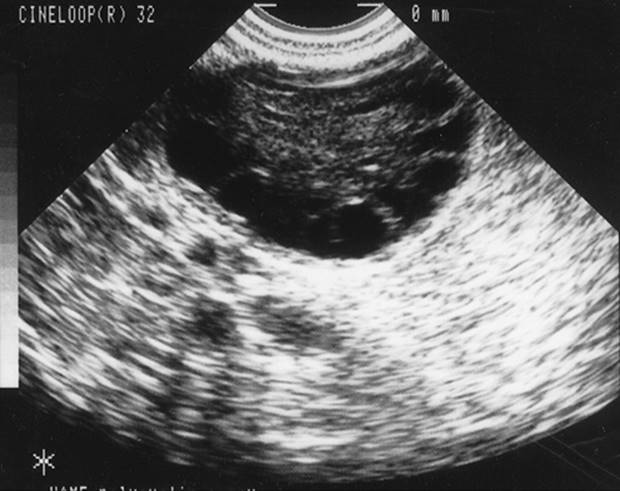
Other diagnostic criteria for PCOS have been proposed, including the finding of a polycystic ovary morphology on ultrasound, with multiple 2-to 8-mm subcapsular preantral follicles forming a “black pearl necklace” sign (Fig. 37.5). However, it should be noted that “polycystic ovaries” on sonography or at pathology are simply a sign of androgen excess and possibly PCOS. For example, this ovarian morphology is frequently seen in patients with adrenal hyperplasia (Table 37.2), and up to 25% of unselected women have polycystic ovaries on ultrasound, many of which are normoandrogenic with regular menstrual cycles. Hence, we consider the appearance of polycystic ovaries, particularly on sonographic exam, to be a sign, albeit nondiagnostic, of androgen excess and PCOS.
|
|
|
FIG. 37.5. Transvaginal ultrasound visualization of a polycystic ovary. Note the string of subcapsular follicles measuring 2 to 8 mm in diameter, with increased central stromal mass. |
|
|
|
TABLE 37.2. Syndromes or diseases entities associated with polycystic ovaries |
Clinical, Biochemical, and Metabolic Features of PCOS
Approximately 70% to 80% of women with PCOS demonstrate frank elevations in circulating androgens, particularly free testosterone, and 25% to 50% will have elevated levels of the adrenal androgen metabolite, DHEA-S. Prolactin levels are usually normal, although they may be slightly elevated (generally <40 ng/mL) in a small fraction of patients. The luteinizing hormone/follicle-stimulating hormone (LH/FSH) ratio is greater than 2 or 3 to 1 in approximately 60% of these patients. As noted, the ovaries of about 70% of patients with PCOS usually contain intermediate and atretic follicles measuring 2 to 8 mm in diameter, resulting in a “polycystic appearance” at sonography (see Fig. 37.4, Fig. 37.5). About 60% of patients with PCOS are obese, although significant fractions are nonobese.
While not part of the diagnostic criterion, many women with PCOS appear to be uniquely insulin-resistant. Approximately 50% to 70% of patients with PCOS demonstrate profound insulin resistance and secondary hyperinsulinemia, independent of body weight. In PCOS, insulin resistance usually refers to the impaired action of insulin in stimulating glucose transport and in inhibiting lipolysis in adipocytes. Insulin resistance in PCOS appears to be due to an intracellular defect of insulin signaling.
The compensatory hyperinsulinemia, resulting from the underlying insulin resistance, augments the stimulatory action of LH on the growth and androgen secretion of ovarian thecal cells, while inhibiting the hepatic production of SHBG. Treatment of patients with PCOS with insulin sensitizers may result in lower circulating levels of LH suggesting that insulin resistance, or more likely hyperinsulinemia, is in part responsible for the gonadotropic abnormalities observed in many women with PCOS. Since insulin is also a mitogenic hormone, the extremely elevated insulin levels may lead to hyperplasia of the basal layers of the epidermis, resulting in the development of acanthosis nigricans (a velvety, hyperpigmented change of the crease areas of the skin) (Fig. 37.6), and acrochordons. Overall, insulin resistance and secondary hyperinsulinemia affects a large fraction of patients with PCOS, and may cause or augment the androgen excess of these patients.
|
|
|
FIG. 37.6. Acanthosis nigricans on the nape of the neck: a sign of hyperinsulinemia. |
Sequelae of PCOS
Clinically evident PCOS tends to develop shortly after menarche and persists through most of the reproductive life. Nonetheless, some patients may present initially in the prepuberty with premature adrenarche, with affected girls displaying hyperinsulinemia, elevated DHEA-S levels, and postmenarche oligomenorrhea. At the other end of the reproductive spectrum, both menstrual irregularity and hyperandrogenemia appear to normalize as women with PCOS approach perimenopause. Although the endocrine and reproductive features of the disorder may improve with age, the associated metabolic abnormalities, particularly glucose intolerance, may actually worsen with age.
Infertility-Chronic Anovulation
One of the most common reasons that women with PCOS present to the gynecologist is due to infertility secondary to chronic anovulation. These women are not sterile, but subfertile due to the infrequency and unpredictability of their ovulation. As a general rule, women with PCOS represent one of the most difficult groups in which to safely induce ovulation. Many women with PCOS are unresponsive or resistant to ovulation induction with clomiphene citrate and may have an inappropriate or exaggerated response to the administration of human menopausal gonadotropins (menotropins). In part, the abnormal response to ovulatory agents of patients with PCOS relates to their degree of hyperinsulinemia and obesity, and to abnormalities of the intraovarian hormonal milieu and the increased number of preantral follicles. Women with PCOS are especially at increased risk for multiple gestation and developing the hyperstimulation syndrome—a syndrome of massive enlargement of the ovaries, development of rapid and symptomatic ascites, intravascular contraction, hypercoagulability, and systemic organ dysfunction. These complications generally occur following treatment with gonadootropins, although ovarian hyperstimulation has even been reported in women with PCOS conceiving a singleton pregnancy spontaneously, or after clomiphene citrate or pulsatile GnRH use.
Gynecologic Cancers
Many gynecologic cancers have been reported to be more common in women with PCOS including ovarian, breast, and endometrial carcinomas. However, the best case of an association between PCOS and cancer can be made for endometrial cancer, as many risk factors for this cancer, including obesity, chronic anovulation, and hyperinsulinemia, are present in the patient with PCOS. Patients with endometrial cancer have significantly higher body mass index (BMI) and frequency of diabetes hypertension, hirsutism, nulliparity, and infertility than controls.
Type II Diabetes Mellitus
The inherent insulin resistance present in many with PCOS, aggravated by the high prevalence of obesity in these individuals, places these women at increased risk for impaired glucose tolerance and type 2 DM. About 30% to 40% of obese women of reproductive age with PCOS have been found to have impaired glucose tolerance (IGT), and about 10% have frank type 2 DM based on a 2-hour glucose level greater than 200 mg/dL. Of note is that only a small fraction of women with PCOS with either IGT or type 2 DM display fasting hyperglycemia consistent with diabetes by the 1997 American Diabetes Association criterion (fasting glucose ≥126 mg/dL) (Fig. 37.7). The conversion rate to glucose intolerance over time is unknown, which makes recommendations for frequency of screening for glucose intolerance difficult. However, the level of insulin resistance found in women with PCOS based on dynamic measures of insulin action has in other populations (i.e., children of parents with diabetes) been associated with a marked increased risk of developing type 2 DM.
|
|
|
FIG. 37.7. Scatterogram of fasting blood glucose levels versus 2-hour postchallenge glucose levels in 254 women with polycystic ovary syndrome (PCOS). Points on the graph are coded to reflect the World Health Organization status based on postchallenge glucose levels. A, 140 to 199 mg/dL = impaired glucose tolerance. The dashed vertical line is the threshold (110 mg/dL) for the diagnosis of impaired fasting glucose according to the 1997 American Diabetes Association criteria. The solid vertical line (126 mg/dL) is the threshold for the diagnosis of type 2 diabetes mellitus by the same criteria. (From Legro RS, Kunselman AR, Dodson WC, et al. Prevalence and predictors of risk for type 2 diabetes mellitus and impaired glucose tolerance in polycystic ovary syndrome: a prospective, controlled study in 254 affected women. J Clin Endocrinol Metab 1999;84:165–169, with permission.) |
Cardiovascular Disease
Many women with PCOS appear to form a subset of the metabolic syndrome first described by Reaven (i.e., syndrome X or insulin-resistance syndrome) consisting of insulin resistance, hypertension, dyslipidemia, glucose intolerance, and CVD. The mechanisms underlying the relationship between the insulin resistance/hyperinsulinemia and hypertension are unclear, but may include sodium retention, overactivity of the sympathetic nervous system, impairment of membrane transport, and stimulation of vascular smooth muscle cells. Other effects of hyperinsulinemia and insulin resistance include the development of an atherogenic plasma lipid profile due to insulin enhancement of synthesis of very-low-density lipoproteins and hypertriglyceridemia. Insulin also causes the proliferation of smooth muscle cells and stimulation of collagen synthesis in small arterioles. These effects, in combination with insulin-induced growth factor elaboration and inhibition of regression of lipid plaques, contribute to the development of atherosclerosis and the resultant CVD.
Many women with PCOS have significant dyslipidemia, with lower high-density lipoprotein (HDL), and higher triglyceride and low-density lipoprotein (LDL) levels than age, sex, and weight-matched controls. Studies have documented other interim risk factors, such as increased carotid artery intimal thickness, to suggest that these women have evidence of atherosclerotic disease earlier and at a more advanced stage than control women. However, an increase in actual cardiovascular events in women with PCOS, such as myocardial infarction, has not been observed, although a higher number of nonfatal cerebrovascular disease events have been noted.
We should note that there are a number of features of PCOS that do not fully fit the definition of the metabolic syndrome. For example, although dyslipidemia is part of the metabolic syndrome, the elevation in LDL levels noted in PCOS is not one of the characteristic findings. Additionally, although the risk of developing hypertension later in life appears to be higher in patients with PCOS, hypertension has not been associated with PCOS in women of reproductive age.
OTHER ANDROGEN EXCESS DISORDERS
Other major androgen excess disorders include the HAIRAN syndrome, 21-hydroxylase (21-OH) deficient NCAH, and very rarely, ovarian or adrenal androgen-secreting neoplasms.
The HAIRAN Syndrome
The HAIRAN (hyperandrogenic-insulin resistant-acanthosis nigricans) syndrome is an inherited disorder of severe insulin resistance, distinct from PCOS, and which actually includes many different genetic syndromes. Although single-point mutations of the insulin receptor are rare among these patients, some patients with this syndrome may actually suffer from a form of the familial lipodystrophy syndrome. Approximately 3% of hyperandrogenic women suffer from these disorders. These patients are diagnosed by having extremely high circulating levels of basal (>80 µU/mL) or glucose-stimulated (>300–500 µU/mL) insulin levels, although exact diagnostic criteria are lacking, in part due to the heterogeneity of the syndrome. Patients can be severely hyperandrogenemic, with testosterone levels reminiscent of patients with androgen-secreting neoplasms, resulting in the development of severe hirsutism and even virilization. In addition to significant hirsutism, patients also develop the characteristic dermatologic finding of acanthosis nigricans (see Fig. 37.6). Because of their high androgen levels, gonadotropin levels in these patients may be somewhat suppressed resulting in persistent endometrial atrophy and amenorrhea in spite of the administration of a cyclic progestogen or an oral contraceptive.
Because of the mitogenic effect of insulin on ovarian theca cells, the ovaries of many patients with the HAIRAN syndrome will become hyperthecotic. On ultrasound and histology the ovaries morphologically have a paucity of cortical cysts, and demonstrate a thickened and enlarged theca/stroma compartment. In addition, these patients are at significant risk for dyslipidemia, type 2 DM, hypertension, and CVD. These patients can be particularly difficult to treat although the selected use of long-acting GnRH analogs has been promising. Some patients may necessitate surgery, either ovarian wedge resection or oophorectomy.
Nonclassic Adrenal Hyperplasia
Nonclassic adrenal hyperplasia (NCAH), also referred to as late-onset congenital adrenal hyperplasia, is a homozygous recessive disorder due to mutations in the CYP21 gene which results in an abnormal (or absent) cytochrome P450c21 with relatively deficient 21-OH activity. Overall, between 1% and 8% of women with androgen excess have 21-OH–deficient NCAH depending on ethnicity, with the highest rates reported among Ashkenazi Jews. Due to the lack of 21-hydroxylation the progestogenic precursors to cortisol, 17 α-hydroxyprogesterone (17 α-HP) and to a certain degree 17 α-hydroxypregnenolone, accumulate in excess. These steroids are then metabolized to C19 products, principally androstenedione and testosterone (see Fig. 37.1, Fig. 37.2). However, clinically and biochemically these patients are very difficult to distinguish from other hyperandrogenic patients, particularly patients with PCOS. Patients with NCAH may present only with persistent acne or may have moderate degrees of hirsutism and oligoamenorrhea. Frank virilization or even severe hirsutism is relatively rare. The levels of the exclusive adrenal androgen metabolite DHEA-S are not any higher than that of other hyperandrogenic women.
Although the frequency is relatively low, all patients with unexplained androgen excess should be screened for NCAH due to CYP21 mutations, as this diagnosis has a different prognosis, a different treatment regimen, and requires genetic counseling regarding the risks of congenital transmission. The measurement of a basal 17 α-HP in the follicular phase and in the morning can be used to screen for this disorder. In addition, these patients can be treated with corticosteroid replacement, although many of the patients who are diagnosed in adulthood tend to also demonstrate a PCOS-like pattern requiring additional ovarian suppression.
While it is theoretically possible that NCAH may be due to mutations in other genes determining steroidogenic enzymes, such CYP11B1 (encoding for P450c11a and 11β-hydroxylase (CYP11B1) or HSD3B (for 3β-hydroxysteroid dehydrogenase), mutations in the genes coding for these enzymes are rarely noted in adult women presenting with androgen excess.
Androgen-Secreting Tumors
Androgen-secreting neoplasms are relatively rare, affecting between 1 of 300 and 1 of 1,000 hirsute patients. These tumors usually originate in the ovary, and rarely the adrenal cortex. The most common androgen-producing tumor in a premenopausal woman is a Sertoli-Leydig cell tumor, with thecomas and hilus cell tumors being less frequent. Hilus cell tumors are often small, and can be less than 1 cm in diameter, theoretically below the range of ultrasound visualization. Another rare neoplasm may be a granulosa cell tumor, 10% of which primarily secrete androgens instead of estrogens. Also, any large tumor within the body of the ovary (i.e., benign cystic teratomas, dysgerminomas, epithelial tumors) can produce androgens indirectly by causing hyperplasia of the surrounding normal stroma. Fortunately, the vast majority of ovarian androgen-secreting neoplasms are benign.
Alternatively, adrenal androgen-secreting tumors are generally malignant (i.e., adrenocortical carcinoma) and associated with a high mortality rate, although androgen-secreting adrenal adenomas have also been reported. Fortunately, adrenal androgen-secreting neoplasms are rare with an estimated incidence of two cases per one million persons per year. The age of onset in adults peaks in the fifth decade. Virilization can accompany both tumors primarily producing androgens and tumors primarily producing cortisol (Cushing syndrome). A long history of symptoms, as in the case with an ovarian tumor does not exclude the presence of an adrenocortical neoplasm.
The presence of androgen-secreting neoplasms should be suspected when the onset of androgenic symptoms is rapid and sudden, when androgen excess initially presents in late life, when they lead to virilization and masculinization, or are associated with cushingoid features. Nonetheless, it should be remembered that some of younger patients with virilization suspected of having an androgen-secreting neoplasm actually suffer from the HAIRAN syndrome (discussed earlier). Suppression and stimulation tests can be misleading and are not encouraged for the diagnosis of these neoplasias. Overall, the single best predictor of an androgen-producing tumor is clinical presentation, and not biochemical markers. For example, in one study we noted that the positive predictive value of a repeat total testosterone above 250 ng/dL was only 9%, as most women with total testosterone levels above this cutoff have other abnormalities, such as the HAIRAN syndrome or PCOS.
Rare Causes of Androgen Excess
Clinical signs of androgen excess may also result from the presence a corticotropin-secreting tumor, due to either a pituitary adenoma (i.e., Cushing's disease) or an extrapituitary (ectopic) source, although patients are usually also cushingoid. In one study of young women with Cushing's syndrome all had stigmata of hyperandrogenism (hirsutism, acne, or balding), but only 70% had oligomenorrhea. However, as Cushing's syndrome has an extremely low prevalence in the population (1 in a million) and screening tests do not have 100% sensitivity/specificity, routine screening of all women with androgen excess for Cushing's syndrome is not indicated. Nonetheless, clinical signs more commonly found in Cushing's syndrome, such as ecchymoses, proximal muscle weakness, centripetal reddened striae, facial rubor and swelling, and perhaps hypertension and glucose intolerance should signal screening tests. Screening for cortisol excess can be conducted with a 24-hour urine collection for free cortisol.
The use of exogenous androgens, often found among body builders, or the excessive use of androgens among postmenopausal women should also figure in the differential diagnosis. Severe hirsutism, and even virilization, that begins during pregnancy has its own unique differential including benign ovarian sources such as hyperreactio luteinalis (i.e., gestational ovarian theca–lutein cysts) or luteomas, and extremely rare fetoplacental sources, such as aromatase deficiency resulting in androgen excess due to the placental inability to convert precursor androgens into estrogens. Finally, gonadal dysgenesis associated with an abnormal Y chromosome can initially present as peripubertal androgenization.
IDIOPATHIC HIRSUTISM
Although idiopathic hirsutism (IH) is not actually an androgen excess disorder, it is included among them and should be considered in the differential diagnosis of androgen excess. Overall, approximately 5% to 15% of hirsute women will have the diagnosis of “idiopathic” hirsutism. The diagnosis of IH is, by exclusion, made in a patient who is obviously hirsute, but in whom the circulating androgens and ovulatory function appear to be normal, and no other disorders can be identified. While some clinicians use regular menses as diagnostic of normal ovulatory function, we should note that approximately 40% of eumenorrheic hirsute women are actually anovulatory, and hence do not suffer from IH. Although biochemically these patients do not have an obvious elevation in circulating androgen levels, it also is likely that many of these patients simply demonstrate degrees of hyperandrogenemia that may not be detectable with routine clinical androgen assays. Nonetheless, in some of these women the 5α-reductase activity in the skin and hair follicles is probably overactive and leads to hirsutism in spite of normal circulating androgen levels.
EVALUATION OF ANDROGEN EXCESS
The history and physical examination are essential to making a diagnosis of the underlying cause of androgen excess. History taking should focus on the onset and duration of the various stigmata of androgen excess, should include a thorough menstrual history, and a discussion of concomitant medications. The physical examination should carefully assess the patient for cushingoid features, acanthosis nigricans, balding, acne, and degree and type of body hair distribution. One commonly used scale to score the degree of excess hair growth is based on a modification of the Ferriman-Gallwey scale (Fig. 37.8). Signs of virilization and masculinization should be sought, although their recognition is usually obvious. Clitoromegaly is usually defined as a clitoral index greater than 35 mm2, where the clitoral index is the product of the sagittal and transverse diameters of the glans of the clitoris in millimeters. In normal women these diameters are in the range of 5 mm.
|
|
|
FIG. 37.8. Modified Ferriman-Gallwey scale for assessing hirsutism. (Modified from Hatch, et al. Hirsutism: implications, etiology, and management. Am J Obstet Gynecol 1981;140:815–830.) |
Evidence and risk factors for insulin resistance should be sought out as they are critical to the long-term health of the women affected. Family history of diabetes and CVD, especially of first-degree relatives with premature-onset heart disease (males <55 years and females <65 years), is an important part of the assessment. Lifestyle factors such as smoking, alcohol consumption, and diet and exercise histories should be recorded. Signs of insulin resistance on physical exam such as obesity, particularly with a centripetal fat distribution, and the presence of acanthosis nigricans and acrochordons should be recorded. Acanthosis nigricans is principally noted in most crural areas of the body, including the back of the neck, the axilla, underneath the breasts, and even on the vulva.
Weight should be corrected for height by using the BMI (divide weight in kilograms by the square of the height in meters, i.e., kg/m2). However, the pattern of fat distribution rather than BMI seems to be most predictive of morbidity and mortality. In women a central or abdominal distribution of adiposity fat (the so-called apple-shaped or android obesity) has been associated with an increased risk for CVD and with an increased index of death from all causes. Fat distribution is most easily assessed using the waist-to-hip ratio (WHR), obtained by measuring the circumferences at the narrowest point of the abdomen (i.e., the abdominal measure), generally immediately above the umbilicus, and the widest part of the hips (i.e., the hip measures). Values greater than 0.72 (primarily android fat distribution) are considered abnormal but increased morbidity and mortality becomes more noticeable at values greater than 1.0. In women, the android pattern of obesity has been associated with abnormal lipid profiles and increased insulin resistance compared to the gynecoid pattern.
The laboratory evaluation should have the objective of excluding related and specific disorders and, if necessary, providing confirmation of androgen excess. As well, the presence of metabolic abnormalities, particularly in patients with PCOS or the HAIRAN syndrome, should be sought out.
Laboratory Evaluation of Androgen Excess
Specific related disorders that should be excluded prior to diagnosis of androgen excess are thyroid dysfunction, hyperprolactinemia, NCAH, the HAIRAN syndrome, and androgen-secreting tumors. Thyroid dysfunction and NCAH are ruled out by measuring a third generation thyroid-stimulating hormone (TSH) and a basal 17- αHP level, the latter measured in the follicular phase of the menstrual cycle. If the 17-HP level is greater than 2 ng/mL, the patient should undergo an acute adrenal stimulation test to exclude 21-hydroxylase deficient NCAH. If the 17-HP level 60 minutes following corticotropin administration is greater than 10 ng/mL, the diagnosis of 21-OH–deficient NCAH is established.
As noted previously, hirsute women claiming to have regular menstrual cycles should be evaluated for ovulatory dysfunction, most simply by obtaining a basal body temperature chart and a serum progesterone in the luteal phase (days 20–24) of the menstrual cycle. If the patient has ovulatory dysfunction, as evidenced by either a luteal progesterone level less than 4 ng/mL in a eumenorrheic patient, or because she has overt menstrual abnormalities, the diagnosis of PCOS should be entertained. In women with ovulatory dysfunction, serum prolactin and fasting insulin levels should also be obtained to exclude hyperprolactinemia and the HAIRAN syndrome, respectively. Androgen-secreting neoplasms are generally excluded by the history and physical exam. Rarely, a 24-hour urine free cortisol is required in a patient with features suggestive of Cushing's syndrome.
The measurement of circulating androgen levels, including total and free testosterone, and DHEA-S is useful, primarily in the minimally or nonhirsute oligoovulatory patient, to exclude the presence of androgen excess as the cause of the ovulatory dysfunction. However, these measurements have limited diagnostic use in the patient who is frankly hirsute, and have a low positive predictive value for adrenal or ovarian androgen-secreting neoplasms.
Inappropriate gonadotropin secretion has been one of the characteristic signs of PCOS, and many clinicians have used elevated LH/FSH ratios of 2:1 or 3:1 as diagnostic criteria for PCOS. Nonetheless, because up to 50% of patients with PCOS may have a normal ratio, the LH and FSH are episodically secreted, and the LH/FSH ratios tend to normalize with increasing BMI, or alternatively with insulin-sensitizer therapy. Measuring gonadotropins is an insensitive marker of PCOS.
Laboratory Assessment for Metabolic Abnormalities
Metabolic abnormalities are common among women with PCOS, and occur in all patients with the HAIRAN syndrome. In patients with the HAIRAN syndrome the presence of insulin resistance is obvious, although this may not be the case in women with PCOS. Unfortunately, there are no accurate, inexpensive, or reproducible tests for clinically assessing insulin sensitivity. The standards are dynamic tests such as the euglycemic clamp and the frequently sampled intravenous glucose tolerance test (FSIVGTT). These assessments primarily determine insulin-mediated glucose uptake in the skeletal muscle, the primary tissue using circulating insulin. Insulin-mediated glucose uptake, however, is just one aspect of insulin's protean actions. While these methods are the most accurate for determining decreased sensitivity to insulin (or insulin resistance), they are too laborious and expensive to use for the routine clinical evaluation of insulin resistance.
A fasting glucose-to-insulin ratio of less than 4.5 may be a good marker of insulin resistance in obese women with PCOS. Although a fasting insulin level alone is often clinically useful in other non-PCOS populations, insulin levels, either fasting or postprandial, have only a moderate correlation with more detailed studies of insulin action, and are less useful in patients with impaired glucose tolerance or frank diabetes, due to deficiencies in insulin secretion. Most important, it is key to understand that there is a wide range of insulin sensitivities in normal populations, and that substantial overlap of even the most precise measure of insulin action between insulin-resistant and control populations is always present. As such, about 50% of obese women with PCOS may not have clinically recognizable insulin resistance on the basis of either fasting values or more dynamic test results. On an individual basis, therefore, it is often not possible to diagnose insulin resistance and a diagnostic trial with an insulin sensitizer should be considered.
Given the high prevalence of abnormalities in glucose tolerance, patients with PCOS or HAIRAN syndrome should be screened for IGT or frank diabetes with an oral glucose tolerance test (oGTT) using a 75-g glucose load. Considering the World Health Organization criteria, IGT is diagnosed by a 2-hour glucose of between 140 mg/dL and 199 mg/dL, while frank diabetes is diagnosed by 2-hour glucose during the oGTT ≥200 mg/dL. Abnormal oGTT results need to be repeated for confirmation. The development of glucose intolerance heralds β-cell dysfunction with inadequate insulin secretion for the degree of reduced peripheral insulin sensitivity. The development of IGT is a clear risk factor for developing type 2 DM and may also be an independent risk factor for CVD, such as myocardial infarction and stroke.
Although no firm guidelines exist, obtaining a plasma lipid profile in patients with PCOS or the HAIRAN syndrome should be considered, particularly in women over the age of 35, those with a strong family history of diabetes or CVD, and those with significant insulin resistance.
Radiologic Imaging in the Evaluation of the Patient with Androgen Excess
Routine ultrasound of the pelvis in patients with androgen excess is probably not indicated. Polycystic ovaries can be noted with many of the androgen excess disorders and remain nonspecific (see Fig. 37.8). Nonetheless, the appearance of polycystic ovaries may be useful as a potential predictor for the development of ovarian hyperstimulation syndrome during ovulation induction. Furthermore, a transvaginal sonogram in obese individuals to properly examine the ovaries for pathologic masses may be appropriate. In cases where an androgen-secreting tumor is suspected, a computed tomography (CT) scan or magnetic resonance imaging (MRI) of the adrenals should be considered to exclude adrenal masses as small as 5 mm in diameter and detect bilateral adrenal hyperplasia in the event of a corticotropin-secreting tumor. However, about 2% of the population harbor a clinically insignificant adrenal adenoma (i.e., incidentaloma) making the finding of such an adrenal mass not diagnostic for an androgen-secreting adrenal tumor. Functional radiologic techniques such as selective venous catheterization or scintigraphy with iodomethyl-norcholesterol are rarely used for the localization of such tumors.
TREATMENT OF ANDROGEN EXCESS
Treatment of androgen excess tends to be symptom-based. In general, we can consider four different reasons for treatment including:
1. regulation of uterine bleeding, reducing the risk of endometrial hyperplasia and cancer, dysfunctional uterine bleeding (DUB), and secondary anemia
2. improvement of dermatologic abnormalities such as hirsutism, acne, or alopecia
3. amelioration and prevention of associated metabolic abnormalities, particularly insulin resistance and hyperinsulinemia
4. treatment of the associated anovulatory infertility.
Overall, avenues of treatment include the suppression of androgen production, the blockade of peripheral androgen action, the improvement of any associated insulin resistance and dyslipidemia, and topical, mechanical, or cosmetic measures for improving the dermatologic manifestations of these disorders (Fig. 37.9). The vast majority of patients with androgen excess will require, and benefit the most from, combination therapy.
|
|
|
FIG. 37.9. Spironolactone (100 mg/d), flutamide (250 mg/d), and finasteride (5 mg/d) were compared for the treatment of hirsutism in a randomized, double-blind, placebo-controlled trial including 40 affected women. (Modified from Moghetti P, Tosi F, Tosti A, et al. Comparison of spironolactone, flutamide, and finasteride efficacy in the treatment of hirsutism: a randomized, double blind, placebo-controlled trial. J Clin Endocrinol Metab 2000;85:89–94, with permission.) |
Regulation of Uterine Bleeding
In general, the regulation of uterine bleeding can be achieved with oral contraceptives, cyclic or continuous progestogens, and in some patients with PCOS, with insulin-sensitizing agents, and rarely surgery.
Oral Contraceptives
Oral contraceptive medications generally act by suppressing circulating LH and FSH leading to a decrease in ovarian androgen production. In addition, the estrogen in the birth control pill increases SHBG, decreasing free testosterone levels. Furthermore, the progestin in the birth control pill can lead to a beneficial antagonism of 5α-reductase activity and androgen binding to the androgen receptor. Finally, these medications may also decrease adrenal androgen production by a mechanism not yet clear. Oral contraceptives are very effective at regulating uterine bleeding and decreasing the associated risk of endometrial hyperplasia or carcinoma in the hyperandrogenic patient, regardless of etiology. It is preferable, although not critical, to select a pill containing a progestin with low androgenic activity (e.g., norethindrone acetate, ethynodiol diacetate, desogesterol, gestodene, or norgestimate). There is some evidence that oral contraceptives can worsen insulin resistance and glucose tolerance in women with PCOS. However, there is no evidence that these effects of the oral contraceptive pill on glucose metabolism result in an increased risk for developing type 2 DM in the general population, and their use in patients with androgen excess patients not be limited by this concern.
Cyclic or Continuous Progestogens
Cyclic progestogen administration is also useful to regulate uterine bleeding in patients with androgen excess, particularly in amenorrheic women. It is not known how many progestin-induced withdrawal bleeds per year are necessary to adequately prevent the development of endometrial cancer in women with PCOS, although extrapolating from the postmenopausal hormone replacement literature, monthly treatment for a minimum of 12 days or longer is optimal. Because progestogen administration can occasionally stimulate an ovulatory event, and because not all these patients are completely anovulatory, it may be preferable to treat sexually active at risk for pregnancy women with continuous oral micronized progesterone, 100 to 200 mg twice daily.
Glucocorticoids
Glucocorticoid therapy may be beneficial in regulating menstrual bleeding in the patient with NCAH. However, we have found that NCAH monotherapy is effective primarily in those women whose treatment is initiated in adolescence and that many older patients will require combination therapy. The lowest effective dose of glucocorticoids should be used (e.g., dexamethasone 0.25 mg daily or every other day). Glucocorticoids should be administered with caution as they have the potential of producing weight gain and cushingoid features, and worsening existing insulin resistance.
Insulin-Sensitizing Agents
Drugs developed initially to treat type 2 DM have now also been used to treat PCOS. These include metformin and thiazolidinediones. Other drugs such as acarbose have also been studied in PCOS, and beneficial effects have been reported. However, we should note that none of these agents are currently approved by the Food and Drug Administration (FDA) for the treatment of PCOS or for related symptoms such as anovulation, hirsutism, or acne.
Metformin
Metformin was approved for the treatment of type 2 DM by the FDA in 1994, but was used clinically for close to 20 years before that in other parts of the world. Metformin is a biguanide that works primarily by suppressing hepatic gluconeogenesis, but it also improves insulin sensitivity peripherally. Gastrointestinal symptoms (diarrhea, nausea, vomiting, abdominal bloating, flatulence, and anorexia) are the most common reactions to metformin, occurring in about 30% of treated women. There is a small risk of lactic acidosis among women taking this medication, which may be triggered by exposure to intravenous iodinated radiocontrast agents in susceptible individuals, although this adverse effect occurs primarily in patients with poorly controlled diabetes and impaired renal function. Metformin therapy has also been found to improve uterine bleeding and menstrual function.
Thiazolidinediones
Thiazolidinediones are peroxisome proliferator activating receptor γ-agonists, and are thought to improve insulin sensitivity through a postreceptor mechanism. In a large multicenter trial, troglitazone has been shown to have a dose-response effect in improving ovulation and menstrual dysfunction in women with PCOS. This appears to be mediated through decreases in hyperinsulinemia and decreases in free testosterone levels. Troglitazone has subsequently been removed from the worldwide market due to its potential for inducing hepatotoxicity. Newer thiazolidinediones such as rosiglitazone and pioglitazone appear to be safer in terms of hepatotoxicity, but have also been associated with embryotoxicity in animal studies (both are pregnancy category C). Furthermore, there are limited studies on their effectiveness in PCOS. When using these drugs, liver function tests should be monitored on a regular basis. Combination treatment with other insulin sensitizers, primarily metformin, has been shown to further improve insulin sensitivity and glycemic control in type 2 DM, although the effect of such combination therapy in women with PCOS is not yet clearly delineated.
Surgery
In patients with intractable uterine bleeding who have completed their childbearing, consideration may be given to a hysterectomy and bilateral salpingo-oophorectomy. In addition, ovulatory function may also improve following ovarian wedge or laparoscopic ovarian drilling procedures, although the long-term effect of these therapies on menstrual function is unclear.
Weight Loss
Multiple studies in hyperandrogenic or women with PCOS have shown that weight loss can lower circulating androgen levels and cause spontaneous resumption of menses. These changes have been reported with a weight loss as small as 5% of the initial weight. In patients with massive obesity, consideration may be given to surgical means of inducing weight loss, such as laparoscopic gastric bypass.
Treatment of Hirsutism, or Androgen Excess-Associated Acne or Alopecia
The treatment of hirsutism can be complex, and has recently been reviewed. Optimum therapy usually requires a combination of approaches, including suppression of circulating androgens (with oral contraceptives, insulin sensitizers, or even long-acting GnRH analogs or glucocorticoids), peripheral androgen blockade (with spironolactone, flutamide, cyproterone acetate, or finasteride); and the use of a topical hair growth inhibitor (eflornithine hydrochloride) or mechanical methods of hair reduction or destruction (electrology or lasers), and appropriate cosmetic measures (bleaching or chemical depilating). Acne in patients with androgen excess can usually be treated with oral contraceptives, accompanied by topical or antibiotic therapies. The treatment of androgenic alopecia may require the use of androgen suppression in combination with androgen blockade and topical means of stimulating hair regrowth (e.g., minoxidil).
Most medical methods alone, while improving hirsutism do not produce the dramatic results that many patients desire. In general, combination therapies appear to produce better results than single agent approaches. Response with medical therapies often take 3 to 6 months to notice improvement, and adjunctive mechanical removal methods are often necessary. However, the majority of women appear to experience improvement in their hirsutism. Unfortunately, there are no universally accepted techniques for assessing the response of hirsutism to treatment. Some of the depilatory treatments used by patients are so effective that it is difficult to ever determine baseline hirsutism and response to treatment such that the patient's subjective assessment guides therapy.
Androgen Suppression
Women with documented hyperandrogenemia would theoretically benefit most from androgen-suppression therapy, although in actual practice, the clinical response to this type of therapy does not correlate with androgen levels. Suppression of ovarian androgen secretion has been achieved with oral contraceptives, progestins, or GnRH analog treatment. Oral contraceptives improve hirsutism, although the response of hirsutism to this therapy alone is generally modest. Treatment with a long-acting GnRH agonist may result in a greater lowering of circulating androgens, and theoretically a greater degree of hair growth suppression, although comparative trials against other agents and combined agent trials have yielded mixed results. It should be noted that a long-acting GnRH agonist given alone results in unacceptable bone loss. Glucocorticoid suppression of the adrenal glands also offers theoretical benefits, but deterioration in glucose tolerance is problematic for women with PCOS, and long-term effects such as osteoporosis are a significant concern. Furthermore, glucocorticoid suppression produces only minimal improvements in hair growth in hirsute women.
Androgen Receptor Antagonists
Androgen receptor antagonists antagonize the binding of testosterone and other androgens to the androgen receptor. As a class therefore, they are teratogenic, and pose risk of feminization of the external genitalia in a male fetus, should the patient conceive. Spironolactone, a diuretic and aldosterone antagonist, binds to the androgen receptor with 67% of the affinity of DHT. It has other mechanisms of action, including inhibition of ovarian and adrenal steroidogenesis, and direct inhibition of 5-α-reductase activity. Hirsutism and acne have been successfully treated with spironolactone. The usual dose for the treatment of hirsutism is 25 to 100 mg twice a day, and the dose should be titrated to minimize the potential for side effects. About 20% of women given spironolactone will experience increased menstrual frequency, a reason for combining this therapy with oral contraceptive. Other side effects include polyuria, orthostatic hypotension, nausea, dyspepsia, and fatigue. Because spironolactone can cause and exacerbate hyperkalemia, it should be used cautiously in patients with renal impairment, and should not be combined with other potassium-sparing diuretics. The medication also has potential teratogenicity as an anti-androgen, although exposure has rarely resulted in ambiguous genitalia in male infants.
Flutamide is another nonsteroidal anti-androgen that has been shown to be effective against hirsutism. The most common side effect is dry skin, but its use has rarely been associated with hepatitis. A dose of 250 mg per day is generally used. Because there is greater risk of teratogenicity with this compound, contraception should be used.
5α-Reductase Inhibitors
There are two forms of the enzyme 5α-reductase: type 1, predominantly found in the skin and type II, predominantly found in the prostate and reproductive tissues. However, both forms are found in the pilosebaceous unit (PSU) and may contribute to the development of hirsutism, acne, and alopecia. Finasteride inhibits both forms of 5α-reductase, and is available as a 5-mg tablet for the treatment of prostate cancer and a 1-mg tablet for the treatment of male alopecia. It has been found to be effective for the treatment of hirsutism in women. Finasteride is better tolerated than other antiandrogens, but has the highest and clearest risk for teratogenicity in a male fetus and adequate contraception must be used. Overall, randomized trials have found that spironolactone, flutamide, and finasteride have similar efficacy in improving hirsutism (see Fig. 37.9).
Insulin-Sensitizing Agents and Lifestyle Modification
It is difficult to separate the effects of an improvement in insulin sensitivity from that of androgen suppression, as any improvement in insulin sensitivity can raise SHBG and thus lower bioavailable androgen. Given the long onset of action for improving hirsutism and acne, longer periods of observation are needed. In the largest and longest randomized trial to date of these agents, troglitazone, in a dose-response fashion, was found to significantly improve hirsutism in women with PCOS without lowering total testosterone, a benefit presumably achieved through the decrease in free testosterone. Weight loss in the obese patient may also benefit hirsutism and acne, albeit modestly. Increases in SHBG through improved insulin sensitivity from weight loss may lower bioavailable androgen levels. About 50% of the women who lose weight experience an improvement in their hirsutism.
Topical Hair Growth Suppression—Ornithine Decarboxylase Inhibitors
Ornithine decarboxylase is necessary for the production of polyamines, and is also a sensitive and specific marker of androgen action in the prostate. Inhibition of this enzyme limits cell division and function, including that of the PSU. A potent inhibitor of this enzyme, eflornithine, has been found to be effective as a facial cream for the treatment of unwanted facial hair. It is available as a 13.9% cream of eflornithine hydrochloride, and is applied to affected areas twice daily. In clinical trials, 32% of patients had marked improvement after 24 weeks compared to 8% of women given placebo, and the benefit was first noted at 8 weeks. It is pregnancy category C. The cream appears to be well tolerated, with only about 2% of patients developing skin irritation or other adverse reactions.
Mechanical and Cosmetic Means of Hair Reduction and Destruction
Mechanical hair removal methods (shaving, plucking, waxing, depilatory creams, electrolysis, and laser vaporization) can assist in controlling hirsutism, and often are the front line of treatment used by women. Shaving, bleaching, or chemical depilation may be useful temporary treatments for unwanted hairs. Although shaving can lead to a blunt hair end that may feel “stubble-like,” it does not lead to a worsening of hirsutism. Depilating agents, while useful, can result in chronic skin irritation and worsening of hirsutism if used excessively or indiscriminately. The use of plucking or waxing in androgenized skin areas should be discouraged since these techniques not only do not kill the hair follicles, but also can induce folliculitis and trauma to the hair shaft with subsequent development of ingrown hairs and further skin damage.
Electrolysis (i.e., electroepilation) results in long-term hair destruction, but this therapy is time consuming. Side effects may include temporary skin irritation, and rarely burning and scarring of the skin. Hair reduction via lasers can also be useful for selected patients. The main objective of laser therapy for hair removal is to selectively cause thermal damage of the hair follicle without destroying adjacent tissues, a process termed selective photothermolysis. Selective photothermolysis relies on the selective absorption of a brief radiation pulse to generate and confine heat at specific pigmented targets. Lasers useful in hair removal may be grouped into three categories based on the type of laser or light source each employs:
1. red light systems (694 nm ruby)
2. infrared light systems (755 nm alexandrite, 800 nm semiconductor diode, or 1,064 nm neodymium: yttrium-aluminum-garnet [Nd:YAG])
3. intense pulsed light (IPL) sources (590–1200 nm).
In general, laser hair removal is most successful in patients with fairer skin, who have dark color hairs. However, repeated therapies are necessary, and complete alopecia is rarely achieved.
In general, treatment with the ruby, alexandrite, or diode lasers, or the IPL results in similar success rates, although it appears to be somewhat lower for the Nd:YAG laser. After laser-assisted hair removal, most patients experience erythema and edema lasting no more than 48 hours. Blistering or crusting may occur in 10% to 15% of patients. Temporary hyperpigmentation occurs in 14% to 25% of patients and hypopigmentation occurs in 10% to 17% of patients. Dyspigmentation is less common with the use of longer wavelengths, as in the alexandrite or diode lasers, and longer pulse durations. Overall, laser hair removal is a promising technique for the treatment of the hirsute patient. Nonetheless, we should note that most studies have been uncontrolled and included fewer than 50 patients, none have been blinded, and all have used a variety of treatment protocols, equipment, skin types, and hair colors studied.
Treatment of Associated Metabolic Abnormalities
The treatment of any associated metabolic abnormalities includes lifestyle alterations and weight reduction, and the use of insulin sensitizers and possibly lipid-lowering agents. Multiple studies have shown that improving insulin sensitivity, through lifestyle modifications or by pharmacologic intervention, can result in lowered circulating androgens (primarily mediated through a reduction in bioavailable androgen), and an increase in the spontaneous ovulation and pregnancy rates. In addition, it is possible that a decrease in insulin resistance may decrease the risk that patients with PCOS will develop type 2 DM. In support of this, data from the Diabetes Prevention Program, which enrolled men and women at risk for diabetes, demonstrate that lifestyle interventions have a more profound effect on preventing the development of subsequent type 2 DM than does the sole use of metformin, although both interventions were superior to placebo. However, long-term studies documenting a decrease in such sequelae as endometrial cancer, type 2 DM, or CVD with improvements in insulin sensitivity are still lacking among women with PCOS.
The gold standard for improving insulin sensitivity in obese women with PCOS should be weight loss, diet, and exercise. Obesity has become epidemic in our society and contributes substantially to reproductive and metabolic abnormalities in PCOS. Unfortunately there are no effective treatments that result in permanent weight loss, and it is estimated that 90% to 95% of patients who experience a weight decrease will relapse. In addition to improving the endocrine aspects of PCOS, weight loss can decrease circulating insulin levels. Unfortunately there have been few studies on the effect of exercise alone on insulin action in women with PCOS, although it is reasonable to assume that exercise would have the same beneficial effects in women with PCOS as women with type 2 DM.
Treatment of Hyperandrogenic Oligoovulatory Infertility
A full discussion of the treatment of oligoovulatory infertility is beyond the scope of this chapter. In brief, ovulation induction for the treatment of androgen-excess–related infertility typically includes the use of clomiphene citrate and human gonadotropins. In women with PCOS there are a variety of methods available to improve ovulatory function that act through a different mechanism (Fig. 37.10). For example, in patients with PCOS, insulin-sensitizer therapy, either alone or in combination, may be useful for improving ovulatory function and pregnancy rate. One of the more commonly used drugs for this purpose is metformin.
|
|
|
FIG. 37.10. Treatment strategies in the patient with androgen excess. |
SUMMARY POINTS
· Androgen excess (i.e., hyperandrogenism) is one of the most frequent, albeit heterogeneous, endocrine disorders of women; the most common etiology of androgen excess, polycystic ovary syndrome (PCOS), affects 4% to 6% of unselected women of reproductive age.
· Androgen excess results in various clinical signs and symptoms including abnormalities of the pilosebaceous unit (hirsutism, acne, and androgenic alopecia), the hypothalamic–pituitary–ovarian axis (i.e., ovulatory and menstrual dysfunction), and the hypothalamic–pituitary–adrenal axis (adrenal androgen excess). If the androgen excess is very severe, virilization or masculinization can also be apparent.
· Common signs of androgen excess are hirsutism and acne, although in up to 15% of such patients, no obvious ovulatory or endocrine abnormality may be observed (i.e., idiopathic hirsutism). Alternatively, not all women with androgen excess will have hirsutism or acne.
· The vast majority of androgen excess is due to PCOS, with 21-OH–deficient nonclassic adrenal hyperplasia (NCAH), the hyperandrogenic-insulin resistant-acanthosis nigricans (HAIRAN) syndrome, androgen-secreting tumors, and drug-induced hyperandrogenism generally accounting for less than 10% of such patients.
· PCOS is a diagnosis by exclusion, basically defined as the presence of androgen excess (hyperandrogenemia or clinical evidence of androgen excess) combined with chronic oligoanovulation, after the exclusion of related disorders (e.g., 21-OH–deficient NCAH, thyroid dysfunction, hyperprolactinemia). Polycystic-appearing ovaries alone are insufficient for the diagnosis of PCOS, and their absence is not exclusionary.
· Approximately 50% to 60% of patients with PCOS suffer from insulin resistance and hyperinsulinism, and are at higher risk for the development of endometrial cancer, glucose intolerance, and type 2 diabetes mellitus. They also have an adverse cardiovascular risk profile.
· The history and physical examination are essential to making a diagnosis of the underlying cause of androgen excess. The laboratory evaluation should have the objective of excluding related and specific disorders and, if necessary, providing confirmation of androgen excess. As well, the presence of metabolic abnormalities, particularly in patients with PCOS or the HAIRAN syndrome, should be sought out.
· Treatment of androgen excess tends to be symptom-based, with reasons for treatment including: (a) regulation of uterine bleeding and reducing the risk of endometrial hyperplasia and cancer, dysfunctional uterine bleeding, and secondary anemia; (b) improvement of dermatologic abnormalities such as hirsutism, acne, or alopecia; (c) amelioration and prevention of associated metabolic abnormalities, particularly insulin resistance and hyperinsulinemia; and (d) treatment of anovulatory infertility.
· Overall, avenues of treatment of androgen excess include the suppression of androgen production, the blockade of peripheral androgen action, the improvement of any associated insulin resistance and dyslipidemia, and topical, mechanical, or cosmetic measures for improving the dermatologic manifestations of these disorders. The vast majority of patients with androgen excess will require and benefit the most from combination therapy.
SUGGESTED READINGS
Normal Androgen Metabolism
Azziz R, Gay F. The treatment of hyperandrogenism with oral contraceptives. Semin Reprod Endocrinol 1989;7:246–254.
Azziz R, Koulianos G. Adrenal androgens and reproductive aging in females. Semin Reprod Endocrinol 1991;9:249–260.
Carmina E, Gonzalez F, Chang L, et al. Reassessment of adrenal androgen secretion in women with polycystic ovary syndrome. Obstet Gynecol1995;85:971–976.
Dunaif A. Do androgens directly regulate gonadotropin secretion in the polycystic ovary syndrome? J Clin Endocrinol Metab 1986;63:215–221.
Horton R, Tait JF. In vivo conversion of dehydroisoandrosterone to plasma androstenedione and testosterone in man. J Clin Endocrinol Metab 1967;27:79.
Ludwig E. Classification of the types of androgenetic alopecia (common baldness) occurring in the female sex. Br J Dermatol 1977;97:247–254.
Moran C, Knochenhauer E, Boots LR, et al. Adrenal androgen excess in hyperandrogenism: relation to age and body mass. Fertil Steril 1999;71:671–674.
Muhlemann MF, Carter GD, Cream JJ, et al. Oral spironolactone: an effective treatment for acne vulgaris in women. Br J Dermatol 1986;115:227.
Nader S, Rodriguez-Rigau LJ, Smith KD, et al. Acne and hyperandrogenism: impact of lowering androgen levels with glucocorticoid treatment. J Am Acad Dermatol 1984;11:256.
Randall VA. Androgens and human hair growth. Clin Endocrinol 1994;40:439–457.
Redmond GP, Olson WH, Lippman JS, et al. Norgestimate and ethinyl estradiol in the treatment of acne vulgaris: a randomized, placebo-controlled trial.Obstet Gynecol 1997;89:615–622.
Slayden SM, Moran C, Sams WM Jr, et al. Hyperandrogenemia in patients presenting with acne. Fertil Steril 2001;75:889–892.
Spinder T, Spijkstra JJ, van den Twell JG, et al. The effects of long-term testosterone administration on pulsatile luteinizing hormone secretion and on ovarian histology in eugonadal female to male transsexual subjects. J Clin Endocrinol Metab 1989;69:151.
Uno H. Biology of hair growth. Semin Reprod Endocrinol 1986;4:131–141.
Vermeulen A. Androgen secretion by adrenals and gonads. In: Mahesh VB, Greenblatt RB, eds. Hirsutism and virilism. Boston: John Wright/PSG Inc, 1983:17.
The Polycystic Ovary Syndrome
Azziz R, Ehrmann D, Legro RS, et al. PCOS/Troglitazone Study Group. Troglitazone improves ovulation and hirsutism in the polycystic ovary syndrome: a multicenter double blind, placebo-controlled trial. J Clin Endocrinol Metab 2001;86:1626–1632.
Dahlgren E, Friberg LG, Johansson S, et al. Endometrial carcinoma: ovarian dysfunction—a risk factor in young women. Eur J Obstet Gynecol Reprod Biol1991;41:143–150.
Deplewski D, Rosenfield RL. Role of hormones in pilosebaceous unit development. Endocrin Rev 2000;21:363–392.
Dunaif A. Insulin resistance and the polycystic ovary syndrome: mechanism and implications for pathogenesis. Endo Rev 1997;18:774–800.
Ek I, Arner P, Bergqvist, et al. Impaired adipocyte lipolysis in nonobese women with the polycystic ovary syndrome: a possible link to insulin resistance? J Clin Endocrinol Metab 1997;82:1147–1153.
Falsetti L, Eleftheriou G. Hyperinsulinemia in the polycystic ovary syndrome: a clinical, endocrine and echographic study in 240 patients. Gynecol Endocrinol 1996;10:319–326.
Farquhar CM, Birdsall M, Manning P, et al. The prevalence of polycystic ovaries on ultrasound scanning in a population of randomly selected women. Aust N Z J Obstet Gynecol 1994;34:67–72.
Knochenhauer ES, Key TJ, Kahsar-Miller M, et al. Prevalence of the polycystic ovary syndrome in unselected black and white women of the Southeastern United States: a prospective study. J Clin Endocrinol Metab 1998;83:3078–3082.
Legro RS, Finegood D, Dunaif A. A fasting glucose to insulin ratio is a useful measure of insulin sensitivity in women with polycystic ovary syndrome. J Clin Endocrinol Metab 1998;83:2694–2698.
Legro RS, Kunselman AR, Dodson WC, et al. Prevalence and predictors of risk for type 2 diabetes mellitus and impaired glucose tolerance in polycystic ovary syndrome: a prospective, controlled study in 254 affected women. J Clin Endocrinol Metab 1999;84:165–169.
Nestler JE, Powers LP, Matt DW, et al. A direct effect of hyperinsulinemia on serum sex hormone-binding globulin levels in obese women with the polycystic ovary syndrome. J Clin Endocrinol Metab 1991;72:83–89.
O'Driscoll JB, Mamtora H, Higginson J, et al. A prospective study of the prevalence of clear-cut endocrine disorders and polycystic ovaries in 350 patients presenting with hirsutism or androgenic alopecia. Clin Endocrinol 1994;41:231–236.
Pierpoint T, McKeigue PM, Isaacs AJ, et al. Mortality of women with polycystic ovary syndrome at long-term follow-up. J Clin Epidemiol 1998;51:581–586.
Rebar R, Judd HL, Yen SS, et al. Characterization of the inappropriate gonadotropin secretion in polycystic ovary syndrome. J Clin Invest 1976;57:1320–1329.
Talbott E, Clerici A, Berga SL, et al. Adverse lipid and coronary heart disease risk profiles in young women with polycystic ovary syndrome: results of a case-control study. J Clin Epidemiol 1998;51:415–422.
Talbott EO, Guzick DS, Sutton-Tyrrell K, et al. Evidence for association between polycystic ovary syndrome and premature carotid atherosclerosis in middle-aged women. Arteriosclerosis, Thrombosis & Vascular Biology 2000;20:2414–2421.
Winters SJ, Talbott E, Guzick DS, et al. Serum testosterone levels decrease in middle age in women with the polycystic ovary syndrome. Fertil Steril2000;73:724–729.
Zawadzki JK, Dunaif A. Diagnostic criteria for polycystic ovary syndrome: towards a rational approach. In: Dunaif A, Givens JR, Haseltine F, et al, eds. Polycystic ovary syndrome. Boston: Blackwell Scientific Publications, 1990:377–384.
Other Androgen Excess Disorders
Azziz R. The hyperandrogenic-insulin resistant-acanthosis nigricans (HAIRAN) syndrome: therapeutic response. Fertil Steril 1994;61:570–572.
Azziz R, Carmina E, Sawaya ME. Idiopathic hirsutism. Endocrin Rev 2000;21:347–362.
Azziz R, Dewailly D, Owerbach D. Non-classic adrenal hyperplasia: current concepts. J Clin Endocrinol Metab 1994;78:810–815.
Azziz R, Hincapie LA, Knochenhauer ES, et al. Screening for 21-hydroxylase deficient non-classic adrenal hyperplasia among hyperandrogenic women: a prospective study. Fertil Steril 1999;72:915–925.
Moller DE, Cohen O, Yamaguchi Y, et al. Prevalence of mutations in the insulin receptor gene in subjects with features of the type A syndrome of insulin resistance. Diabetes 1994;43:247–255.
Reaven GM. 1988 Banting lecture. Role of insulin resistance in human disease. Diabetes 1988;37:1595–1607.
Tsigos C, Chrousos GP. Differential diagnosis and management of Cushing's syndrome. Annu Rev Med 1996;47:443–461.
Waggoner W, Boots LR, Azziz R. Total testosterone and DHEAS levels as predictors of androgen-secreting neoplasms: a populational study. Gynecol Endocrinol 1999;13:1–7.
Evaluation of Androgen Excess
Alberti K, Zimmet P, WHO Consultation Group. Definition, diagnosis and classification of diabetes mellitus and its complications. Part 1: diagnosis and classification of diabetes mellitus, provisional report of a WHO consultation. Diabet Med 1998;15:539–553.
Derksen J, Nagesser SK, Meinders AE, et al. Identification of virilizing adrenal tumors in hirsute women. N Engl J Med 1994;331:968–973.
Folsom AR, Kaye SA, Sellers TA, et al. Body fat distribution and 5-year risk of death in older women [Published erratum appears in JAMA 1993;269:1254]. JAMA 1993;269:483–487.
Lapidus L, Bengtsson C, Larsson B, et al. Distribution of adipose tissue and risk of cardiovascular disease and death: a 12-year follow-up of participants in the population study of women in Gothenburg, Sweden. Br Med J 1984;289:1257–1261.
Surrey ES, de Ziegler D, Gambone JC, et al. Preoperative localization of androgen-secreting tumors: clinical, endocrinologic and radiologic evaluation of ten patients. Am J Obstet Gynecol 1988;158:1313–1322.
Treatment of Androgen Excess
Azziz R, Ochoa TM, Bradley EL Jr, et al. Leuprolide and estrogen versus oral contraceptive pills for the treatment of hirsutism: a prospective randomized study. J Clin Endocrinol Metab 1995;80:3406–3411.
Braun B, Zimmermann MB, Kretchmer N. Effects of exercise intensity on insulin sensitivity in women with non-insulin-dependent diabetes mellitus. J Appl Physiol 1995;78:300–306.
Carr BR, Breslau NA, Givens C, et al. Oral contraceptive pills, gonadotropin-releasing hormone agonists, or use in combination for treatment of hirsutism: a clinical research center study. J Clin Endocrinol Metab 1995;80:1169–1178.
Cumming DC, Yang JC, Rebar RW, et al. Treatment of hirsutism with spironolactone. JAMA 1982;247:1295–1298.
Franks S, Kiddy D, Sharp P, et al. Obesity and polycystic ovary syndrome. Ann N Y Acad Sci 1991;626:201–206.
Guzick DS, Wing R, Smith D, et al. Endocrine consequences of weight loss in obese, hyperandrogenic, anovulatory women. Fertil Steril 1994;61:598–604.
Kiddy DS, Hamilton-Fairley D, Seppala M, et al. Diet-induced changes in sex hormone binding globulin and free testosterone in women with normal or polycystic ovaries: correlation with serum insulin and insulin-like growth factor-I. Clin Endocrinol 1989;31:757–763.
Knowler WC, Barrett-Connor E, Fowler SE, et al. Diabetes Prevention Program Research Group. Reduction in the incidence of type 2 diabetes with lifestyle intervention or metformin. N Engl J Med 2002;346:393–403.
Korytkowski MT, Mokan M, Horwitz MJ, et al. Metabolic effects of oral contraceptives in women with polycystic ovary syndrome. J Clin Endocrinol Metab1995;80:3327–3334.
McCann PP, Pegg AE. Ornithine decarboxylase as an enzyme target for therapy. Pharm Therap 1992;54:195–215.
Moghetti P, Castello R, Negri C, et al. Metformin effects on clinical features, endocrine and metabolic profiles and insulin sensitivity in polycystic ovary syndrome: a randomized, double-blind, placebo-controlled 6-month trial, followed by open, long-term clinical evaluation. J Clin Endocrinol Metab2000;85:139–146.
Moghetti P, Tosi F, Tosti A, et al. Comparison of spironolactone, flutamide, and finasteride efficacy in the treatment of hirsutism: a randomized, double blind, placebo-controlled trial. J Clin Endocrinol Metab 2000;85:89–94.
Nestler JE, Jakubowicz DJ. Lean women with polycystic ovary syndrome respond to insulin reduction with decreases in ovarian p450c17 alpha activity and serum androgens. J Clin Endocrinol Metab 1997;82:4075–4079.
Pasquali R, Antenucci D, Casimirri F, et al. Clinical and hormonal characteristics of obese amenorrheic hyperandrogenic women before and after weight loss. J Clin Endocrinol Metab 1989;68:173–179.
Peereboom-Wynia JD. Effect of various methods of depilation on density of hair growth in women with idiopathic hirsutism. Arch Derm Forsch1972;272:164–176.
Rosenbaum M, Leibel RL, Hirsch J. Obesity. N Engl J Med 1997;337:396–407.
Sanchez LA, Perez M, Azziz R. Laser hair reduction in the hirsute patient: a critical assessment. Hum Reprod Update 2002;8:169–181.
Velazquez E, Acosta A, Mendoza SG. Menstrual cyclicity after metformin therapy in polycystic ovary syndrome. Obstet Gynecol 1997;90:392–395.