D. Ware Branch
James R. Scott
The term spontaneous abortion, which has a negative connotation to many patients, is gradually being replaced by the word miscarriage. Both terms originally defined pregnancy losses prior to 20 weeks gestation, but they are more commonly used by physicians to describe first-trimester losses. These arbitrary time limits have become less useful with advances in developmental biology and diagnostic sonography. The preembryonic period is defined as conception through the first 5 weeks of pregnancy from the first day of the last menstrual period. The embryonic period encompasses 6 to 9 weeks gestation, and the fetal period is from 10 weeks until delivery.
EPIDEMIOLOGY
Human reproduction is relatively inefficient, and miscarriage is the most common complication of pregnancy, with an overall incidence of approximately 15% among clinically recognized pregnancies. The rate of miscarriage is, however, heavily dependent upon the past obstetric history, being higher among women with prior miscarriages and lower among women whose past pregnancy(ies) ended in live births.
Histologically defective ova found in hysterectomy specimens (Fig. 4.1) and data on early pregnancies detected with sensitive β-human chorionic gonadotropin (β-hCG) assays indicate that the very early and often unrecognized pregnancy loss rate is two to three times higher than that of recognized pregnancy. The prevalence of miscarriage also increases with maternal age from 12% in women younger than 20 years of age to over 50% in women older than 45 years of age (Fig. 4.2).
|
|
|
FIG. 4.1. Histologic comparison of (A) a morphologically normally implanted human ovum estimated to be about 11 to 12 days of age with (B) an abnormal conceptus, showing a defective trophoblast with pathologically large lacunae and an empty chorionic sac that is destined to abort. (From Hertig AT, Rock J, Adams EC. Am J Anat 1956;98:435, with permission.) |
|
|
|
FIG. 4.2. Relation of maternal age to the risk of spontaneous abortion. (Data from Warburtin D, Kline J, Stein Z, et al. Cytogenetic abnormalities in spontaneous abortions of recognized conceptions. In: Porter IH, ed. Perinatal genetics: diagnosis and treatment. New York: Academic Press, 1986:133.) |
ETIOLOGY
In view of the complicated genetic, hormonal, immunologic, and cellular events requiring precise integration for fertilization, implantation, and embryonic development, it is remarkable that successful pregnancy occurs so often. When early pregnancy loss occurs, it can be due to a number of embryonic and parental factors.
Embryonic Factors
Most single, sporadic miscarriages are caused by nonrepetitive intrinsic defects in the developing conceptus, such as abnormal germ cells, chromosomal abnormalities in the conceptus, defective implantation, defects in the developing placenta or embryo, accidental injuries to the fetus, and probably other causes as yet unrecognized. Fifty percent of women presenting with spotting or cramping already have a nonviable conceptus by sonogram, and many of these embryos are morphologically abnormal. About one-third of abortus specimens from losses occurring before 9 weeks gestation are anembryonic. Some cases of empty gestational sacs or “blighted ova” actually represent pregnancy failures with subsequent embryonic resorption. The high proportion of abnormal aborted concepti is apparently the result of a selective process that eliminates about 95% of morphologic and cytogenetic errors.
The frequency of chromosomally abnormal spontaneously aborted products of conception in the first trimester is approximately 60% and decreases to 7% by the end of week 24 (Fig. 4.3). The rate of genetic abnormalities is even higher in anembryonic miscarriages. Autosomal trisomies are the most common (51.9%) and arise de novo as a result of meiotic nondisjunction during gametogenesis in parents with normal karyotypes. The relative frequency of each type of trisomy differs considerably. Trisomy 16, which accounts for about one-third of all trisomic abortions, has not been reported in live-born infants and is therefore uniformly lethal. Trisomy 22 and 21 follow in frequency. The next most common chromosomal abnormalities, in decreasing order, are monosomy 45,X (the single most common karyotypic abnormality), triploidy, tetraploidy, translocations, and mosaicism.
|
|
|
FIG. 4.3. The frequencies of chromosomal anomalies among 3040 spontaneously aborted fetuses related to the duration of pregnancy. For comparison, the frequency of chromosomal anomalies among 54,749 newborn infants is shown. (Data from Shiota K, Uwabe C, Nishimaura H. High prevalence of defective human embryos at the early implantation period. Teratology 1987;35:309; Boue J, Boue A, Lazar P. Retrospective and prospective epidemiological studies of 1500 karyotyped spontaneous human abortions. Teratology 1975;12:11; Lauritsen JG. Aetiology of spontaneous abortion: a cytogenetic and epidemiological study of 288 abortuses and their parents. Acta Obstet Gynecol Scand Suppl 1976;52:1; and Creasy MR, Crolla JA, Alberman ED. A cytogenetic study of human spontaneous abortions using banding techniques. Hum Genet 1976;31:177.) |
Parental Factors
In a small percentage of cases, one member of the couple is the carrier of a balanced translocation, and the offspring of these parents may be repeatedly aborted. Media publicity tends to give the impression that a variety of agents such as infections, video display terminals, cigarette smoking, coffee, ethanol, chemical agents, and drugs (see Chapter 7) markedly increase the risk of miscarriage. In reality, there is little credible evidence that extrinsic factors account for anything other than a very small proportion of early pregnancy loss.
PATHOLOGY
Most miscarriages occur within a few weeks after the death of the embryo or rudimentary analog. Initially, there is hemorrhage into the decidua basalis, with necrosis and inflammation in the region of implantation. The gestational sac is partially or entirely detached. Uterine contractions and dilation of the cervix usually result in expulsion of most or all of the products of conception. When the sac is opened, fluid is often found surrounding a small macerated embryo, or there may be no visible embryo in the sac. Histologically, hydropic degeneration of the placental villi caused by retention of tissue fluid is common.
CLINICAL FEATURES AND TREATMENT
An unrecognized pregnancy episode should always be considered a possibility in any woman of reproductive age with abnormal bleeding or pain. Each new pregnant patient should also be instructed to notify her physician promptly about vaginal bleeding or uterine cramps. Since management depends on a number of factors, it is convenient to consider the clinical aspects of miscarriage under the following subgroups.
Threatened Miscarriage
Any bloody vaginal discharge or uterine bleeding that occurs during the first half of pregnancy has traditionally been assumed to be a threatened miscarriage. Because as many as 25% of pregnant women have some degree of spotting or bleeding during the early months of gestation, it is a common diagnosis.
Bleeding associated with threatened miscarriage is typically scanty, varies from a brownish discharge to bright red bleeding, and may occur repeatedly over the course of many days. It usually precedes uterine cramping or low backache. On pelvic examination, the cervix is closed and uneffaced, and no tissue has passed. The differential diagnosis includes ectopic pregnancy, molar pregnancy, vaginal ulcerations, cervicitis with bleeding, cervical erosions, polyps, and carcinoma.
A viable conceptus can be detected with modern ultrasound as early as 5.5 weeks of gestation. The ability to visualize the embryo and embryonic heart motion has made evaluation and management of threatened miscarriage more precise. However, accurate knowledge of gestational age is necessary for proper interpretation. Ultrasound findings are unreliable at 3 to 4 weeks gestation, and what appears to be an empty uterus can be misinterpreted as an abnormal intrauterine or ectopic pregnancy when it is actually a normal early gestation. If there is any doubt of normalcy, it is best to perform serial β-hCG measurements and a follow-up sonogram. From 5 to 6 weeks, the yolk sac and gestational sac are visible by transvaginal ultrasound, and the embryo with cardiac activity is seen soon after that. Abnormal gestational sac and yolk sac size, an embryo small for dates, and slow embryonic heart rates suggest impending pregnancy loss. The presence of an appropriately sized embryo with a normal cardiac rate is encouraging, even in the setting of uterine bleeding, and more than two thirds of these will survive. In the absence of signs of miscarriage, more than 95% of pregnancies continue if a live embryo is demonstrated ultrasonically at 8 weeks gestation. These embryos have a very low mortality rate during the next few weeks, and the subsequent pregnancy loss rate is only 1% if a live fetus is seen at 14 to 16 weeks gestation.
Although there is no convincing evidence that any treatment favorably influences the course of threatened miscarriage, a sympathetic attitude by the physician along with continuing support and follow-up are important to patients. This includes a tactful explanation about the pathologic process and favorable prognosis when the pregnancy is viable. An optimistic but cautious approach is prudent, since a few of these women will have a later embryonic or fetal death. It is reasonable to advise patients to remain available to medical care until it can be determined whether the symptoms will persist or cease. Continued observation is indicated as long as bleeding and cramping are mild, the cervix remains closed, quantitative β-hCG levels are increasing normally, and a normal embryo or fetus is evident on follow-up sonogram. If the bleeding and cramping progressively increase, the prognosis becomes worse. An unfavorable outcome is also associated with negative or falling β-hCG values, sonographic evidence of an embryo or fetus decreasing in size (Fig. 4.4), a slow heart rate, and a uterus that is not increasing in size on pelvic examination. If careful clinical evaluation indicates that the conceptus is no longer viable, the treatment options are expectant management or evacuation of the uterus. In women with minimal intrauterine tissue by ultrasound, waiting for spontaneous passage of the products of conception is possible. The complication rate may be decreased by elective uterine curettage in patients with significant amounts of tissue (see “Missed Miscarriage” later in the chapter).
|
|
|
FIG. 4.4. Ultrasonic comparison of (A) an anembryonic pregnancy with no fetal tissue that is destined to abort with (B) a normal gestational sac with a transonic area, echogenic rim, and fetal pole. |
Inevitable and Incomplete Miscarriage
Early pregnancy loss is a process rather than a single event. Previously classified as different entities, inevitable and incomplete miscarriages present a similar clinical picture and are treated in the same way. A miscarriage is inevitable when bleeding or gross rupture of the membranes is accompanied by pain and dilation of the cervix. The miscarriage is incomplete when the products of conception have partially passed from the uterine cavity, are protruding from the external os, or are in the vagina with persistent bleeding and cramping. Placental tissue is more likely to be retained when this occurs in the second trimester. Bleeding can be profuse and occasionally produces hypovolemia. A careful vaginal examination usually establishes the diagnosis. Rarely, one conceptus is aborted, and a normal retained twin proceeds to delivery at term. This unusual situation can be diagnosed by ultrasound at the time of first-trimester bleeding. There is otherwise no fetal survival in inevitable or incomplete miscarriages. Evacuation of the uterus is advisable to prevent maternal complications from further hemorrhage or infection.
In most cases, suction curettage can be performed promptly and safely in an outpatient setting using analgesia, a paracervical block, and an intravenous infusion of normal saline containing 10 to 20 U of oxytocin. Often, the cervix is dilated, and the products of conception can be removed from the cervical canal and lower uterine segment with ring forceps to facilitate uterine contractions and hemostasis. Suction curettage is performed using a plastic curette and vacuum pressure. As the curettage proceeds, tissue can be seen as it flows through the curette and suction tubing. The curette is rotated 360 degrees clockwise as it is withdrawn, and the procedure is repeated in a counterclockwise direction. When a grating sensation is noted and no more tissue is obtained, the endometrial cavity has been emptied.
Preparation is necessary to anticipate any problems such as allergic reactions to medication, uterine atony, uterine perforation, seizure, or cardiac arrest. In selected cases, a hemoglobin level should be obtained, and blood replacement may be necessary if hemorrhage occurs. If measures taken in the emergency room fail to promptly control bleeding, the patient should be transferred to the operating room for an examination under anesthesia and evacuation of the uterus. After curettage, the patient is observed for several hours. When stable, she is discharged and followed as an outpatient.
Medical management of incomplete miscarriage has been explored in well-designed trials. Misoprostol may be used instead of surgical evacuation of the uterus in clinically stable patients, though the need for uterine curettage in the misoprostol-treated patients is high (50%). In spite of this, misoprostol treatment of incomplete miscarriage is associated with lower rates of short- and long-term complications compared to surgical evacuation.
Rh-negative women with miscarriage should receive 50 g (in the first trimester) or the standard 300-g (in the second trimester) dose of Rh immune globulin to prevent Rh immunization. The tissue obtained should be examined to confirm the presence of products of conception and rule out the possibility of ectopic pregnancy.
Complete Miscarriage
Patients followed for a threatened miscarriage are instructed to save all tissue passed, so it can be inspected.
When the entire products of conception have passed, pain and bleeding soon cease. If the diagnosis is certain, no further therapy is necessary. In questionable cases, ultrasound is useful to determine that the uterus is empty. In some circumstances, curettage may be necessary to be sure that the uterus is completely evacuated. Removal of remaining necrotic decidua decreases the incidence of bleeding and shortens the recovery time.
Missed Miscarriage
In this situation, expulsion of the conceptus does not occur despite a prolonged period after embryonic death. The reason that some dead embryos do not abort spontaneously is not clear. Typically, the patient's symptoms of pregnancy regress, the pregnancy test becomes negative, and no fetal heart motion is detected by ultrasound. Most patients do eventually abort spontaneously, and coagulation defects due to retention of a dead fetus are rare in the first half of pregnancy. However, expectant management is emotionally trying, and many women prefer to have the uterus evacuated. During the first trimester, this is done by suction curettage preceded by cervical preparation using misoprostol or insertion of laminaria if the cervix is closed. The procedure is often performed in a hospital setting with intravenous fluids and blood available in case significant bleeding occurs.
Evacuation of the uterus by medical means is also an acceptable approach in the first trimester. In one randomized, controlled trial, an 80% complete abortion rate was achieved using 800-µg of misoprostol (four 200-µg tablets) per vagina every four hours, and the need for dilation and curettage (D&C) was reduced to 28%. Most patients responded to the first dose of misoprostol. Combination regimens that include methotrexate or RU-486 with misoprostol appear even more promising, but have not yet been tested in missed abortion.
In the second trimester, the uterus can be emptied by dilation and evacuation (D&E) or induction of labor with intravaginal prostaglandin E2 (PGE2) or misoprostol. D&E is an extension of the traditional D&C and vacuum curettage. It is especially appropriate at 13 to 16 weeks gestation, although many proponents use this procedure through 20 weeks. The cervix is usually first prepared using misoprostol or passively dilated with laminaria to avoid trauma, and the fetus and placenta are mechanically removed with suction and instruments.
If induction of labor is chosen, vaginal PGE2 is used, one 20-mg suppository is placed high in the posterior vaginal vault every 4 hours until the fetus and placenta are expelled. Between 2.5 and 5 mg of diphenoxylate given orally and 10 mg of prochlorperazine given intramuscularly can control diarrhea and nausea, and narcotics or epidural anesthesia can be used to control pain. In this situation, a retained placenta is relatively common and may require manual removal and uterine curettage. Misoprostol, 200-µg tablets placed high in the vagina every 4 hours, is equally effective. This regimen may cause less nausea, vomiting, diarrhea, and fever than PGE2.
Septic Miscarriage
Septic abortion, once a leading cause of maternal mortality, has become a less frequent occurrence, primarily because changes in abortion laws have made pregnancy terminations by physicians available to women with unwanted pregnancies. However, any type of spontaneous miscarriage can also be complicated by infection. The infection is most commonly endometritis but can progress to parametritis and peritonitis. These patients present with fever, abdominal tenderness, and uterine pain. In severe cases, local infection progresses to septicemia and septic shock. The polymicrobial infection mirrors the endogenous vaginal flora and includes Escherichia coli and other aerobic, enteric, Gram-negative rods, group B-hemolytic streptococci, anaerobic streptococci, Bacteroidesspecies, staphylococci, and microaerophilic bacteria.
The initial evaluation and management of septic abortion should include several steps:
· physical and pelvic examination
· complete blood cell count and determination of electrolyte, blood urea nitrogen, and creatinine levels
· type and screen or crossmatch of blood
· smears from cervix for Gram stain
· aerobic and anaerobic cultures of endocervix, blood, and available products of conception
· indwelling Foley catheter
· intravenous fluids (e.g., saline, Ringer lactate) through a large-bore angiocatheter
· administration of 0.5 mL of tetanus toxoid, given subcutaneously for immunized patients, or 250 U of tetanus immune globulin, administered deep within the muscle
· supine and upright radiographs of the abdomen to detect free air or foreign bodies.
Optimal therapy consists of evacuation of the uterus and aggressive use of parenteral antibiotics before, during, and after removal of necrotic tissue by curettage (Table 4.1). Prompt removal of the infected tissue is important and should be performed within a few hours after beginning intravenous antibiotics. Numerous antibiotic regimens have been recommended, but high-dose, broad-spectrum coverage as outlined in Table 4.1 is essential. Although most patients with septic abortions respond favorably to treatment, septic shock syndrome is a serious complication that requires aggressive management in an intensive care setting (see Chapter 25).
|
|
|
TABLE 4.1. Antibiotic regimens for septic abortion |
RECURRENT MISCARRIAGE
Recurrent miscarriage (RM), traditionally defined as three or more consecutive first-trimester spontaneous losses, affects up to 1% of couples. Primary recurrent miscarriage is diagnosed in women who have never had a successful pregnancy, and secondary recurrent miscarriage in those whose repetitive losses follow a live birth. There is no specific classification for women who have multiple miscarriages interspersed with normal pregnancies. It is generally agreed that a workup for possible causes of RM is indicated in most patients after two or three consecutive miscarriages.
The management of couples with RM is controversial. This clinical entity has received much attention in the lay and medical literature during the past decade. A definite cause is established in no more than 50% of couples, and several alleged causes of RM are controversial. Despite publicity to the contrary, there is little evidence that poor nutrition, infections, unrecognized diabetes, toxic agents, or psychological trauma are significant etiologic factors. Some alleged experts and Internet sites inappropriately emphasize unproven hypotheses and results from poorly designed clinical studies. Seeking a solution, some patients and physicians may explore less well-accepted etiologies and empirical or alternative treatments. Moreover, new diagnostic tests for RM are continually being proposed to replace those that have been disproved and discarded. For example, antithyroid antibodies, elevated follicular-phase luteinizing hormone levels, circulating maternal embryotoxic factor, and abnormal lymphocyte subset ratios (elevated CD56+ levels) have been touted within the last decade. It is beyond the scope of this chapter to critically analyze each new “treatment” or assay, but the mechanism of pregnancy loss and potential relationship to each of these remains largely theoretical. Until effective treatments are identified and proven by properly designed studies, these screening tests have little use in the routine evaluation of patients with RM.
Typically, investigation of anatomic, hormonal, genetic, and infectious factors has been recommended. Even these may be criticized because the derivation of their diagnostic use and treatments advocated are empirical and have come under scrutiny because they were never submitted to properly designed study. Importantly, evidence has mounted that the average women with RM has a fairly good prognosis for a successful next pregnancy without any specific treatment.
Known and Suspected Causes of Recurrent Miscarriage
Structural Uterine Defects
Hysterosalpingography, magnetic resonance imaging, hysteroscopy, sonohysteroscopy, and laparoscopy can be used to diagnose septate uterus, other müllerian anomalies, uterine defects associated with diethylstilbestrol exposure, submucous myomas, and intrauterine synechiae. The prognosis for successful pregnancies in patients with müllerian anomalies is related to the type of malformation, with asymmetric fusion defects carrying the worst prognosis and septate, bicornuate, and didelphic uteri carrying increasingly better prognoses. In patients with RM, the prevalence of these anatomic defects is approximately 10% to 15%. The cause of pregnancy loss in women with uterine anomalies is uncertain. A diminished blood supply interfering with normal implantation and placentation and the reduced size of the uterine cavity are often cited as possible causes, but these reasons seem unlikely or insubstantial in the setting of the arcuate uterus.
Abdominal metroplasty has been replaced in most cases by the hysteroscopic removal of uterine septa. This procedure can be accomplished in an outpatient setting and eliminates the need for cesarean delivery. Noncontrolled, retrospective studies suggest the subsequent live-birth rate is greater than 80%. Removal of synechiae and submucous myomas can also be performed hysteroscopically (see Chapter 47).
Endocrine Problems
The luteal phase defect (LPD) has long been thought to be a cause of spontaneous abortion, but the evidence linking LPD to recurrent abortion is subject to criticism. It is traditionally thought that women with LPD have short menstrual cycles, postovulatory intervals less than 14 days, and secondary infertility. LPD was initially thought to be due to failure of the corpus luteum to make enough progesterone to establish a mature endometrial lining suitable for placentation. This theory has evolved to implicate poor follicular-phase oocyte development, which results in disordered estrogen secretion, inadequate ovarian steroidogenesis, and subsequent maldevelopment of endometrial receptors. In turn, these effects could result from excess luteinizing hormone or hyperandrogenic states.
Some investigators claim that LPD accounts for over one fourth of cases of RM, but studies of this disorder have not included concurrently tested controls. Also, there is little agreement on the criteria necessary to make the diagnosis. Endometrial biopsy or luteal-phase serum progesterone levels are the most widely accepted diagnostic tests. Both are timed for the late luteal phase of the cycle. The endometrial biopsy is histologically dated, and a lag greater than 2 to 3 days is considered suspect. However, this should be confirmed by repeat biopsy, because delayed endometrial histology can occur sporadically in women with no reproductive problems. To further confuse the issue, normal women have endometrial histology suggestive of LPD in up to 50% of single menstrual cycles and 25% of sequential cycles.
Though the association between LPD and RM remains speculative, many clinicians have treated women with RM with progesterone in their next pregnancy. One commonly advocated treatment is a 25-mg progesterone suppository inserted into the vagina twice a day (morning and night) beginning after ovulation and continuing until menses begin or through the first 8 to 10 weeks of pregnancy. Comparable doses of oral micronized progesterone have also been used. No properly designed studies have evaluated the role of progesterone treatment in women with RM with LPD. Older studies and meta-analyses are difficult to interpret because of marked differences in inclusion criteria and how LPD was diagnosed, the use of various progesterone compounds, and the small number of women studied. In a more recent randomized trial, a subgroup of women with polycystic ovary syndrome (PCOS) and three or more miscarriages were randomized to treatment with either progesterone or placebo pessaries. There was no difference in the pregnancy outcomes. Clomiphene and other ovulatory agents have been tried to improve follicular development and corpus luteum function, but the results have been variable. Human chorionic gonadotropin has been used in an attempt to stimulate the corpus luteum support of pregnancy in women with RM. One placebo-controlled, multicentered trial found no significant difference in the successful pregnancy rates (83% vs. 79%).
In summary, the relationship between the LPD and recurrent pregnancy loss remains a subject of controversy. It has not been shown conclusively that progesterone treatment or corpus luteum support influences pregnancy outcome in women with recurrent pregnancy loss. PCOS has been found in one third or more of women with RM. However, the diagnosis of PCOS in women with RM does not predict a worse pregnancy outcome than in women with RM without PCOS. There is no known effective therapy for women with PCOS and RM.
Genetic Abnormalities
Parental chromosomal anomalies are found in approximately 3% to 5% of couples with RM. Cytogenetic examination of both partners is helpful to predict recurrence and forms the basis for genetic counseling. Most abnormalities are balanced translocations, with two-thirds being reciprocal translocations and one-third robertsonian translocations. Couples with balanced translocations have spontaneous loss rates ranging from 50% for reciprocal translocations to 25% for robertsonian translocations. All couples with a parental chromosomal abnormality deserve counseling about genetic amniocentesis or chorionic villus sampling in any future pregnancy to exclude a serious fetal chromosomal abnormality. Parental chromosomal abnormalities do not usually preclude further attempts at pregnancy, because most couples eventually have normal offspring. For the rare homologous robertsonian translocation that prevents successful pregnancy, therapeutic possibilities include artificial donor insemination, in vitro fertilization with donor oocytes, and adoption.
Chromosomal analysis of the products of conception is also clinically useful, particularly in the evaluation of the reason for failure of a treatment regimen.
Molecular mutations that may be shown in the future to cause recurrent miscarriages include lethal, single-point mutations, possibly linked to MHC genes; mutations in genes that code for products critical for normal development; mutations in homeobox genes that control transcriptional regulation; mutations that lead to severe metabolic errors and embryonic death; and disorders of protooncogenes and oncogenes. One group has shown that certain polymorphisms of the HLA-G gene are associated with significantly higher rates of miscarriage among couples presenting with RM. Also, marked skewing of the normal 50:50 distribution of X chromosome inactivation in the mother, a condition termed highly skewed X-chromosome inactivation, may be associated with otherwise unexplained RM. Before testing is recommended, confirmation of these molecular genetic associations in different populations is required. For now, commercially available tests for these conditions are not widely available, and there are no proven treatment options.
Autoimmune Disorders
Antiphospholipid Syndrome
Antiphospholipid syndrome (APS) has been recognized as a proven cause of pregnancy loss for over a decade. Approximately 5% to 15% of women with RM have lupus anticoagulant (LA), anticardiolipin (aCL), or both. These acquired antiphospholipid autoantibodies are induced by as yet unknown stimuli in the setting of aberrant immunoregulation. Low levels of immune globulin G or immune globulin M aCL are of questionable significance.
Though women with APS may present with RM in the first trimester, fetal death in the second or early third trimesters may be more specific for the condition. Patients with high levels of aCL or a history of prior fetal death are at greatest risk of another fetal loss. The cause of fetal death appears to be a decidual vasculopathy that results in decidual infarction and insufficient blood flow to the placenta. Intervillous thrombosis has also been described. However, these lesions are nonspecific, and the degree of pathology is not always sufficient to explain the fetal death. The mechanisms by which aCL may cause decidual vasculopathy and fetal death are unknown. A number of pathophysiologic mechanisms have been proposed, including an imbalance of local prostacyclin and thromboxane production, enhanced platelet aggregation, decreased activation of protein C, increased tissue factor, and decreased trophoblast annexin V production or availability. Most recently, the complement system has been invoked as having a major role in antiphospholipid syndrome-related pregnancy loss.
Maternally administered heparin is widely considered the treatment of choice for APS pregnancies, both to improve embryo-fetal outcome and protect the mother from thrombotic events (Table 4.2). Treatment is usually initiated in the early first trimester after ultrasonographic demonstration of a live embryo. The dose of heparin required for safe and effective treatment, however, is debated. Some experts use relatively low doses of heparin (e.g., 5000 U of standard heparin b.i.d.), particularly when treating women with recurrent preembryonic or embryonic losses. However, higher doses of heparin are recommended for patients with APS with prior thrombosis, and some experts urge full anticoagulation. The optimal dose of heparin is controversial for women whose APS is diagnosed because of prior fetal loss or neonatal death after delivery before 34 weeks gestation due to severe preeclampsia or placental insufficiency, but who do not have a history of thromboembolism. These women are at risk for thromboembolic disease, and it is our opinion that these cases should receive sufficient thromboprophylaxis. Low molecular-weight heparins (LMWHs) are widely used in Europe for the treatment of APS pregnancy, and there is little reason to suspect that the appropriate use of LMWHs differs from that of standard heparin with regard to efficacy. In most case series and trials, daily low-dose aspirin is included in the treatment regimen. One important caveat deserves mention—a small, placebo-controlled trial found that otherwise healthy women with RM and low titers of antiphospholipid antibodies do not require treatment.
|
|
|
TABLE 4.2. Subcutaneous heparin regimens used in the treatment of antiphospholipid syndrome during pregnancy |
Intravenous immune globulin has also been used during pregnancy, usually in conjunction with heparin and low-dose aspirin, especially in women with particularly poor past histories or recurrent pregnancy loss during heparin treatment. However, a randomized, controlled, pilot study of intravenous immune globulin treatment during pregnancy in unselected APS cases proved negative.
Anticoagulant coverage of the postpartum period in women with APS and prior thrombosis is critical. We prefer switching the patient to warfarin thromboprophylaxis as soon as she is clinically stable from delivery. In most cases, an international normalized ratio of 3.0 is desirable, and postpartum coverage should extend for 6 to 8 weeks after delivery. Because of their risk for thrombosis, the same strategy is recommended in women without prior thrombosis but in whom APS is diagnosed because of prior fetal loss or neonatal death after delivery at or before 34 weeks gestation for severe preeclampsia or placental insufficiency. Both heparin and warfarin are safe for nursing mothers. The need for postpartum anticoagulation in women with primary APS diagnosed solely on the basis of recurrent preembryonic and embryonic losses is unclear.
Other Autoimmune Disorders
Autoantibodies to thyroid antigens are associated with a modest increased rate of pregnancy loss if identified in early pregnancy or immediately before pregnancy. Some investigators have found a significant proportion of women with RM to have antithyroid antibodies; others have not. Even if antithyroid antibodies are associated with RM, no treatment options have proven beneficial.
Approximately 15% of women with RM have detectable antinuclear antibodies (ANA). Subsequent pregnancy outcomes among women with a positive ANA test result are similar to those among women with a negative ANA test result. A randomized treatment trial of women with recurrent pregnancy loss and a positive autoantibody result, including ANA, found no benefit to treatment with prednisone and low-dose aspirin and treatment with placebo. Thus, currently available data do not support testing women with recurrent pregnancy loss for ANA.
Thrombophilic Disorders
The relationship between inherited thrombophilic disorders and recurrent miscarriage has been the subject of intense study within the last several years. The most common inherited thrombophilic disorders are factor V Leiden and prothrombin G20210A mutation, found in approximately 8% and 3%, respectively, of Caucasian women in the United States. These mutations are associated with approximately 25% of isolated thrombotic events and approximately 50% of familial thrombosis. Other less common thrombophilias include deficiencies of the anticoagulants protein C, protein S, and antithrombin III. Hyperhomocysteinemia, most commonly due to the C677T polymorphism of the methylenetetrahydrofolate reductase (MTHFR) gene, is also associated with venous thrombosis. Data regarding the association of these thrombophilic abnormalities and RM do not allow clear and consistent conclusions. The rather obvious fact that most women with common thrombophilic mutations, such as factor V Leiden, the prothrombin G20210A mutation, or the MTHFR C677T mutation, do not have RM further confounds the picture. One prospective study of next pregnancies in women with RM found a significantly lower successful pregnancy rate among those with the factor V Leiden mutation compared to those without (37.5% vs. 69.3%).
Various studies are more consistent in finding an association between thrombophilias and second- or third-trimester fetal loss. The odds ratio for stillbirth is significantly higher in women with combined thrombophilic defects.
Some women with RM have evidence of ongoing, perhaps chronic, thrombin generation or the formation of thrombosis-related microparticles. These studies underscore the potential importance of prothrombotic states to pregnancy loss, but more research is required to bring the current findings into the clinical realm.
Despite the recent interest in this field, no treatment trials have been performed. Thus, which therapy, if any, is effective in promoting successful pregnancy among women with recurrent pregnancy loss and thrombophilia is uncertain.
Cervical Incompetence
Incompetent cervix, also called premature cervical dilation, is an important cause of second-trimester pregnancy loss. It is characterized by gradual, painless dilation of the cervix with bulging and rupture of the membranes and subsequent expulsion of a fetus too immature to survive. Pregnancy loss from this cervical abnormality usually occurs in the second trimester and is thought to be an entirely different and distinct entity from a first-trimester miscarriage or premature labor in the third trimester. It results from different factors, presents a distinctive clinical picture, and requires different management. Moreover, miscarriage and premature labor are common, but mid-trimester premature cervical dilation is relatively rare.
Unlike the rest of the uterus, the cervix is fundamentally a connective tissue structure. The cause of cervical incompetence is obscure, and various etiologic factors have been proposed. Previous surgery or trauma to the cervix, such as D&C, amputation, conization, cauterization, loop electrosurgical excision procedure (LEEP), or traumatic delivery, seem to be factors in some cases. In other instances, congenital cervical structural defects, uterine anomalies, or abnormal cervical development associated with in utero diethylstilbestrol exposure appear to play a role.
Little agreement can be found regarding the diagnosis of cervical incompetence, except that it is one of exclusion that requires careful evaluation to rule out other potential causes of mid-trimester pregnancy loss. Other causes of very early delivery include abruptio placentae, chorioamnionitis, and uterine anomalies, but they usually present different clinical pictures. Whether or not the condition can be diagnosed during the nonpregnant state by methods designed to calibrate the diameter of the endocervical canal or during early pregnancy by sonographic findings is questionable. The absolute diagnosis of cervical incompetence can be made only by seeing the fetal membranes bulging through the partially dilated cervix of a patient in the second trimester of pregnancy who is not in labor. More typically, a presumptive diagnosis is made from the characteristic history of apparently silent dilation of the cervix followed by rupture of the membranes and a relatively painless, rapid labor with delivery of an immature infant. Also, the fetus is typically alive at the time of presentation to the hospital; delivery of a dead, macerated fetus makes the diagnosis of cervical competence questionable. Upon inspection in the nonpregnant state, the cervix may be shortened with a patulous os or may be deformed with lacerations that sometimes extend to the vaginal fornix.
Although bed rest, various intravaginal devices, and pharmacologic agents have been used with some success, the generally accepted treatment for incompetent cervix is surgical. Various methods have been described, but the McDonald or Shirodkar procedures (Fig. 4.5) are most commonly employed prophylactically. These are techniques performed vaginally, usually under regional anesthesia, designed to reinforce the cervix close to the level of the internal os. If there is insufficient cervical tissue to allow placement of a cerclage vaginally, an abdominal approach is sometimes used. The reinforcement suture is usually placed toward the end of the first trimester after ultrasound documentation of a live fetus, after the risk of miscarriage has passed, and before the cervix starts to dilate.
|
|
|
FIG. 4.5. Incompetent cervix can be treated by three procedures. A: In the McDonald cerclage procedure, a multiple-bite suture using large, monofilament nylon is placed around the cervix and tied securely to reduce the diameter of the cervical canal to a few millimeters. B: In the Shirodkar procedure, Merseline tape encircling the cervix is passed under the mucosa and anchored to the cervix anteriorly and posteriorly with interrupted sutures. C: With transabdominal cervicoisthmic cerclage, a Merseline band is placed in an avascular space medial to the uterine vessels at the level of the cervicouterine junction. |
Placement of the cerclage in the second trimester after cervical change has occurred is sometimes necessary but appears less effective. The procedure should not be used if the diagnosis is in doubt, if membranes are ruptured, or if vaginal bleeding and cramping are part of the clinical picture. There is no evidence that postoperative antibiotics, progesterone, or tocolytic agents are useful adjuvants. If membranes rupture or labor ensues at any time, removal of the cerclage should be strongly considered to prevent chorioamnionitis, sepsis, cervical laceration, and rupture of the uterus. Otherwise, the suture is removed when fetal maturity is achieved, usually after 37 weeks gestation, which is often followed by the onset of labor and a relatively rapid delivery. If the patient desires further pregnancies, some physicians leave the cerclage in place and deliver by cesarean section.
Often, the history is not typical, and it is difficult to determine whether or not premature cervical dilation will occur in a subsequent pregnancy. These patients are usually followed with frequent vaginal examinations and serial sonograms to diagnose potential cervical changes. Transvaginal ultrasound can be used to accurately assess cervical length and may be useful in deciding for or against cerclage in women with an unclear history.
The effectiveness of cerclage has often been questioned, even in women with a classic clinical picture. Nevertheless, when patients are carefully selected, this type of management is 80% to 90% successful in preventing delivery of an immature fetus. There is little difference in the fetal survival rates between the McDonald and Shirodkar techniques. The procedure has also been used prophylactically for patients with previous preterm deliveries with less convincing evidence for cervical incompetence. Results from prospective randomized studies have led to conflicting conclusions.
Idiopathic Causes
Because most cases of RM have no discernible cause, alloimmune factors have long been suspected. These have yet to be proven, largely because little is known about the mechanisms that prevent immunologic rejection of the conceptus in successful pregnancies (see Chapter 18). Early reports proposed that HLA compatibility between couples, the absence of maternal leukocytotoxic antibodies, or the absence of maternal blocking antibodies were related to RM. The importance of these factors has not been substantiated, and these expensive tests are no longer clinically indicated. Contemporary research is focused on local decidual or trophoblast immunosuppressive factors such as cytokines, growth factors, hormones, enzymes, and endometrial proteins. Some of these immunoactive factors appear to be necessary for implantation and growth and development of the early placenta and embryo, and others may cause abortion, when expressed. There are, however, no practical clinical tests available for these factors and no proven treatment if they were found abnormal.
Although no alloimmune mechanism has been unequivocally shown to cause RM in humans, several types of immunotherapy have been advocated. Originally, the attempt to improve maternal immunotolerance in recurrent aborters was based on evidence that pretransplant blood transfusions decreased rejection of organ allografts and that the rate of resorptions or abortions in animal models was reduced by prior immunization with spleen cells from a paternally related strain. The most popular regimen involves injections of the father's leukocytes. Though proponents persist, this treatment is questionable at best and harmful at worst. Most randomized trials have proven negative, and the largest and only multicenter randomized trial found that treated pregnancy outcomes were worse in the women who received leukocyte immunization. Based largely on this trial, the U.S. Food and Drug Administration has stated that the administration of this therapy for RM may only be done as part of a clinical investigation, and then only if there is an investigational new drug application in effect. Participating women should be counseled that immunization using viable leukocytes carries the risks of any blood transfusion, such as hepatitis, human immunodeficiency virus, and cytomegalovirus infections. Reactions have been uncommon but include soreness and redness at the injection site, cutaneous graft-versus-hostlike reaction, fever, maternal platelet and leukocyte alloimmunization, and blood group sensitization.
Intravenous immune globulin has been proposed as an alternative therapy in patients with idiopathic RM. A number of randomized trials have been reported, and the results are conflicting. Nevertheless, this treatment seems to be no more successful than paternal cell immunization, and intravenous immune globulin is not recommended outside of a research protocol by either the American College of Obstetricians and Gynecologists or the American Society of Reproductive Medicine.
It is imperative for physicians to recognize that the prognosis for idiopathic RM is by no means dismal. Numerous studies and several meta-analyses indicate that the average next pregnancy live-birth rate for placebo-treated women with idiopathic RM is 60% to 70%. Many couples see this modestly favorable prognosis in a somewhat positive light, and it compares favorably with the prognosis for conditions such as APS or parental karyotype abnormalities. Understanding this prognosis may allow the couple to choose against an expensive unproven treatment. One caveat—as expected, increasing maternal age and increasing number of miscarriages are negative variables.
Recommendations for Recurrent Miscarriage
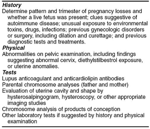
The scheme for a reasonable and cost-effective evaluation of women with RM shown in Table 4.3 is based on current guidelines published by the American College of Obstetricians and Gynecologists and the Royal College of Obstetricians and Gynaecologists. A sympathetic attitude by the physician is crucial—establishment of trust and rapport and a sincere appreciation of the distress and grief experienced by these couples permit tactful and thorough discussions with patient and partner. It is reasonable to institute an evaluation after two consecutive miscarriages in anxious women or if the patient has few reproductive years remaining or has had an infertility problem. Couples interested in an investigational protocol are perhaps best referred to legitimate research centers.
|
|
|
TABLE 4.3. Proposed evaluation for recurrent miscarriage |
SUMMARY POINTS
· Miscarriage is the most common complication of pregnancy, and the most frequent etiology is a chromosomal abnormality of the conceptus.
· Ultrasound is helpful in determining whether or not the embryo is viable, and appropriate modern management may be either observation or evacuation of the uterus.
· Recurrent early pregnancy loss is sometimes associated with underlying maternal abnormalities that can be detected with standard tests. Physicians should be aware of unproven tests and controversial treatments in order to best counsel their patients.
SUGGESTED READINGS
Aarts JM, Brons JTJ, Bruinse HW. Emergency cerclage: a review. Obstet Gynecol Surv 1995;50:459.
Aldrich CL, Stephenson MD, Karrison T, et al. HLA-G genotypes and pregnancy outcome in couples with unexplained recurrent miscarriage. Mol Hum Reprod2001;7:1167–1172.
Althuisius SM, Dekker GA, Hummel P, et al. Final results of the Cervical Incompetence Prevention Randomized Cerclage Trial (CIPRACT): therapeutic cerclage with bed rest versus bed rest alone. Am J Obstet Gynecol 2001;185:1106–1112.
American College of Obstetrician and Gynecologists. Management of recurrent early pregnancy loss. ACOG Practice Bulletin 2001;24.
Branch DW, Khamashta MA. Antiphospholipid syndrome: obstetric diagnosis, management, and controversies. Obstet Gynecol 2002 (in press).
Branch DW, Silver RM, Pierangelli SS, et al. Antiphospholipid antibodies other than lupus anticoagulant and anticardiolipin antibodies in women with recurrent pregnancy loss, fertile controls, and antiphospholipid syndrome. Obstet Gynecol 1997;89:549.
Brent RL, Beckman DA. The contribution of environmental teratogens to embryonic and fetal loss. Clin Obstet Gynecol 1994;37:646.
Chung TK, Lee DT, Cheung LP, et al. Spontaneous abortion: a randomized, controlled trial comparing surgical evacuation with conservative management using misoprostil. Fertil Steril 1999;71:1054–1059.
Clifford K, Rai R, Watson H, et al. Does suppressing luteinising hormone secretion reduce the miscarriage rate? Results of a randomised controlled trial. BMJ 1996;312:1508–1511.
Davis OK, Berkeley AS, Naus GJ, et al. The incidence of luteal phase defect in normal, fertile women determined by serial endometrial biopsies. Fertil Steril1989;51:582–586.
Daya S. Efficacy of progesterone support for pregnancy in women with recurrent miscarriage. A meta-analysis of controlled trials. Br J Obstet Gynaecol1989;96:275–280.
Erkan D, Merrill JT, Yazici Y, et al. High thrombosis rate after fetal loss in antiphospholipid syndrome: effective prophylaxis with aspirin. Arthritis Rheum2001;44:1466–1467.
Fantel AG, Shepard TH. Morphological analysis of spontaneous abortuses. In: Bennett MJ, Edmunds DK, eds. Spontaneous and recurrent abortion. Oxford: Blackwell Scientific Publications, 1987:8.
Ginsberg JS, Greer I, Hirsh J. Use of antithrombotic agents during pregnancy. Chest 2001;119[Suppl 1]:122S–131S.
Goldstein P, Berrier J, Rosen S, et al. A meta-analysis of randomized control trials of progestational agents in pregnancy. Br J Obstet Gynaecol1989;96:265–274.
Goldstein SR. Embryonic death in early pregnancy: a new look at the first trimester. Obstet Gynecol 1994;84:294.
Holers VM, Girardi G, Mo L, et al. Complement C3 activation is required for antiphospholipid antibody-induced fetal loss. J Exp Med 2002;195:211–220.
Hurd WW, Whitfield RR, Randolph JF, et al. Expectant management versus elective curettage for the treatment of spontaneous abortion. Fertil Steril1997;68:601.
Jeng GT, Scott JR, Burmeister LF. A comparison of meta-analytic results using literature vs. individual data. JAMA 1995;274:830.
Kelly S, Pollock M, Maas B, et al. Early transvaginal ultrasonography versus early cerclage in women with an unclear history of incompetent cervix. Am J Obstet Gynecol 2001;184:1097–1099.
Kutteh WH. Antiphospholipid antibody-associated recurrent pregnancy loss: treatment with heparin and low-dose aspirin is superior to low-dose aspirin alone. Am J Obstet Gynecol 1996;35:1584.
Lanasa MC, Hogge WA, Kubic C, et al. Highly skewed X-chromosome inactivation is associated with idiopathic recurrent spontaneous abortion. Am J Hum Genet 1999;65:252–254.
Lanasa MC, Hogge WA, Kubik CJ, et al. A novel X chromosome-linked genetic cause of recurrent spontaneous abortion. Am J Obstet Gynecol 2001;185:563–568.
Laskin CA, Bombardier C, Hannah ME, et al. Prednisone and aspirin in women with autoantibodies and unexplained recurrent fetal loss. N Engl J Med1997;337:148–153.
Levine JS, Branch DW, Rauch J. The antiphospholipid syndrome. N Engl J Med 2002;346:752–763.
MacNaughton MC, Chalmers IG, Dubowitz V, et al. Final report of the Medical Research Council/Royal College of Obstetricians and Gynaecologists multicentered randomized trial of cervical cerclage. Br J Obstet Gynaecol 1993;100:516.
Ober C, Karrison T, Odem RR, et al. Mononuclear-cell immunisation in the prevention of recurrent miscarriages: a randomised trial. Lancet 1999;354:365–369.
Patton PE. Anatomic uterine defects. Clin Obstet Gynecol 1994;37:705.
Preston FE, Rosendaal FR, Walker ID, et al. Increased fetal loss in women with heritable thrombophilia. Lancet 1996;348:913–916.
Rai R, Cohen H, Dave M, Regan L. Randomized controlled trial of aspirin and aspirin plus heparin in pregnancy in women with recurrent miscarriage associated with phospholipid antibodies. BMJ 1997;314:253.
Rai R, Backos M, Elgaddal S, et al. Factor V Leiden and recurrent miscarriage—prospective outcome of untreated pregnancies. Hum Reprod 2002;17:442–445.
Rai R, Backos M, Rushworth F, Regan L. Polycystic ovaries and recurrent miscarriage—a reappraisal. Hum Reprod 2000;15:612–615.
Scientific Advisory Committee of the Royal College of Obstetricians and Gynaecologists. The management of recurrent miscarriage. RCOG ‘Green-top’ Guideline No. 17, 2001.
Wilcox AJ, Weinberg CR, O'Connor JF, et al. Incidence of early pregnancy loss. N Engl J Med 1988;319:189.
Woelfer B, Salim R, Banerjee S, et al. Reproductive outcomes in women with congenital uterine anomalies detected by three-dimensional ultrasound screening. Obstet Gynecol 2001;98:1099–1103.
Wood SL, Brain PH. Medical management of missed abortion: a randomized clinical trial. Obstet Gynecol 2002;99:563–566.