Natalie S. Gould
Joan L. Walker
Robert S. Mannel
Vulvar and vaginal cancer represent uncommon gynecologic cancers that occur most often in older women. Squamous lesions are the most frequent histology and risk factors are similar for both disease sites. This chapter describes the epidemiology, clinical presentation, patterns of spread, and treatment of squamous cell carcinomas of the vulva and vagina. Less common histologic subtypes including Paget disease, melanoma, adenocarcinomas, and sarcomas are also reviewed.
VULVAR CARCINOMA IN SITU
Vulvar carcinoma theoretically results from malignant transformation of a vulvar carcinoma in situ as is seen with cervical squamous lesions. Unlike squamous lesions of the cervix, the natural history of vulvar intraepithelial neoplasia (VIN) is less well understood. The incidence of vulvar dysplasia has increased over the last 20 years, particularly among younger women. A report from Austria demonstrated a 307% increase in the overall incidence of high-grade VIN and a 394% increase among women under 50 years of age between 1985 and 1998. In a review of Surveillance, Epidemiology, and End Results (SEER) data, Sturgeon found that the incidence of VIN III nearly doubled between 1973 and 1976 and 1985 and 1987, from 1.1 to 2.1 per 100,000 woman years. Factors implicated in this increase include increased human papillomavirus (HPV) infection, increased surveillance, tobacco use, and immunosuppression, either with human immunodeficiency virus (HIV), organ transplant, or diabetes.
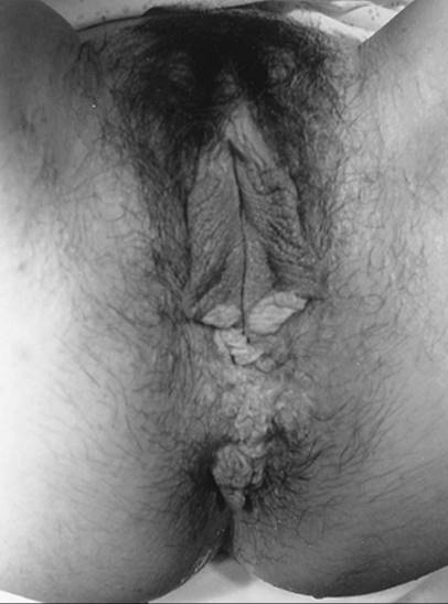
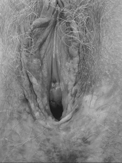
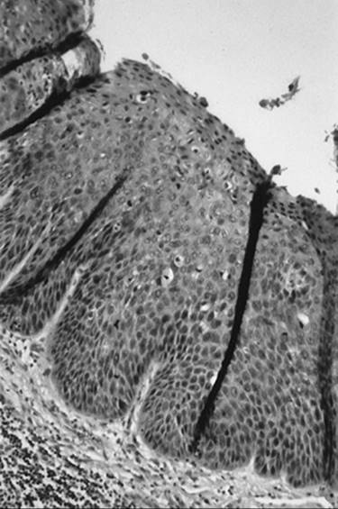
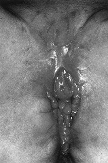
Patients with VIN most commonly present with pruritis and vulvar lesions. These lesions may appear scaly, white, red, or hyperpigmented (Fig. 51.1, Fig. 51.2, Fig. 51.3 and Fig. 51.4). Careful inspection with 5% acetic acid and liberal use of punch biopsy are the cornerstones of diagnosis. An underlying malignancy may be present in 7% to 22% of patients who undergo surgical excision for vulvar carcinoma in situ. Wide local excision with at least a 5-mm margin is the preferred management option as it allows pathologic confirmation and is associated with less morbidity than skinning vulvectomy. Skinning vulvectomy with split thickness skin graft may be an option in patients with widespread disease. Laser ablation is also an effective nonmutilating option in patients with multifocal or clitoral disease. Recurrences are frequent (10%–50%) despite negative surgical margins and therapy should be tailored to symptom control and ruling out underlying malignancy. Patients should be followed every few months with careful visual inspection of the vulva and taught self-exam skills as well.
|
|
|
FIG. 51.1. Vulvar carcinoma in situ presenting as white or hyperpigmented lesion. See color figure 51.1. |
|
|
|
FIG. 51.2. Vulvar carcinoma in situ before application of 5% acetic acid. See color figure 51.2. |
|
|
|
FIG. 51.3. Vulvar carcinoma in situ after application of 5% acetic acid. See color figure 51.3. |
|
|
|
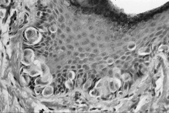
FIG. 51.4. Vulvar carcinoma in situ with full thickness involvement of dysplastic cells. |
Epidemiology
Vulvar carcinoma is the fourth most common genital tract malignancy in women, representing 3% to 5% of gynecologic malignancies and affecting an estimated 3,600 women in the United States in 2001. The majority of cancers are squamous in origin with occasional cases of basal cell carcinoma, melanoma, adenocarcinoma, and Paget disease.
Vulvar cancer tends to be a disease of older women with a mean age at diagnosis of approximately 65 years. As the population ages, more older women will be at risk for vulvar carcinoma. Evidence exists that there are two distinct types of vulvar carcinoma with different etiologies. Tumors in older women are often unifocal and may be associated with chronic vulvar inflammation of long-standing duration such as lichen sclerosus or hyperplastic dystrophy. These keratinizing squamous cell carcinomas have associated HPV changes in only 6% of cases. While retrospective studies indicate that up to 50% of vulvar carcinomas are related to hyperplastic dystrophies and lichen sclerosus, prospective studies have demonstrated only 5% of women with vulvar dystrophies develop invasive carcinoma. Cancers found in younger women tend to be multifocal with adjacent VIN and have a basaloid or warty histology. Nearly 90% are associated with HPV infection, particularly HPV 16. Thirty-eight percent of women with HPV-associated lesions in a series by Trimble and colleagues were under age 55, compared with only 17% of those with classic keratinizing squamous cancers. Other associated risk factors include immunosuppression from chronic steroid use, diabetes or HIV, smoking, and a history of other lower genital tract dysplasia or neoplasia.
Presentation
Most women present with pruritis and an identifiable lesion (Fig. 51.5, Fig. 51.6). Less common symptoms include pain and bleeding. Unfortunately, there is often a delay of many years between onset of symptoms and diagnosis. This is in part because patients self-medicate with a variety of over-the-counter preparations rather than seeking care and also because physicians may not biopsy liberally. The cornerstone for diagnosis of vulvar malignancy is a low threshold for a punch biopsy as it may be extremely difficult to distinguish between dysplasia, chronic vulvar dystrophy, and carcinoma. Any patient with symptoms lasting for longer than 2 weeks deserves a thorough exam and a biopsy. In order to adequately evaluate a patient with a vulvar lesion, 5% acetic acid is applied to the vulva for 5 minutes and then the area is examined either with the naked eye or a handheld magnifying glass. The entire vulva including the hair-bearing, perianal, and periclitoral regions should be examined for suspicious ulcerations and hyperpigmented, acetowhite, or gross warty lesions. Up to 5% of patients will have multifocal disease and may require multiple punch biopsies.
|
|
|
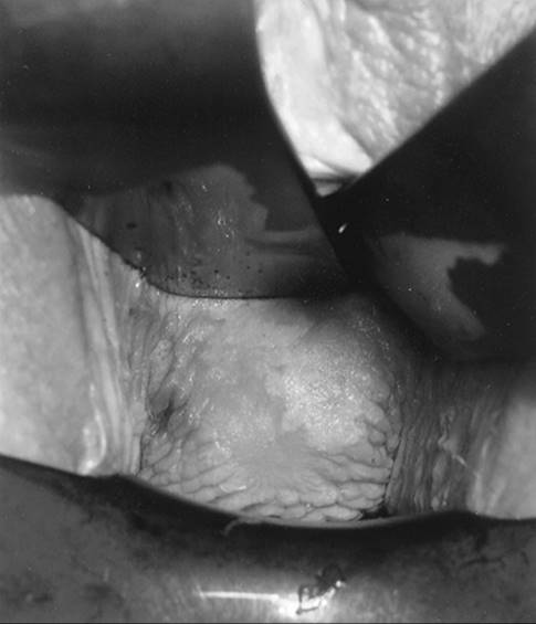
FIG. 51.5. Exophytic vulvar carcinoma involving posterior fourchette. |
|
|
|
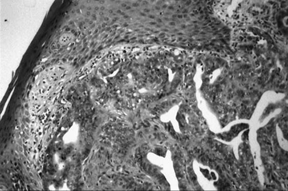
FIG. 51.6. Well-to-moderately differentiated invasive squamous cell carcinoma of the vulva. |
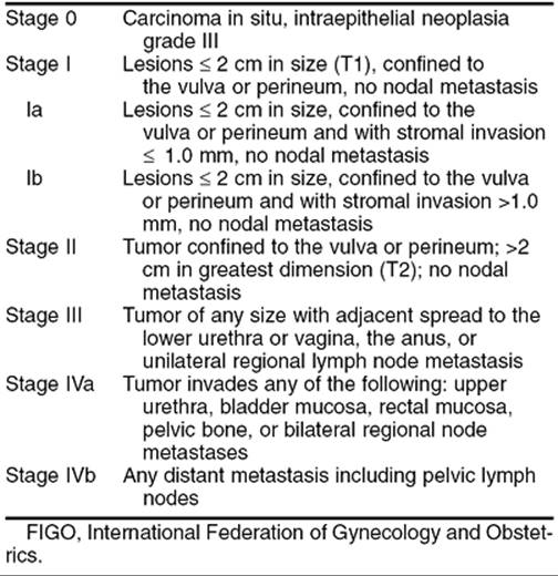
Staging/Spread Patterns
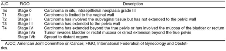
An International Federation of Gynecologists and Obstetricians (FIGO) surgical classification system replaced clinical staging for vulvar cancer in 1989 (Table 51.1). By relying on histopathologic classification of the lymph nodes, the new system more accurately reflects prognosis since the previous system provided an inaccurate assessment of groin node involvement in one-fourth of cases.
|
|
|
TABLE 51.1. Vulvar carcinoma: FIGO staging 1994 |
Local spread occurs to contiguous structures such as the vagina, urethra, and rectum. Metastatic spread via lymphatics is common and has been well characterized by anatomic studies. Ipsilateral lesions tend to be characterized by spread to the ipsilateral groin nodes first, followed by contralateral groin, and then pelvic nodes. Lesions that cross the midline may have lymphatic drainage to both groins. Metastasis to the contralateral groin or pelvic nodes is highly unusual in the absence of ipsilateral groin node involvement. The superficial inguinal nodes above the cribriform fascia are believed to be involved prior to the deep femoral nodes below the cribriform fascia. Risk of lymphatic spread is related to tumor size, tumor thickness, older patient age, tumor grade, presence of lymphovascular space invasion, and clinically suspicious groin nodes. Groin node metastasis was found in 19% of patients with lesions ≤2 cm and in 42% of patients with lesions greater than 2 cm. The Gynecologic Oncology Group (GOG) surgicopathologic staging analysis demonstrated that lymph node dissection could be safely omitted in patients with less than 1 mm of stromal invasion. Hematogenous metastasis to distant organs such as bone, liver, and lungs is rare as a primary event but may be seen in patients with recurrent disease.
Treatment
Historically vulvar cancer has been treated with en-bloc radical vulvectomy and bilateral inguinofemoral lymphadenectomy or “longhorn incision” (Fig. 51.7, Fig. 51.8 and Fig. 51.9). This technique resulted in a median hospitalization of 30 days, 70% to 90% wound breakdown, and chronic debilitating lymphedema in nearly 9% of women. Significant disruption in self-image and sexual function also occurred. Attempts to decrease morbidity first led to the use of separate incisions for groin node dissection while continuing to use radical vulvectomy. In 1981, Hacker and colleagues reported a series of 100 patients who underwent radical vulvectomy and lymphadenectomy through three separate incisions. The incidence of major wound breakdown was only 14% with a mean hospitalization shortened to 19 days. This procedure lead to the shortest length of hospitalization reported at that time and produced a dramatic decrease in complications. The majority of local recurrences were salvaged with repeat excision. Overall survival was not compromised by deviating from en-bloc dissection and the authors concluded that this technique is appropriate for patients with stage I and II vulvar carcinoma.
|
|
|
FIG. 51.7. A “longhorn” incision. Radical excision of the vulva from genitocrural fold to genitocrural fold, deep to the inferior fascia of the urogenital diaphragm along with en-bloc lymphadenectomy. |
|
|
|
FIG. 51.8. Surgical specimen after en-bloc radical vulvectomy and inguinofemoral lymphadenectomy via longhorn incision. |
|
|
|
FIG. 51.9. Healed vulva after en-bloc resection via longhorn incision. |
DiSaia and associates first proposed conservative vulvar surgery in 1979 as a way of preserving sexual function in young patients. Requirements for conservative surgery were an invasive cancer 1 cm or less in diameter confined to the vulva with less than a 5-mm depth of invasion. Patients were treated with radical wide excision ensuring a 3-cm margin and lymphadenectomy was performed through separate groin incisions. There were no recurrences at a mean follow-up of 32 months and sexual function was deemed preserved. The authors concluded that there existed a subset of patients with vulvar cancer who could be treated with less radical vulvar procedures.
Berman and co-workers expanded the experience with conservative surgery in 1989 with a report of 50 patients with T1 lesions treated with radical local excision, again providing a 3-cm margin and superficial lymphadenectomy. No patient had a resection margin positive for carcinoma. Median hospital stay was shortened to 7 days and only 12% required wound debridement compared to 50% or more with historical controls. There were five recurrences (10%), four of them local. All local recurrences were salvaged with a second local resection.
In 1990, Burke and colleagues provided the first report of a conservative vulvar surgery in patients with both T1 and T2 lesions. Thirty-two patients (15 with T2 lesions) underwent radical wide excision removing a 1- to 2-cm margin of normal tissue in addition to selective inguinal dissection. The mean lesion diameter was 23 mm with a mean depth of invasion of 4.1 mm. No patients had invasive cancer at the surgical margins but 19% were positive for VIN. The authors reported only 15.5% wound separation with a mean hospital stay of 10 days. Three patients (10%) developed local recurrence and two were salvaged with repeat excision. The authors concluded that radical wide excision appears to be an acceptable surgical option for patients with resectable vulvar carcinomas.
As these retrospective trials used differing criteria to identify patients suitable for less radical approaches, the GOG conducted a prospective trial beginning in 1983 which evaluated modified radical hemivulvectomy and ipsilateral superficial inguinal lymphadenectomy in 155 patients with clinical stage I vulva carcinoma limited to a depth of invasion of less than 5 mm. A 2-cm margin of normal skin around the lesion was excised. There were 19 recurrences and 7 deaths among 122 patients available for evaluation. Recurrences were evenly distributed between local and distant failures. Acute and long-term morbidity was decreased significantly compared to historical controls, but at the cost of increased risk of local recurrence. The authors concluded that although there was no consensus regarding the characteristics that would make a patient with early vulvar carcinoma a candidate for a limited surgical approach, this approach appeared to be an alternative to traditional radical surgery for a highly selected group of patients with stage I vulvar carcinoma (Fig. 51.10, Fig. 51.11).
|
|
|
FIG. 51.10. Modified radical vulvectomy. Radical excision of the vulva deep to the inferior fascia of the urogenital diaphragm with a 2-cm margin around the lesion. |
|
|
|
FIG. 51.11. Surgical specimen after modified radical vulvectomy. Lymphadenectomy is performed through separate incisions. |
A need for establishing a clear margin for treatment of patients with vulvar cancer may be more important than the particular treatments. Heaps and colleagues reviewed surgicopathologic factors predictive of local recurrence in 135 patients treated with radical vulvectomy or radical local excision. Twenty-one developed local recurrence after radical resection. No patient with a surgical tumor-free margin ≥ 8 mm suffered from local recurrence but 48% with less than an 8-mm margin recurred locally. As an 8-mm margin on fixed tissue corresponds to 10-mm margin with fresh tissue, the authors concluded that use of a 1-cm margin should successfully prevent local recurrence and reduce the currently used standard margin by over 50%. This would allow further modification of the procedure in order to decrease morbidity and avoid disfigurement and loss of organ function without sacrificing survival.
Other modifications in the surgical management of vulvar carcinoma over the last 30 years include the elimination of routine pelvic lymphadenectomy and elimination of contralateral groin node dissection in patients with lateral T1 lesions and negative ipsilateral nodes.
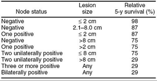
Overall survival in vulvar carcinoma is related to groin node status and lesion diameter with nodal status being the most important factor. Five-year survival is 98% for patients with negative nodes and a T1 lesion. Patients with one to two positive ipsilateral nodes have over 70% to 80% 5-year survival while those with three or more positive nodes or bilaterally positive nodes have survival in the range of 12% to 36% (Table 51.2). Postoperative adjuvant groin radiotherapy is indicated in patients with more than one positive node or clinically evident groin nodes.
|
|
|
TABLE 51.2. Survival by groin node status and lesion size in vulvar carcinoma |
Radiotherapy for vulvar carcinomas is indicated in patients with unresectable vulvar tumors or unresectable groin nodes. Boronow popularized treatment of locally advanced tumors with radiotherapy followed by resection of the primary tumor in order to preserve urethral, bladder, or rectal function and to avoid the morbidity of pelvic exenteration. In recent years, concurrent radiotherapy and chemotherapy has been shown to improve both local control and overall survival. The GOG has demonstrated high rates of resectability and local control of the lymph nodes in patients with clinically suspicious, fixed, or ulcerated nodes treated preoperatively with radiotherapy, concurrent cisplatin/5-fluorouracil (5-FU) chemotherapy, and followed by tailored surgery.
New approaches to decrease morbidity of groin node dissection include the use of lymphatic mapping and sentinel lymph node biopsy. Intraoperative mapping with vital dye with or without lymphoscintigraphy may identify 56% to 100% of sentinel nodes. While this technique is widely used for breast cancer and melanoma, it is being prospectively evaluated by the GOG and other investigators. If the sentinel node can predict lymph node involvement with sufficiently high negative predictive value, then more extensive lymphadenectomy may be avoided. This may decrease one of the major morbidities of vulvar cancer surgery.
Recurrence after definitive therapy for vulvar carcinoma may occur locally in the vulva or in the groin. The risk of local recurrence depends on prior surgical technique with higher rates of recurrence seen with less aggressive vulvar approaches. Vulvar recurrences may be characterized as new primaries due to field effect or recurrence at the prior site of excision. They are usually salvageable with radical excision though patients who have had prior radiotherapy to the groin will require some type of reconstructive flap to ensure adequate vascular supply and healing. Groin recurrences are much more difficult to treat. If the patient has not been irradiated, surgical excision of the nodes with postoperative radiotherapy is the treatment of choice. If radiotherapy has already been administered, no satisfactory therapy exists and survival is dismal.
Other Histologic Subtypes
Rare histologic types of vulvar carcinoma exist, representing about 10% of vulvar malignancies. These include melanoma, invasive Paget disease, basal cell carcinoma, verrucous carcinoma, Merkel cell carcinoma, adenocarcinoma, adenosquamous carcinoma, transitional cell carcinoma, sarcomas, and metastatic disease from other sites.
Melanoma
Melanoma is the second most common vulvar malignancy, accounting for about 6% of vulvar lesions and 1.3% of all melanomas among women. It is highly aggressive and characterized by both local recurrence and distant metastasis through hematogenous spread. The median age at presentation is 66 years, several decades older than for cutaneous melanomas. It is more common in white women with a relative risk of 2.6 compared to other races. Overall 5-year survival ranges from 35% to 50%.
Patients frequently present with pruritis and visible lesions but many may be asymptomatic. The differential diagnosis of a pigmented lesion includes melanoma, lentigo, vulvar melanosis, squamous dysplasia, hemangioma, Paget disease, various nevi, and acanthosis nigricans. Biopsy should be considered of any pigmented lesion, particularly if the borders are indistinct or spreading or the lesion is raised. Excisional biopsy is preferred to better delineate depth of invasion. A punch biopsy of the most nodular area is the next best option. Three histologic subtypes of vulvar melanoma have been described. The most common is superficial spreading melanoma followed by nodular and acral lentiginous melanoma. Immunohistochemical testing may be necessary to distinguish between melanoma and Paget disease. Paget disease is typically positive for carcinoembryonic antigen (CEA) and melanoma is positive for S-100.
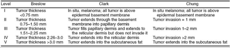
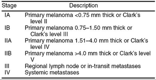
Unlike squamous lesions, vulvar melanoma is staged using classification systems for cutaneous melanomas. The Clark system is based on level of invasion, while the Breslow system is based on vertical thickness of the lesion from the surface of intact epithelium to the deepest point of invasion. Other staging systems include Chung's modification of the Clark's system and the American Joint Committee on Cancer (AJCC) staging system (Table 51.3). While the Breslow and Chung microstaging systems are more accurate than the Clark system, prospective evaluation by the GOG found that the AJCC staging system is most predictive of survival (Table 51.4).
|
|
|
TABLE 51.3. Melanoma staging: Breslow, Clark, and Chung microstaging levels |
|
|
|
TABLE 51.4. American Joint Committee on Cancer staging for vulvar melanoma |
As with squamous lesions, more conservative vulvar surgery is now being advocated for melanoma. Excision should include a 1- to 2-cm lateral margin and at least a one centimeter deep margin. The presence of capillary lymphatic space involvement and central tumor location has been correlated with positive groin node status. The role of lymphadenectomy in vulvar melanoma remains unresolved. Prospective evaluations have failed to show a survival benefit with lymphadenectomy.
Adjuvant therapy for advanced disease includes the use of chemotherapy, immune modulators, and tumor vaccines. Given the small number of patients, no trials specific to vulvar melanoma have evaluated the role of adjuvant therapy.
Paget Disease
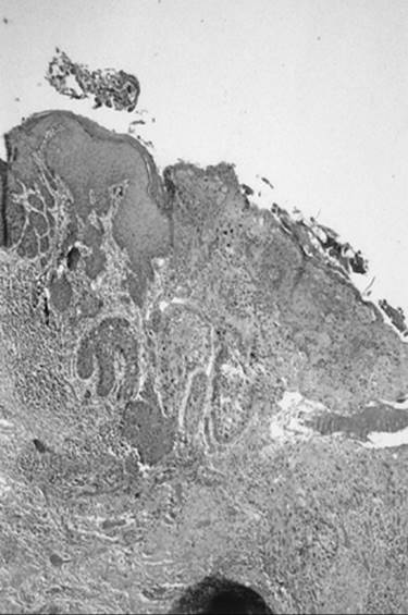
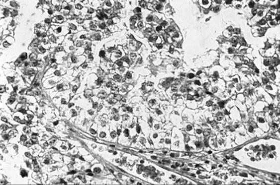
Paget disease accounts for 1% to 2% of vulvar malignancies. It occurs most frequently in postmenopausal white women and is characterized by red eczematous lesions with a superficial white coating and intense pruritis. As with other vulvar malignancies, there may be a long delay between initial symptoms and diagnosis. Histologic disease may spread well beyond the visible lesion. Biopsy reveals characteristic large eosinophilic cells in the basal layer of the epithelium (Fig. 51.12). Paget disease of the vulva may exist as four clinical entities, the first of which is noninvasive or intraepithelial Paget disease. This represents 60% of cases and is cured with local excision. In invasive Paget disease, the Paget cells penetrate the basement membrane and invade the dermis. Intraepithelial Paget disease may also be associated with an underlying adenocarcinoma of sweat gland origin or a coexistent cancer. Underlying malignancy is seen in approximately 20% to 30% of patients, a rate lower than that seen with mammary Paget disease. Parker and co-workers reported poor survival in patients with invasive Paget disease and in patients with underlying malignancy. Patients with clitoral disease also had a poorer prognosis. Therapy for Paget disease involves wide excision. A skinning vulvectomy with split thickness skin graft is often needed to remove large areas of involved skin. Recurrences tend to be local and range from 7% to 58% with an average of about 30%. Some authors recommend sending margins for frozen section to ensure complete resection while others have not demonstrated a benefit to this approach with respect to recurrence. Radical surgery is reserved for patients with underlying malignancy.
|
|
|
FIG. 51.12. Paget disease of the vulva. Large eosinophilic cells with large nuclei and prominent cytoplasm in basal cells represent Paget cells. |
Basal Cell Carcinoma
Basal cell carcinoma accounts for 2% to 4% of vulvar malignancies. It is typically seen in older women and tends to be well circumscribed, firm, and typically less than 2 cm in diameter. Metastasis is extremely rare and wide local excision is the preferred therapy.
Verrucous Carcinoma
Verrucous carcinoma is a variant of squamous cell carcinoma with distinctive pathologic and clinical characteristics. It is characterized by a large condylomatous or cauliflower-like lesion with minimal cellular atypia, a pushing border, and rare metastasis. Unless biopsies are deep enough, it may be confused with benign condyloma or papilloma. Wide local excision is the preferred therapy. Radiotherapy is contraindicated given the potential for anaplastic transformation.
Merkel Cell Carcinoma
Merkel cell tumors are neuroendocrine tumors of the skin and resemble small cell carcinomas. They are associated with lymphatic and distant metastases and have a very poor prognosis.
Adenocarcinoma
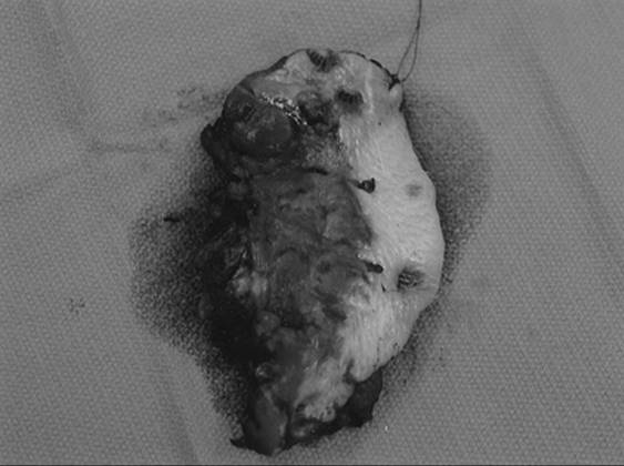
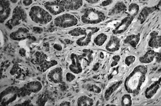
Adenocarcinoma of the vulva tends to arise in the Bartholin gland, although it may also arise from the sweat glands or Skene glands of the vulva (Fig. 51.13). Adenoid cystic carcinoma also arises from the Bartholin gland and is characterized by slow growth and a tendency for local and perineural invasion (Fig. 51.14). Distant metastasis is most commonly seen in the lung. Therapy involves radical excision, ipsilateral lymphadenectomy, and postoperative radiotherapy.
|
|
|
FIG. 51.13. Moderately differentiated adenocarcinoma arising from vulva. |
|
|
|
FIG. 51.14. Adenoid cystic carcinoma arising from Bartholin gland. |
Adenosquamous Carcinoma
Adenosquamous carcinoma is a rare cause of Bartholin gland carcinoma and is composed of both malignant squamous and glandular components.
Transitional Cell Carcinoma
Transitional cell carcinoma may arise within the Bartholin gland but is more commonly seen as metastatic spread from a urethral or bladder carcinoma.
Sarcoma
Sarcomas of the vulva are extremely rare, representing only 1% to 3% of vulvar malignancies. While leiomyosarcoma is the most common histologic subtype, malignant fibrous histiocytoma, and various other sarcomas have been reported. Radical surgery is the primary therapy, although the behavior of these neoplasms is not well understood.
Metastatic Disease
Metastatic disease to the vulva usually occurs from other lower genital tract malignancies, most commonly cervix. Rare causes include malignancies arising from the vagina, endometrium, ovary, breast, kidney, stomach, or lung.
VAGINAL CARCINOMA
Primary vaginal cancer is the fifth most common gynecologic malignancy, with an expected 2,000 new cases in the United States in 2001. Only fallopian tube carcinomas are a less common gynecologic malignancy than vaginal carcinoma. The vast majority of vaginal neoplasms are metastatic from other sites, particularly direct extension from the cervix or endometrium. Lymphatic or hematogenous spread to the vagina is also possible. Primary vaginal neoplasms are typically squamous in origin and occur in postmenopausal women. Risk factors for vaginal dysplasia or carcinoma include a history of prior lower genital tract dysplasia or carcinoma, tobacco use, low socioeconomic status, history of genital warts or HPV infection, prior abnormal Pap smear, prior radiotherapy of the genital tract, immunosuppression, vaginal discharge or irritation, and early hysterectomy. History of diethylstilbestrol (DES) exposure is associated with development of clear cell adenocarcinoma of the vagina.
Vaginal Intraepithelial Neoplasia
The vagina is the least common site for lower genital tract dysplasia, occurring 100 times less frequently than cervical dysplasia. A review by Dodge and associates of 121 patients with biopsy-proven vaginal intraepithelial neoplasia (VAIN) showed that 65% of women with a uterus in place had associated cervical intraepithelial neoplasia and 10% had associated VIN. The upper third of the vagina is the most common site of involvement and over 60% of these women will have multifocal disease. The mean age for patients with VAIN III is approximately 40 years.
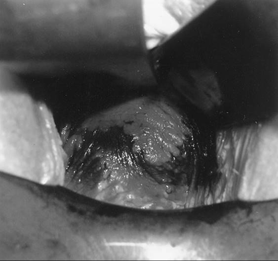
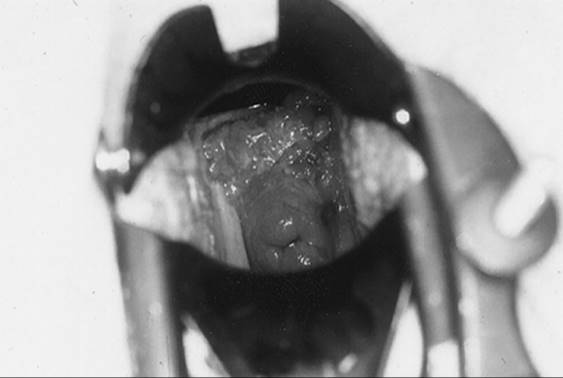
Diagnosis is through careful colposcopic inspection after application of 5% acetic acid and directed biopsy (Fig. 51.15, Fig. 51.16). Due to the rugated nature of the vagina, colposcopic evaluation may be a challenge. It is important to remember to colposcopically evaluate the vagina in patients with cytologic abnormalities, particularly those with normal appearing cervices. Acetowhite epithelium is the most common colposcopic feature. Application of half-strength Lugol solution allows for identification of nonstaining areas, which may represent dysplasia. Most patients with vaginal dysplasia are completely asymptomatic, fewer than 5% will present with bleeding or abnormal vaginal discharge. Postmenopausal patients with atrophy may benefit from a course of vaginal estrogen prior to undergoing colposcopy and directed biopsies.
|
|
|
FIG. 51.15. Vaginal intraepithelial neoplasia III after application of acetic acid. Note acetowhite lesions. See color figure 51.15. |
|
|
|
FIG. 51.16. Vaginal intraepithelial neoplasia III after application of Lugol solution. Note nonstaining lesions. |
Various treatment options have been employed for therapy of VAIN. These include laser ablation, partial or total vaginectomy, topical 5% 5-FU cream, cavitron ultrasonic aspiration, and loop electrosurgical excision procedure (LEEP). Choice of therapy for VAIN depends on the number and severity of lesions and their location, whether the patient is sexually active, and prior radiotherapy, as well as physician and patient preference. Hoffman and associates found invasive carcinoma in 28% of patients undergoing upper vaginectomy for VAIN III. Overall recurrence rates after therapy range from 10% to 42%. While partial vaginectomy is well suited to unifocal lesions and those involving the upper aspect of the vagina, it may lead to vaginal shortening and sexual dysfunction. Total vaginectomy with split thickness skin graft is reserved for patients who have failed more conservative approaches. Laser ablation to a depth of 2 to 3 mm is associated with minimal blood loss and is well suited for multifocal lesions. The challenge with laser therapy is ensuring that no lesions have been missed among the vaginal rugae. While 5-FU allows patients to treat themselves medically, it may be associated with significant skin desquamation, chronic ulceration, and be poorly tolerated. Dodge and colleagues found that recurrence is highest after laser ablation (38%) and 5-FU (59%) as well among those patients with multifocal disease (45%). Recurrence is lowest after partial vaginectomy (0%). Overall 2% to 12% of patients progress to vaginal cancer after therapy for vaginal dysplasia so careful cytologic and colposcopic surveillance is critical.
Squamous Carcinoma
Squamous carcinoma accounts for 80% to 90% of vaginal carcinomas with a mean age at presentation of 60 to 65 years. As with vaginal dysplasia, the most common location will be the posterior upper third of the vagina but the entire vaginal cylinder is at risk for malignant transformation. Abnormal vaginal bleeding occurs in over half of patients. Less common symptoms include abnormal vaginal discharge, pain, and dysuria (Fig. 51.17).
|
|
|
FIG. 51.17. Vaginal carcinoma located in upper third of the vagina. |
Vaginal carcinoma may spread by local extension, lymphatic spread to pelvic or inguinal nodes, and hematogenously. Lesions involving the upper two-thirds of the vagina tend to spread to pelvic nodes while lesions involving the lower third of the vagina tend to spread to inguinal nodes as would be seen with vulvar malignancies. To be considered primary vaginal carcinoma, a patient must have no evidence of disease in either the vulva or cervix. Staging of vaginal cancers is clinical and based on either the FIGO or AJCC systems (Table 51.5).
|
|
|
TABLE 51.5. Vaginal carcinoma: FIGO and AJCC clinical staging |
Therapy for vaginal carcinoma typically involves both external irradiation and brachytherapy. Surgical resection is typically reserved for small stage I tumors confined to the posterior aspect of the vaginal apex. Complications from therapy include fistula formation and stenosis. Overall prognosis is poorer than for cancer of the cervix. Recurrent disease tends to occur locally within 2 years of primary therapy. Extenterative procedures are required to adequately treat locally recurrent or persistent disease but are associated with considerable morbidity. Chemotherapy does not yet have a significant role in treatment of vaginal carcinoma.
Clear Cell Adenocarcinoma
Clear cell adenocarcinoma of the vagina is a rare malignancy most commonly seen in young women whose mothers received DES during pregnancy for prevention of miscarriage (Fig. 51.18). This association was first noted in 1970, leading to removal of the drug from the U.S. market in 1971. Approximately 60% of patients with clear cell carcinoma are known to have been exposed to DES or other synthetic estrogens. The risk of developing clear cell adenocarcinoma of the cervix or vagina has been estimated at 1 per 1,000 to 1 per 1,500, with DES exposure in the first trimester conveying the greatest risk. The median age at diagnosis for DES-exposed patients is 19 years. DES-exposed daughters do not appear to have an increased risk of developing any other malignancies but do have an increased risk of cervical dysplasia. This may be due to increased surveillance or congenital abnormalities in the lower genital tract. No increased risk in squamous cancers of the cervix or vagina has been reported in DES-exposed women.
|
|
|
FIG. 51.18. Clear cell adenocarcinoma of the vagina associated with diethylstilbestrol exposure. |
Most patients present with early stage disease, located in the exocervix or upper third of the vagina. Therapy is typically surgical and involves radical hysterectomy with removal of the upper vagina and pelvic lymphadenectomy. While this results in sterility, it may allow ovarian preservation and improved vaginal function over treatment with radiotherapy. Overall 5-year survival for stage I disease is more than 90% and approximately 80% for patients with stage II disease. Favorable factors in survival include early stage disease, older age, a tubulocystic histology, superficial invasion, small tumor diameter, and negative nodal status. Adenocarcinoma unrelated to DES exposure is a rare entity seen in postmenopausal women.
Melanoma
Melanoma is the second most common cancer of the vagina, accounting for less than 5% of vaginal malignancies and 0.3% of all melanomas among women. Nodular lesions are the most frequent histologic subtype. While vulvar melanoma is more common among white women, no association with race is seen for vaginal melanoma. It is a disease of older women and overall 5-year survival is poor at only 19%.
Sarcoma Botryoides
Sarcoma botryoides, or embryonal rhabdomyosarcoma, is a rare sarcoma found in young girls. It presents with vaginal bleeding and a polypoid “grapelike” mass. Historically this was treated with radical surgery, but now multimodality chemotherapy with VAC (vincristine, dactinomycin, and cyclophosphamide) and limited surgery can allow for a cure while preserving reproductive function. Radiotherapy may also be used in conjunction with multi-agent chemotherapy.
Endodermal Sinus Tumor
This is an extremely rare germ cell tumor arising in the vagina. It is seen in infants and is treated with combination chemotherapy as is used for germ cell tumors of the ovary.
Lymphoma
Vaginal involvement with lymphoma may be secondary to systemic disease or may be localized to the vagina. This rare entity presents with bleeding and a vaginal mass. Primary vaginal lymphomas are typically diffuse, large, B-cell type.
Metastatic Disease
Although primary vaginal malignancy is quite rare, metastatic spread to the vagina is common and occurs through direct extension of vulvar, cervical, endometrial, and rectal malignancies. A thorough search for another primary site should be undertaken before concluding that a patient has a primary vaginal malignancy.
SUMMARY POINTS
· The incidence of vulvar intraepithelial neoplasia (VIN) is increasing with younger patients more likely to have human papillomavirus (HPV)-related lesions. The underlying risk for cancer in a field of VINIII is 7% to 22%.
· Vulvar carcinoma in older women tends to be unifocal and is associated with chronic vulvar irritation rather than HPV changes. Carcinomas in younger women tend to be multifocal and HPV changes are seen in 90% of cases (HPV 16 is most common).
· The risk of groin node metastasis with vulvar carcinoma increases with increasing tumor size, depth of invasion, and presence of lymphovascular space involvement.
· Modifications in surgery to reduce the radical nature of vulvar and groin surgery are possible in select patients. Vulvar resection margins should be at least 1 cm. The use of sentinel node biopsy to modify groin node dissections remains investigational.
· Squamous cell carcinoma of the vagina is a rare entity best treated with combined external beam radiation therapy and brachytherapy.
SUGGESTED READINGS
Vulvar Intraepithelial Neoplasia
Joura EA, Lösch A, Haider-Angeler, MG, et al. Trends in vulvar neoplasia: increasing incidence of vulvar intraepithelial neoplasia and squamous cell carcinoma of the vulva in young women. J Reprod Med 2000;45:613–615.
Küppers V, Stiller M, Somville T, et al. Risk factors for recurrent VIN, role of multifocality and grade of disease. J Reprod Med 1997;42:140–144.
Sturgeon SR, Brinton LA, Devesa SS, et al. In situ and invasive vulvar cancer trends (1973 to 1987). Am J Obstet Gynecol 1992;166:1482–1485.
Vulvar Carcinoma: Etiology, Presentation, and Staging
Abramov Y, Elchalal U, Abramov D, et al. Surgical treatment of lichen sclerosus: a review. Obstet Gynecol Surv 1996;51:193–199.
Greenlee RT, Hill-Harmon MB, Murray T, et al. Cancer statistics, 2001. CA Cancer J Clin 2001;51:15–36.
Homesley H, Bundy B, Sedlis A, et al. Prognostic factors for groin node metastasis in squamous cell carcinoma of the vulva (a gynecologic oncology group study). Gynecol Oncol 1993;49:279–283.
Trimble CL, Hildesheim A, Brinton L, et al. Heterogeneous etiology of squamous carcinoma of the vulva. Obstet Gynecol 1996;87:59–64.
Treatment
American College of Obstetricians and Gynecologists. Vulvar cancer. ACOG Technical Bulletin No. 186, Nov 1993.
Berman M, Super J, Creasman W, et al. Conservative surgical management of superficially invasive stage I vulvar carcinoma. Gynecol Oncol 1989;35:352–357.
Boronow RC. Combined therapy as an alternative to exenteration for locally advanced vulvo-vaginal cancer: rationale and results. Cancer 1982;49:1085–1091.
Burke T, Stringer C, Gershenson D, et al. Radical wide excision and selective inguinal node dissection for squamous cell carcinoma of the vulva. Gynecol Oncol 1990;38:328–332.
Cavanagh D, Fiorica J, Hoffman M, et al. Invasive carcinoma of the vulva, changing trends in surgical management. Am J Obstet Gynecol 1990;163:1007–1015.
DiSaia P, Creasman W, Rich W. An alternate approach to early cancer of the vulva. Am J Obstet Gynecol 1979;133:825–830.
Hacker N, Leuchter R, Berek J, et al. Radical vulvectomy and bilateral inguinal lymphadenectomy through separate groin incisions. Obstet Gynecol1981;58:574–579.
Heaps J, Fu Y, Montz F, et al. Surgical-pathologic variables predictive of local recurrence in squamous cell carcinoma of the vulva. Gynecol Oncol1990;38:309–314.
Homesley HD, Bundy BN, Sedlis A, et al. Assessment of current International Federation of Gynecology and Obstetrics staging of vulvar carcinoma relative to prognostic factors for survival (A Gynecologic Oncology Group study). Am J Obstet Gynecol 1991;164:997–1003.
Levenback C, Coleman RL, Burke TW, et al. Intraoperative lymphatic mapping and sentinel node identification with blue dye in patients with vulvar cancer. Gynecol Oncol 2001;83:276–281.
Montana GS, Thomas GM, Moore DH, et al. Preoperative chemo-radiation for carcinoma of the vulva with N2/N3 nodes: a Gynecologic Oncology Group study. Int J Radiat Oncol Biol Phys 2000;48:1007–1013.
Stehman F, Bundy B, Dvoretsky, et al. Early stage I carcinoma of the vulva treated with ipsilateral superficial inguinal lymphadenectomy and modified radical hemivulvectomy: a prospective study of the gynecologic oncology group. Obstet Gynecol 1992;79:490–497.
VULVA MELANOMA
Phillips GL, Bundy BN, Okagaki T, et al. Malignant melanoma of the vulva treated by radical hemivulvectomy: a prospective study of the Gynecologic Oncology Group. Cancer 1994;73:2626–2632.
Weinstock MA. Malignant melanoma of the vulva and vagina in the United States: patterns of incidence and population-based estimated of survival. Am J Obstet Gynecol 1994;171:1225–1230.
Paget Disease
Parker LP, Parker JR, Bodurka-Bevers D, et al. Paget's disease of the vulva: pathology, pattern of involvement, and prognosis. Gynecol Oncol 2000;77:183–189.
Vaginal Intraepithelial Neoplasia
Dodge JA, Eltabbakh GH, Mount SL, et al. Clinical features and risk of recurrence among patients with vaginal intraepithelial neoplasia. Gynecol Oncol2001;83:363–369.
Vaginal Cancer
Hatch EE, Palmer JR, Titus-Ernstoff L, et al. Cancer risk in women exposed to diethylstilbestrol in utero. JAMA 1998;280:630–634.
Hoffman MS, DeCesare SL, Roberts WS, et al. Upper vaginectomy for in situ and occult, superficially invasive carcinoma of the vagina. Am J Obstet Gynecol1992;166:30–33.